. 2025 May 19;16(1):4657.
doi: 10.1038/s41467-025-59871-3.

Wnt/β-catenin activation by mutually exclusive FBXW11 and CTNNB1 hotspot mutations drives salivary basal cell adenoma

Affiliations

Wnt/β-catenin activation by mutually exclusive FBXW11 and CTNNB1 hotspot mutations drives salivary basal cell adenoma

Kim Wong et al. Nat Commun. .

Abstract

Basal cell adenoma (BCA) and basal cell adenocarcinoma (BCAC) of the salivary gland are rare tumours that can be difficult to distinguish from each other and other salivary gland tumour subtypes. Using next-generation sequencing, we identify a recurrent FBXW11 missense mutation (p.F517S) in BCA that is mutually exclusive with the previously reported CTNNB1 p.I35T gain-of-function (GoF) mutation with these mutations collectively accounting for 94% of BCAs. In vitro, mutant FBXW11 is characterised by defective binding to β-catenin and higher protein levels within the nucleus. This is consistent with the increased nuclear expression of β-catenin and activation of the Wnt/β-catenin pathway. The genomic profiles of BCAC are distinct from BCA, with hotspot DICER1 and HRAS mutations and putative driver mutations affecting PI3K/AKT and NF-κB signalling pathway genes. These findings have important implications for the diagnosis and treatment of BCA and BCAC, which, despite histopathologic overlap, may be unrelated entities.


Conflict of interest statement

Competing interests: The authors have no conflicts to declare.

Figures

Fig. 1
Fig. 1. Overview of the salivary gland basal cell adenoma (BCA) and basal cell adenocarcinoma (BCAC) cohorts.
BCA and BCAC cases are shown to the left and right of the dashed line, respectively. Genes shown in the oncoplot are genes known to be mutated in salivary gland tumours and/or are COSMIC Cancer Gene Census (CGC) genes and mutated in at least 1 BCA or BCAC. Also shown is FBXW11, which is not a CGC gene, but shown in this study to be significantly mutated in BCA. Genes that were mutated exclusively in the dMMR case, PD56546c, are not shown. Genes in the Wnt/β-catenin, NF-κB, PI3K/AKT or HRAS pathways are grouped by colour. PD56541a is a carcinoma of salivary gland type in the lung, with unknown origin. TMB is the tumour mutation burden in mutations per megabase (Mb). Genes are grouped by pathway. TMB was calculated using mutations found in coding exons and 2 bp upstream and downstream of exon boundaries to account for splice site mutations. Fusion genes in red text are those found in the Trinity Cancer Transcriptome Analysis Toolkit human fusion library (see “Methods”). Samples marked as “Discovery set only” did not pass strict criteria for transcriptome sequencing quality control (see “Methods”) and may have a higher false discovery rate as a result. The copy number panel indicates the tumours that had copy number loss of chromosome arms 5q and/or 16q, which was significant in the BCAC cohort. IHC indicates samples that were positive or negative for nuclear β-catenin immunohistochemical staining. BCAC/EMC is differential diagnosis of salivary gland basal cell adenocarcinoma and epithelial-myoepithelial carcinoma. Source data are provided as a Source Data file.
Fig. 2
Fig. 2. Tumour mutation burden, mutation spectra and recurrent copy number alterations.
a Tumour mutation burden in salivary gland basal cell adenoma (BCA; n = 32) and basal cell adenocarcinoma (BCAC; n = 11). Boxplot annotations: centre line, median; box limits, upper (75th) and lower (25th) quartiles; whiskers, 1.5x interquartile range; points, tumour mutation burden of samples. b Penetrance plot of somatic copy number gains and losses in BCA (top; n = 32) and BCAC (lower; n = 11). c The single base substitution spectrum (top) and indel spectrum (lower) from PD56546c. Show are the number of mutations occurring in each of the 96 possible trinucleotide contexts for single base substitutions and specific repeat contexts for indels.
Fig. 3
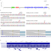
Fig. 3. Validation of mutations in FBXW11.
a The location and frequency of the p.F517S and p.I526M mutations in FBXW11. The amino acid position is shown on the x-axis, and coloured boxes represent protein domains. Beta-TrCP_D is the D domain of beta-TrCP; also shown are the location of an F-box domain and WD40 repeats. b Examples of traces generated from Sanger sequencing of clones derived from PCR amplification of FBXW11 exon 13 in tumour (top) and matched normal (lower) DNA from PD52393a (left) and PD52386a (right). Tumour PD52393a had a p.F517S (NM_001378974.1:c.1550 T > C) and a p.I526M (c.1578 C > G) mutation. c Examples of mutant alleles observed in tumour RNA-seq reads from the patients in (b). d Visualisation of the alignment of reads from (b), patient PD52393, in the UCSC Genome Browser, showing the location of mutant alleles relative to FBXW11 exon 13. The A > G mutation at p.F517S (relative to transcript ENST00000517395.6) in the Genomenom Mastermind Variants track refers to the report of this mutation in the OKAJIMA cell line.
Fig. 4
Fig. 4. Structural analyses.
a Location of the recurrently mutated sites in β-catenin (I35) and FBXW11 (F517) in the FBXW11-SKP1 complex with a β-catenin peptide phosphorylated on S33 and S37 (PDB ID 6WNX). Relevant residues contributing to the intermolecular binding network stabilising the complex are indicated with their lateral chains. b Root mean square fluctuation (RMSF) plot showing the effects of the p.F517S substitution on the conformation of the region of the FBXW11 WDR domain involved in β-catenin binding. The residues at the two highest peaks of RMSF changes are indicated. The F-to-S substitution at position 517 increases the mobility of R468 and a small amino acid stretch adjacent to Y265, which are key residues in the interaction with β-catenin. Source data are provided as a Source Data file.
Fig. 5
Fig. 5. Functional consequences of the BCA-associated FBXW11 p.F517S and β-catenin p.I35T substitutions.
a FBXW11F517S and β-cateninI35T are stable proteins. Western blot (WB) and quantitative analyses of V5-tagged FBXW11 and Flag-tagged β-catenin levels in transfected COS-1 cells, basally and after cycloheximide (CHX) treatment. GAPDH was the loading control. M, protein markers. Representative blots shown; data are expressed as mean ± SEM of three biological replicates. Two-way ANOVA followed by Sidak’s multiple comparison test was used to test significance (*, p = 0.047). b FBXW11F517S has impaired binding to β-catenin. Lysates from transfected HEK293T cells expressing V5-tagged FBXW11WT or FBXW11F517S proteins and serum-starved were immunoprecipitated with anti-β-catenin antibody and assayed by WB (two biological replicates). c Lysates from transfected HEK293T cells co-expressing FBXW11WT or FBXW11F517S with Flag-tagged β-catenin and serum-starved were immunoprecipitated with anti-Flag or anti-V5 antibody and assayed by WB (two biological replicates). d β-cateninI35T has impaired binding ability to FBXW11. Lysates from transfected HEK293T cells co-expressing β-cateninWT or β-cateninI35T with V5-tagged FBXW11WT and serum-starved were immunoprecipitated with anti-Flag or anti-V5 antibody and assayed by WB (two biological replicates). e Relative protein levels and subcellular localisation of serum-starved COS-1 cells co-expressing V5-tagged FBXW11 and Flag-tagged β-catenin revealed by confocal microscopy analysis. Representative images from confocal maximum z-projections of cells co-expressing β-cateninWT and FBXW11WT (top panels), β-cateninWT and FBXW11F517S (middle panels), and β-cateninI35T and FBXW11WT (lower panels). Cells were stained with anti-Flag (green) and anti-V5 (magenta) antibodies. Merged images are shown in the right panels. Scale bar: 20 μm. Bar plots reporting the quantification of the green fluorescent intensity relative to β-catenin expression for the different experimental groups within the entire cell, nucleus or cytoplasm. For each cell, fluorescent intensity is normalised to the cell size (area). Data are expressed as mean ± SEM; n = 17 cells (β-cateninWT and FBXW11WT), n = 26 cells (β-cateninWT and FBXW11F517S), n = 24 cells (β-cateninI35T and FBXW11WT). Brown-Forsythe and Welch one-way ANOVA followed by Dunnett’s multiple comparison post hoc test was used to test statistical significance (*, p = 0.0131; **, p = 0.0014; * p = 0.0194). Source data are provided as a Source Data file.
Fig. 6
Fig. 6. Immunohistochemical patterns of β-catenin in salivary gland basal cell adenoma (BCA) and basal cell adenocarcinoma (BCAC).
a Case PD56510a, a BCA with a CTNNB1 p.I35T mutation, demonstrating patchy nuclear β-catenin expression in both abluminal basal cells and stromal cells. Scale bar: 200 µm. b Case PD52381a, a BCA with a FBXW11 p.517S mutation, exhibiting patchy nuclear β-catenin expression in a random pattern, along with focal nuclear expression in stromal cells. Scale bar: 100 µm. c Normal salivary gland exocrine gland cells from case PD52386a, with membranous β-catenin expression. Scale bar: 50 µm. d Case PD56517a, a BCA without a CTNNB1 or FBXW11 mutation, exhibiting membranous β-catenin expression. Scale bar: 100 µm. e Case PD56543a, a BCAC with a FBXW11 p.517S mutation, showing patchy, random nuclear β-catenin expression. Scale bar: 200 µm. f Case PD56526a, a BCAC showing only membranous β-catenin expression. Scale bar: 200 µm.
Fig. 7
Fig. 7. Schematic drawing representing the mechanism of Wnt/β-catenin pathway activation by FBXW11F517S and β-cateninI35T (CTNNB1I35T).
FBXW11 is part of the Skp1-cullin 1-F-box (SCF) complex that regulates β-catenin levels by promoting its ubiquitination and subsequent proteasomal degradation (left). FBXW11 functions as substrate adaptor and is required for binding of phosphorylated β-catenin to the SCF complex. β-catenin phosphorylation is mediated by the destruction complex, which includes APC, AXIN, GSK3 and CK1. The reduced binding of FBXW11F517S to β-cateninWT (middle) and FBXW11WT to β-cateninI35T (right) inhibits the activity of the SCF complex, leading to accumulation of β-catenin in the cytoplasm and its subsequent translocation to the nucleus.

Similar articles

References

    1. Robinson, R. A. Basal cell adenoma and basal cell adenocarcinoma. Surg. Pathol. Clin.14, 25–42 (2021). - PubMed
    1. Pusztaszeri, M. Does malignant transformation of basal cell adenoma occur? Head. Neck Pathol.17, 279–280 (2023). - PMC - PubMed
    1. WHO Classification of Tumours Editorial Board. Head and Neck Tumours (WHO Classification of Tumours Series, 5th Ed). 9 (International Agency for Research on Cancer, Lyon (France), 2022).
    1. Kazakov, D. V. Brooke-Spiegler Syndrome and Phenotypic Variants: An Update. Head. Neck Pathol.10, 125–130 (2016). - PMC - PubMed
    1. Scott, A. R., Faquin, W. C. & Deschler, D. G. Parotid mass in a woman with multiple cutaneous cylindromas. Head. Neck32, 684–687 (2010). - PubMed

MeSH terms