Review
. 2025 May 19;45(1):45.
doi: 10.1007/s10571-025-01564-y.

The Hallmarks of Ageing in Microglia

Affiliations
Review

The Hallmarks of Ageing in Microglia

Laura Carr et al. Cell Mol Neurobiol. .

Abstract

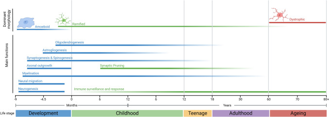
As ageing is linked to the development of neurodegenerative diseases (NDs), such as Alzheimer's Disease and Parkinson's Disease, it is important to disentangle the independent effect of age-related changes from those due to disease processes. To do so, changes to central nervous system (CNS) cells as a function of advanced age need better characterisation. Microglia are of particular interest due to their proposed links with the development and progression of NDs through control of the CNS immune response. Therefore, understanding the extent to which microglial dysfunction is related to phyisological ageing, rather than a disease process, is critical. As microglia age, they are believed to take on a pro-inflammatory phenotype with a distinct dystrophic morphology. Nevertheless, while established hallmarks of ageing have been investigated across a range of other cell types, such as macrophages, a detailed consideration of functional changes that occur in aged microglia remains elusive. Here, we describe the dynamic phenotypes of microglia and evaluate the current state of understanding of microglial ageing, focusing on the recently updated twelve hallmarks of ageing. Understanding how these hallmarks present in microglia represents a step towards better characterisation of microglial ageing, which is essential in the development of more representative models of NDs.

Keywords: Ageing; Immunosenescence; Inflammageing; Microglia; Morphology; Neuroinflammation.


Conflict of interest statement

Declarations. Competing interests: The authors declare no competing interests.

Figures

Fig. 1
Fig. 1
Microglial roles and morphologies during different stages of life. During development and early childhood, microglia have major roles in supporting the development of the CNS. Before birth, microglia display an amoeboid morphology. Ramified morphology begins to become the dominant morphology at the time of birth and remains so until late adulthood. During childhood, microglia maintain developmental roles through synaptic pruning, but transition to their major immune role around adolescence. During ageing, microglia acquire a dystrophic morphology; however, the functional changes at this stage remain unclear. Figure created in BioRender
Fig. 2
Fig. 2
Microglial classifications used throughout the literature and associated morphologies, functions and markers. Green: Phenotypes present under physiological conditions, Yellow: Phenotypes present following induction of immune response (i.e. response to injury/infection), Red: Phenotypes present under dysregulated or pathological conditions a Ramified microglia are in a state of CNS surveillance, with low baseline levels of inflammatory cytokine release. b Hyper-ramified microglia functionally have either a heightened or an impaired response to stimuli. c Reactive microglia initiate the immune response. d Bushy microglia are thought to be transitioning towards immune activation and have some phagocytic and inflammatory functions. e Rod microglia are associated with degeneration and injury, with potential roles in structural support of neurons. f Amoeboid microglia function as macrophages, with main roles in phagocytosis and secretion of pro-inflammatory factors. g Dystrophic microglia are associated with neurodegeneration and ageing and have a diminished capacity to perform normal homeostatic functions. *Markers expressed on microglial cells within each classification. #Factors measured in conjunction with microglia for each classification. Images from BioRender
Fig. 3
Fig. 3
Pathways of microglial responses. a PRR response pathways in microglia. PRRs, such as 1. TLRs, 2. RLRs, and 3. NLRs, recognise PAMPs/DAMPs and trigger signalling cascades, including 4. Exposure of NF-κB subunits by degradation if IκB and translocation of NF-κB to the nucleus. These cascades cause the downstream release of inflammatory cytokines. b DNA-driven immune responses in microglia. 1. cGAS binds to free DNA in the cytosol (mtDNA, nuclear DNA, foreign DNA), which activates the cGAS-STING pathway, causing the transcription of pro-inflammatory genes and release of pro-inflammatory cytokines. Figure was created using BioRender
Fig. 4
Fig. 4
Features of adult (left) and aged (right) microglia. Left: Normal young adult microglia are highly motile with a ramified morphology and can perform homeostatic and immune functions without inducing hostile environmental conditions. Right: Aged microglia display enlarged dystrophic morphology and present with many of the classic hallmarks of ageing

Similar articles

Cited by

References

    1. Abdel-Haq R, Schlachetzki JCM, Glass CK, Mazmanian SK (2019) Microbiome-microglia connections via the gut-brain axis. J Exp Med 216(1):41–59. 10.1084/jem.20180794 - PMC - PubMed
    1. Aird KM, Zhang R (2013) Detection of senescence-associated heterochromatin foci (SAHF). Methods Mol Biol 965:185–196. 10.1007/978-1-62703-239-1_12 - PMC - PubMed
    1. Ajami B, Bennett JL, Krieger C, Tetzlaff W, Rossi FMV (2007) Local self-renewal can sustain CNS microglia maintenance and function throughout adult life. Nat Neurosci 10(12):1538–1543. 10.1038/nn2014 - PubMed
    1. Alliot F, Godin I, Pessac B (1999) Microglia derive from progenitors, originating from the yolk sac, and which proliferate in the brain. Brain Res Dev Brain Res 117(2):145–152. 10.1016/s0165-3806(99)00113-3 - PubMed
    1. Ames BN, Atamna H, Killilea DW (2005) Mineral and vitamin deficiencies can accelerate the mitochondrial decay of aging. Mol Aspects Med 26(4):363–378. 10.1016/j.mam.2005.07.007 - PubMed