

Multiple deep learning models based on MRI images in discriminating glioblastoma from solitary brain metastases: a multicentre study
- PMID: 40389875
- PMCID: PMC12090387
- DOI: 10.1186/s12880-025-01703-3
Multiple deep learning models based on MRI images in discriminating glioblastoma from solitary brain metastases: a multicentre study
Abstract
Objective: Development of a deep learning model for accurate preoperative identification of glioblastoma and solitary brain metastases by combining multi-centre and multi-sequence magnetic resonance images and comparison of the performance of different deep learning models.
Methods: Clinical data and MR images of a total of 236 patients with pathologically confirmed glioblastoma and single brain metastases were retrospectively collected from January 2019 to May 2024 at Provincial Hospital of Shandong First Medical University, and the data were randomly divided into a training set and a test set according to the ratio of 8:2, in which the training set contained 197 cases and the test set contained 39 cases; the images were preprocessed and labeled with the tumor regions. The images were pre-processed and labeled with tumor regions, and different MRI sequences were input individually or in combination to train the deep learning model 3D ResNet-18, and the optimal sequence combinations were obtained by five-fold cross-validation enhancement of the data inputs and training of the deep learning models 3D Vision Transformer (3D Vit), 3D DenseNet, and 3D VGG; the working characteristic curves (ROCs) of subjects were plotted, and the area under the curve (AUC) was calculated. The area under the curve (AUC), accuracy, precision, recall and F1 score were used to evaluate the discriminative performance of the models. In addition, 48 patients with glioblastoma and single brain metastases from January 2020 to December 2022 were collected from the Affiliated Cancer Hospital of Shandong First Medical University as an external test set to compare the discriminative performance, robustness and generalization ability of the four deep learning models.
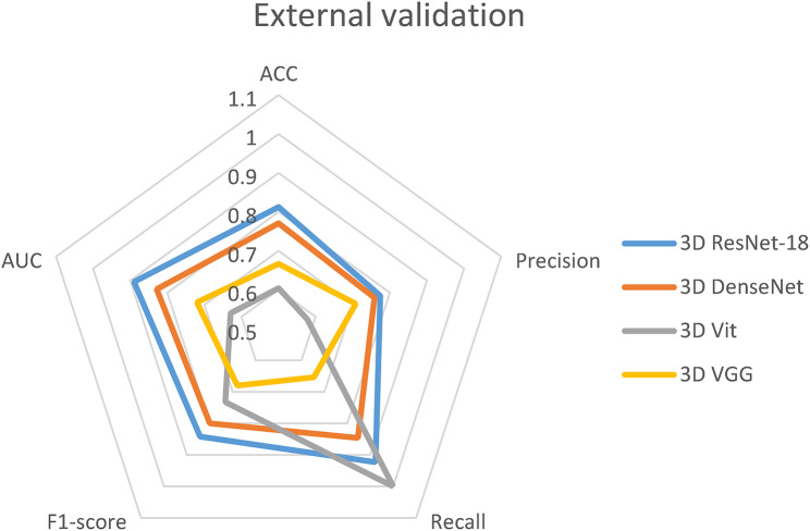
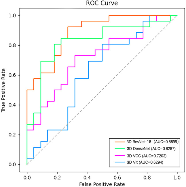
Results: In the comparison of the discriminative effect of different MRI sequences, the three sequence combinations of T1-CE, T2, and T2-Flair gained discriminative effect, with the accuracy and AUC values of 0.8718 and 0.9305, respectively; after the four deep learning models were inputted into the aforementioned sequence combinations, the accuracy and AUC of the external validation of the 3D ResNet-18 model were 0.8125, respectively, 0.8899, all of which are the highest among all models.
Conclusions: A combination of multi-sequence MR images and a deep learning model can efficiently identify glioblastoma and solitary brain metastases preoperatively, and the deep learning model 3D ResNet-18 has the highest efficacy in identifying the two types of tumours.
Keywords: Companion diagnostic system; Deep learning; Glioblastoma; Magnetic resonance imaging; Solitary brain metastasis.
© 2025. The Author(s).
Conflict of interest statement
Declarations. Ethics approval and consent to participate: The protocol for this study was approved by the Institutional Review Committee of the Shandong Provincial Hospital and Shandong First Medical University Affiliated Cancer Hospital Ethics Committee (SDTHEC 2024001002). All experiments were conducted in strict compliance with relevant international and national guidelines and regulations regarding human research, including but not limited to ethical principles, patient rights protection, and data security requirements. As this is a retrospective study and sensitive information of all patients was hidden during the study process, so Shandong First Medical University Affiliated Cancer Hospital Ethics Committee (SDTHEC 2024001002) waived the requirement for informed consent. Consent for publication: Not applicable. Competing interests: The authors declare no competing interests.
Figures


References
Publication types
MeSH terms
LinkOut - more resources
Full Text Sources
Medical
