. 2025 May 19;32(1):49.
doi: 10.1186/s12929-025-01140-y.

Gemcitabine resistance by CITED4 upregulation via the regulation of BIRC2 expression in pancreatic cancer

Affiliations

Gemcitabine resistance by CITED4 upregulation via the regulation of BIRC2 expression in pancreatic cancer

Eun-Jeong Jeong et al. J Biomed Sci. .

Abstract

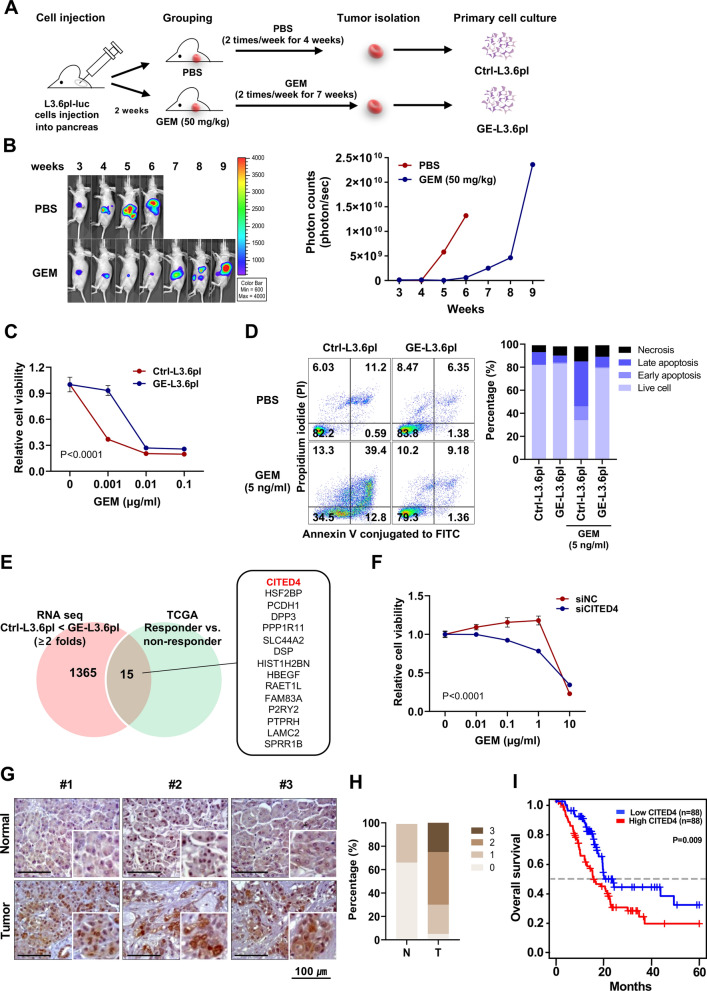
Background: Gemcitabine (GEM) is used as a first-line therapy for patients diagnosed with any stage of pancreatic cancer (PC); however, patient survival is poor because of GEM resistance. Thus, new approaches to overcome GEM resistance in PC are urgently needed. Here, we aimed to establish an in vivo drug-resistant PC model and identify genes involved in GEM resistance. We focused on one of these factors, CITED4, and elucidated its mechanisms of action in GEM resistance in PC.

Methods: L3.6pl, a GEM-sensitive PC cell line, was orthotopically injected into the pancreas of BALB/c nude mice to establish a GEM-resistant PC animal model. Transcriptomic data from control or GEM-resistant tumor-derived cells were analyzed. GEM resistance was evaluated using cell viability, clonogenicity, and apoptosis assays. An apoptosis array was used to identify genes downstream of CITED4. A CITED4 knockout-mediated GEM sensitivity assay was performed in an orthotopic xenograft mouse model using PANC-1 cells, which are GEM-resistant cells.

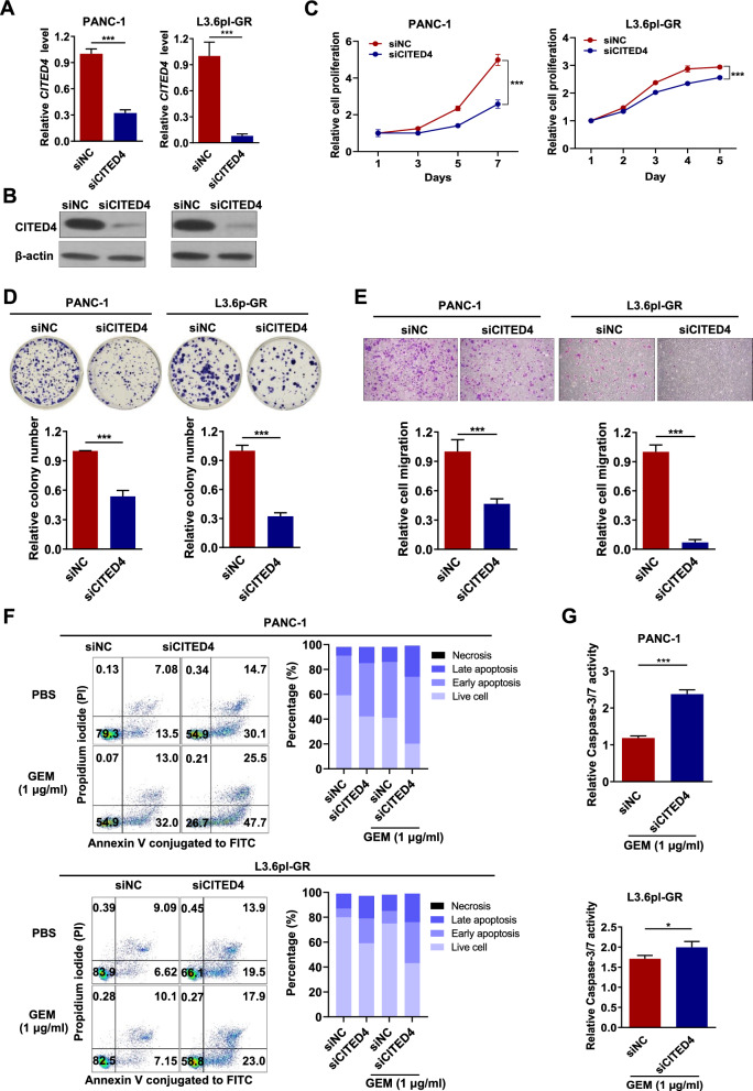
Results: From the RNA sequencing data of isolated GEM-resistant PC cells and The Cancer Genome Atlas dataset, 15 GEM resistance-related genes were found to be upregulated, including CITED4, the gene encoding a type of CBP/p300-interacting transactivator implicated in several cancers. CITED4 knockdown in drug-resistant cells reduced cell proliferation and migration but increased apoptosis. To identify the molecular mechanism underlying CITED4-mediated induction of GEM resistance, alterations in Baculoviral IAP Repeat Containing 2 (BIRC2) levels were observed using an apoptosis array. BIRC2 expression was downregulated following CITED4 knockdown in GEM-resistant PC cell lines. Furthermore, chromatin immunoprecipitation and promoter assays showed that BIRC2 was directly regulated by CITED4. Consistent with the CITED-knockdown experiments, silencing of BIRC2 increased the sensitivity of L3.6pl-GEM-resistant and PANC-1 cell lines to GEM. Furthermore, CITED4 knockout using the CRISPR-Cas9 system in PANC-1 cells increased the sensitivity to GEM in orthotopic mice. Moreover, elevated CITED4 and BIRC2 expression levels were associated with poorer outcomes in human PC clinical samples.

Conclusions: Collectively, these results indicate that CITED4 regulates GEM resistance via inhibition of apoptosis by upregulating BIRC2 expression in PC cells. Therefore, CITED4 may serve as a valuable diagnostic marker and therapeutic target for GEM-resistant PC.

Keywords: BIRC2; CITED4; Drug resistance; Gemcitabine; Pancreatic cancer.


Conflict of interest statement

Declarations. Ethics approval and consent to participate: This study was approved by the Public Institutional Review Board of the Ministry of Health and Welfare (P01-202105-31-011). The animal protocol was approved by the Committee on Animal Experimentation of the Korea Research Institute of Bioscience and Biotechnology. Consent for publication: Not applicable. Competing interests: The authors declare that they have no competing financial interests.

Figures

Fig. 1
Fig. 1
Identification of gemcitabine (GEM)-resistance genes in PC. A Schematic representation of the preparation of a GEM-resistant PC cell line. B Mice were subjected to bioluminescent imaging using an in vivo imaging system. Bioluminescent images were captured once a week post-cell injection, and representative images are shown (left panel). The levels of bioluminescent intensity (total photon flux per second) in the pancreatic regions were quantified and compared between the PBS and GEM treatment groups (right panel). (PBS, n = 6; GEM, n = 6). C Cell viability was measured using a CCK-8 assay. Control cells (GEM-sensitive pancreatic cells from PBS-injected mice) and GE-L3.6pl cells (GEM-resistant pancreatic cells from GEM-injected mice) were seeded at a density of 3 × 103 cells/well in 96-well plates, and treated with various concentrations of GEM for 48 h. D Fluorescence-activated cell sorting analysis of apoptosis. Representative flow cytometry dot plots with double Annexin V-FITC/propidium iodide staining for control and GE-L3.6pl cells exposed to GEM (5 ng/ml) for 48 h (left panel). Percentages of viable (white bars), early apoptotic (light gray bars), late apoptotic (dark gray bars), and necrotic (black bar; right panel) cells. E Venn diagram showing 15 genes correlated with GEM resistance between The Cancer Genome Atlas data and RNA sequencing analysis. F Cell viability was measured using a CCK-8 assay after cells were transfected with siCITED4 and treated with various concentrations of GEM for 48 h. G Immunohistochemical staining of CITED4 in tumorous and non-tumorous pancreatic tissues (scale bar, 100 μm). H Expression pattern of CITED4 in the human pancreatic tumor tissue array. Diagnostic score 0 (beige), score 1 (light brown), score 2 (brown), and score 3 (dark brown). I Overall survival analysis for PC samples from Q-omics (https://qomics.sookmyung.ac.kr/) depending on CITED4 expression
Fig. 2
Fig. 2
CITED4 expression is upregulated in GEM-resistant PC cells. A Schematic representation of stronger GEM resistance in cells (L3.6pl-GR). B Phase-contrast image for control (top) and L3.6pl-GR (bottom). C Cell viability was measured using a CCK-8 assay in control and L3.6pl-GR cells seeded at a density of 3 × 103 cells/well in 96-well plates, and treated with various concentrations of GEM for 48 h. D Cell viability was measured using a CCK-8 assay. PANC-1 (green circle), AsPC-1 (purple circle), L3.6pl-GR (red triangle), control (brown triangle), and L3.6pl (blue triangle) cells were treated with various concentrations of GEM for 48 h. E CITED4 mRNA and protein expression levels in AsPC-1, PANC-1, and L3.6pl PC cell lines. F CITED4 mRNA and protein expression levels in Ctrl-L3.6pl and L3.6pl-GR cells. G Immunofluorescence staining of CITED4 (green), phalloidin (red), and DAPI (blue) was detected in control and L3.6pl-GR. Scale bar, 20 μm. H mRNA expression and I Protein expression after treatment of Trichostatin A (TSA, 0.2 μM) and/or DNA demethylation agent, 5-Aza-2’-deoxycytidine (5-Aza-dC, 5 μM) for 24 h. β-actin was used as an internal control for mRNA and protein expression. J DNA methylation status of CITED4 promoter region by DNA methylation amplicon sequencing for Ctrl-L3.6pl and L3.6pl-GR. K Scatter plots showing the correlation between CITED4 expression and DNA methylation levels at specific CpG sites (cg18812909, left; cg11240320, right). L Kaplan–Meier survival curves show the overall survival of PC patients depending on the DNA methylation status of CITED4 (cg18812909, left; cg11240320, right). ***P < 0.001
Fig. 3
Fig. 3
Oncogenic properties of CITED4 in PC. A CITED4 mRNA expression level in CITED4-knockdown PANC-1 and L3.6pl-GR. ACTB was used as an internal control for mRNA expression analysis. B Protein expression level of CITED4 in CITED4-knockdown PANC-1 and L3.6pl-GR cells. β-actin was used as an internal control for mRNA and protein expression. C Cell viability was measured using a CCK-8 assay. CITED4-knockdown PANC-1 and L3.6pl-GR cells were cultured with 1 µg/ml of GEM for 7 days. D Representative image of the colony-formation assay for PANC-1 and L3.6pl-GR cells and relative quantification of the colony number. E Representative image of migration of PANC-1 and L3.6pl-GR cells and relative quantification of the number of migrated cells. F Comparative analysis of cellular apoptosis between siRNA for negative control (siNC) and siRNA for CITED4 (siCITED4) in PANC-1 (upper) and L3.6pl-GR (bottom) with GEM treatment using flow cytometry. FACS analysis of siNC- and siCITED4-transfected cells treated with or without GEM (1 µg/ml) for 48 h, following which the cells were stained with Annexin V-FITC and propidium iodide (left panel). Percentages of viable, early apoptotic, late apoptotic, and necrotic cells (right panel). G Caspase-3/7 activity was measured using a Caspase-Glo 3/7 Assay kit. CITED4-knockdown PANC-1(upper) and L3.6pl-GR (bottom) cells were treated with 1 µg/ml GEM for 48 h, following the manufacturer’s protocol. *P < 0.05 and ***P < 0.001
Fig. 4
Fig. 4
CITED4 is related to apoptosis and functions through BIRC2. A Proteome profiling of apoptosis-associated proteins. Array images showing cleaved Caspase-3 and BIRC2 expression in CITED4-knockdown L3.6pl-GR cells. B Apoptosis-related gene expression in CITED4-knockdown PANC-1 (left) and L3.6pl-GR (right) cells. C mRNA (top) and protein (bottom) expression levels of CITED4 in CITED4-knockdown PANC-1 and L3.6pl-GR cells. D Schematic representation of the BIRC2 promoter region. Chromatin immunoprecipitation (ChIP)-qPCR was used to amplify chromatin derived from immunoprecipitation with CITED4 antibody, as indicated. E Luciferase reporter assay of BIRC2 promoter activity and schematic representation of the truncated promoter plasmid (upper panel). Relative luciferase activity was determined using the ratio of firefly luciferase/Renilla luciferase activity (bottom panel). F CITED4 and BIRC2 expression levels in normal pancreatic tissues and PC tumor tissues. G Correlation analysis of between CITED4 and BIRC2 expression levels using tissue samples. H Correlation analysis of between CITED4 and BIRC2 expression levels from PAAD-TCGA using GEPIA2 (R = 0.21, P < 0.0001). I Heat map of the differentially expressed genes (DEGs) in BIRC2-knockdown PANC-1 and L3.6pl-GR. Representative genes are shown on the right. J The Database for Annotation, Visualization, and Integrated Discovery (DAVID) tool was used to perform KEGG pathway analysis for DEGs. K The expression of CITED4, BIRC2, phosphorylated p38 MAPK (p-p38), total p38 MAPK (p38), phosphorylated JNK (p-JNK), and total JNK in BIRC2 knockdown PACN-1 and L3.6pl-GR cells. GAPDH were used as an internal control for protein expression. **P < 0.01 and ***P < 0.001
Fig. 5
Fig. 5
BIRC2 mediates GEM-resistance features related to CITED4 in PC. A mRNA and B protein expression levels of BIRC2 in BIRC2-knockdown PANC-1 and L3.6pl-GR cells. β-Actin was used as an internal control for mRNA and protein expression. C Cell viability was measured using a CCK-8 assay. D Representative image of the colony-formation assay in PANC-1 and L3.6pl-GR (top) cells. Relative quantification of the colony number (bottom). E Representative image of the cell-migration assay (top). Relative quantification of the number of migrated cells (bottom). F Comparative analysis of apoptosis after GEM treatment of BIRC2-knockdown PANC-1 and L3.6pl-GR cells using flow cytometry. Fluorescence-activated cell sorting analysis of siNC- and siBIRC2-transfected cells treated with (1 µg/ml) or without GEM for 48 h, following which the cells were stained with Annexin V-FITC and propidium iodide (upper panel). Percentages of viable, early apoptotic, late apoptotic, and necrotic cells. G Caspase-3/7 activity was measured using Caspase-Glo 3/7 Assay kit. BIRC2-knockdown PANC-1 (upper) and L3.6pl-GR (bottom) cells were treated with 1 µg/ml GEM for 48 h. *P < 0.05 and ***P < 0.001
Fig. 6
Fig. 6
Knockout of CITED4 decreases pancreatic tumor growth in an orthotopic mouse model. A Schematic of the experimental procedure. PANC-1 (PBS, n = 5; GEM, n = 6), CITED4 knockout PANC-1 #1 (PBS, n = 8; GEM, n = 9) or #3 (PBS, n = 7; GEM, n = 8) cell were implanted to pancreas of BALB/c nude mice. After 2 weeks, the mice were treated with GEM (50 mg/kg) twice per week for 6 weeks. B Tumors were detected in mice using in vivo bioluminescence imaging (left), and bioluminescent intensities were quantified (right). Two-way ANOVA was used for statistical analysis. C Macroscopic images of each tumor (scale bar, 1 cm). Red circles indicate complete regression. D Average of tumor weight. E Schematic representation of the roles of CITED4 in GEM-resistant PC. *P < 0.05 and ***P < 0.001

References

    1. Twohig PA, Butt MU, Gardner TB, Chahal P, Sandhu DS. Racial and gender disparities among obese patients with pancreatic cancer: a trend analysis in the United States. J Clin Gastroenterol. 2023;57(4):410–6. - PubMed
    1. Kolbeinsson HM, Chandana S, Wright GP, Chung M. Pancreatic cancer: a review of current treatment and novel therapies. J Invest Surg. 2023;36(1):2129884–95. - PubMed
    1. Biancur DE, Kimmelman AC. The plasticity of pancreatic cancer metabolism in tumor progression and therapeutic resistance. Biochim Biophys Acta Rev Cancer. 2018;1870(1):67–75. - PMC - PubMed
    1. Siegel RL, Miller KD, Jemal A. Cancer statistics, 2020. CA Cancer J Clin. 2020;70(1):7–30. - PubMed
    1. Farhangnia P, Khorramdelazad H, Nickho H, Delbandi AA. Current and future immunotherapeutic approaches in pancreatic cancer treatment. J Hematol Oncol. 2024;17(1):40–106. - PMC - PubMed

MeSH terms