. 2025 May 19;18(1):57.
doi: 10.1186/s13045-025-01712-3.

Engineered CRO-CD7 CAR-NK cells derived from pluripotent stem cells avoid fratricide and efficiently suppress human T-cell malignancies

Affiliations

Engineered CRO-CD7 CAR-NK cells derived from pluripotent stem cells avoid fratricide and efficiently suppress human T-cell malignancies

Yunqing Lin et al. J Hematol Oncol. .

Abstract

Background: T-cell malignancies are highly aggressive hematological tumors with limited effective treatment options. CAR-NK cell therapy targeting CD7 has emerged as a promising approach for treating T-cell malignancies. However, conventional CAR-NK cell therapy faces the challenges of cell fratricide due to CD7 expression on both malignant cells and normal NK cells. Additionally, engineering CARs into human tissue-derived NK cells demonstrates heterogeneity, low transduction efficiency, and high manufacturing costs.

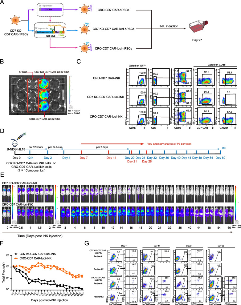
Methods: The human pluripotent stem cells (hPSCs) were genetically modified by knocking out the CD7 gene and introducing the CD7 CAR expression cassette to generate CD7 KO-CD7 CAR-hPSCs. These modified hPSCs were subsequently differentiated into CD7 KO-CD7 CAR-iNK cells using an efficient organoid induction method. The cytotoxicity of CD7 KO-CD7 CAR-iNK cells against CD7+ tumor cells was evaluated. Furthermore, we overexpressed the CXCR4 gene in CD7 KO-CD7 CAR-hPSCs and derived CXCR4-expressing CD7 KO-CD7 CAR-iNK (CRO-CD7 CAR-iNK) cells. The dynamics of CRO-CD7 CAR-iNK cells in vivo were tracked, and their therapeutic efficacy was assessed using human T-cell acute lymphoblastic leukemia (T-ALL) xenograft models.

Results: The CD7 KO-CD7 CAR-iNK cells derived from CD7 KO-CD7 CAR-hPSCs effectively avoided fratricide, demonstrated normal expansion, and exhibited potent and specific anti-tumor activity against CD7+ T-cell tumor cell lines and primary T-ALL cells. CXCR4 overexpression in CRO-CD7 CAR-iNK cells improved their homing capacity and extended their persistence in vivo. The CRO-CD7 CAR-iNK cells significantly suppressed tumor growth and prolonged the survival of T-ALL tumor-bearing mice.

Conclusions: Our study provides a reliable strategy for the large-scale generation of fratricide-resistant CD7 CAR-iNK cells with robust anti-tumor effects from hPSCs, offering a promising cell product to treat T-cell malignancies.

Keywords: CD7 CAR-NK; CXCR4; Fratricide-resistance; Human pluripotent stem cells; Persistence; T-ALL.


Conflict of interest statement

Declarations. Ethics approval and consent to participate: Experiments and handling of mice were conducted under the Institutional Animal Care and Use Committee of the Institute of Zoology, Chinese Academy of Sciences. The studies involving humans were approved by the Biomedical Research Ethics Committee of the Institute of Zoology, Chinese Academy of Sciences. The use of patient samples was conducted in accordance with the provisions of the Declaration of Helsinki. All patient samples were collected with priori patient consent signatures and were reviewed and approved by the Ethics Committee of State Key Laboratory of Experimental Hematology, Institute of Hematology and Blood Disease Hospital, Chinese Academy of Medical Sciences and Peking Union Medical College. Consent for publication: Not applicable. Competing interests: The authors declare no competing interests.

Figures

Fig. 1
Fig. 1
hPSC-derived iNK cells lacking CD7 expression avoid fratricide. A Schematic diagram showing the generation of CD7 CAR-NK cells from UCB. B Flow cytometry analysis showing the infection rate of CD7 CAR in CD3CD56+ NK cells. The infection rate was analyzed 48 h after transduction. C Schematic diagram of evaluating the cell lysis ratios of UCB-derived Ctrl-NK cells (uninfected with retrovirus) cocultured with CD7 CAR-NK cells. D Statistical analysis of the fratricide percentage of CD7 CAR-NK cells against Ctrl-NK cells at indicated E:T ratios (mean ± SD). n = 4 repeats. E Strategy for the knockout of CD7 gene in hPSCs. The exons of the CD7 gene are shown as blue boxes. Two gRNA sequences, gRNA1 and gRNA2, are shown in black. The deleted sequences are marked in red, and the sequences after editing are marked in blue. F Schematic diagram of CD7 KO-iNK cell induction. G Representative flow cytometry plots of CD7 KO-iNK cells (GFPCD45+CD3CD56+CD16±CD7) on day 27. H Schematic diagram of evaluating the in vitro cytotoxic activity of CD7 CAR-NK cells against Ctrl-iNK cells or CD7 KO-iNK cells. I Statistical analysis of the fratricide percentage of CD7 CAR-NK cells against Ctrl-iNK cells or CD7 KO-iNK cells at indicated E:T ratios (two-way ANOVA, mean ± SD). n = 4 repeats. ***p < 0.001
Fig. 2
Fig. 2
Disruption of CD7 expression restores the expansion of CD7 CAR-iNK cells. A Statistics analysis of the expansion folds of NK cells and CD7 CAR-NK cells derived from UCB on indicated expansion days (two-tailed independent t-test, mean ± SD). n= 3 repeats. NS, not significant (p > 0.05), *p < 0.05, **p < 0.01. B Strategy diagram for the acquisition of CD7 KO-CD7 CAR-iNK cells and the detection of their expansion capacity and cytotoxic activity. C Representative flow cytometry plots of Ctrl-iNK, CD7 KO-iNK, and CD7 KO-CD7 CAR-iNK cells (GFPCD45+CD3CD56+CD16±CD7CD7 CAR+) on day 27. D Statistics analysis of the expansion folds of Ctrl-iNK cells, CD7 KO-iNK cells, and CD7 KO-CD7 CAR-iNK cells on indicated expansion days (one-way ANOVA with Tukey’s multiple-comparison test). n = 3 repeats. NS, not significant (p > 0.05). E Flow cytometry histograms showing the expression levels of NKp30, NKp44, CD319, DNAM-1, CD96, NKG2A, CD94, CD69, TRAIL GzmB, and Perforin in Ctrl-iNK, CD7 KO-iNK, and CD7 KO-CD7 CAR-iNK cells. F Schematic diagram of evaluating the in vitro cytotoxic activity of CD7 KO-CD7 CAR-iNK cells against UCB-NK, Ctrl-iNK, and CD7 KO-iNK cells. G Statistical analysis of the cytotoxic activity of CD7 KO-CD7 CAR-iNK cells against UCB-NK, Ctrl-iNK, and CD7 KO-iNK cells at an E:T ratio of 1:1 (one-way ANOVA with Tukey’s multiple-comparison test). n = 4 repeats. ***p < 0.001
Fig. 3
Fig. 3
The cytotoxic activity of CD7 KO-CD7 CAR-iNK cells against CD7+ tumor cell lines in vitro. A Experimental design for the tumor-killing assay. The Ctrl-iNK and CD7 KO-CD7 CAR-iNK cells (effectors, E) were cocultured with Jurkat or CCRF-CEM tumor cells labeled with eFluor™ 670 (targets, T), respectively. The E:T ratios include 0.05:1, 0.1:1, 0.2:1, 0.4:1, 0.8:1, 1.6:1, 3.2:1, and 5:1. B-C Cytotoxicity analysis of Ctrl-iNK cells and CD7 KO-CD7 CAR-iNK cells against Jurkat (B) or CCRF-CEM (C) tumor cells at the indicated E:T ratios after 4-h incubation. Data are represented as mean ± SD (n = 4). Mann-Whitney U test was used for statistics. ***p < 0.001. D Experimental design for the multiple rounds of tumor killing. The Ctrl-iNK and CD7 KO-CD7 CAR-iNK cells were respectively cocultured with Jurkat or CCRF-CEM tumor cells for 12 h per round at the E:T ratio of 1:1. Fresh tumor cells were added to the Ctrl-iNK/CD7 KO-CD7 CAR-iNK cell residues incubated every other 12 h. E Cytotoxicity analysis of three consecutive rounds of Jurkat cell killing by Ctrl-iNK or CD7 KO-CD7 CAR-iNK cells. Data are represented as mean ± SD (n = 4). Two-tailed independent t-test was used for statistics. ***p < 0.001. F Cytotoxicity analysis of three consecutive rounds of CCRF-CEM cell killing by Ctrl-iNK or CD7 KO-CD7 CAR-iNK cells. Data are represented as mean ± SD (n = 4). Two-tailed independent t-test was used for statistics. ***p < 0.001. G-H Assessment of CD107a expression by Ctrl-iNK or CD7 KO-CD7 CAR-iNK cells following 4 h of coculture with Jurkat/CCRF-CEM tumor cells at the ratio of 1:1. Data are represented as mean ± SD (n = 3). Two-tailed independent t-test was used for statistics. **p < 0.01, ***p < 0.001. I-J Measurement of IFN-γ production by Ctrl-iNK or CD7 KO-CD7 CAR-iNK cells in response to Jurkat/CCRF-CEM tumor cells. The Ctrl-iNK or CD7 KO-CD7 CAR-iNK cells were stimulated by Jurkat/CCRF-CEM at the ratio of 1:1 for 4 h. Data are represented as mean ± SD (n = 3). Two-tailed independent t-test was used for statistics. NS, not significant (p > 0.05), ***p < 0.001. K-L Evaluation of TNF-α production by Ctrl-iNK or CD7 KO-CD7 CAR-iNK cells in response to Jurkat/CCRF-CEM tumor cells. The Ctrl-iNK or CD7 KO-CD7 CAR-iNK cells were stimulated by Jurkat/CCRF-CEM at the ratio of 1:1 for 4 h. Data are represented as mean ± SD (n = 3). Two-tailed independent t-test was used for statistics. NS, not significant (p > 0.05), **p < 0.01
Fig. 4
Fig. 4
Anti-tumor activity of CD7 KO-CD7 CAR-iNK cells against primary T-ALL cells. A Experimental design for the T-ALL patient tumor-killing assay by hPSC-derived CD7 KO-CD7 CAR-iNK cells. Mononuclear cells (MNCs) were first isolated from the bone marrow of patients with T-ALL and transplanted into B-NDG immunodeficient mice (1 × 106/mouse) via tail vein injection. Four weeks after transplantation, the proportion of primary tumor cells in the PB of B-NDG mice was monitored weekly using flow cytometry. Once the proportion of huCD45+CD7+ primary tumor cells exceeded 80% (approximately 6-9 weeks post-transplantation), the spleens of the B-NDG recipient mice were harvested and processed into single-cell suspensions. The resulting splenocytes were then cocultured with Ctrl-iNK or CD7 KO-CD7 CAR-iNK cells for tumor cytotoxicity assays. B Surface expression of CD7 in the splenic cells from B-NDG recipient mice measured by flow cytometry. C-E Cytotoxicity analysis of Ctrl-iNK or CD7 KO-CD7 CAR-iNK cells against T-ALL cells isolated from patient 1 (C), patient 2 (D), and patient 3 (E) at the indicated E:T ratios after 6-h incubation. Data are represented as mean ± SD (n = 4). Two-way ANOVA and Mann-Whitney U test were used for statistics. ***p < 0.001. F–H Cytotoxicity analysis of Ctrl-iNK or CD7 KO-CD7 CAR-iNK cells against T-ALL cells isolated from patient 1 (F), patient 2 (G), and patient 3 (H) at the E:T ratio of 1:2 after 3 h-, 6 h-, 9 h-, and 12 h- incubation. Data are represented as mean ± SD (n = 4). Two-way ANOVA and Mann-Whitney U test were used for statistics. ***p < 0.001
Fig. 5
Fig. 5
Dynamic analysis of CRO-CD7 CAR-iNK cells in the B-NDG hIL15 mice after injection. A Strategy for the construction of CXCR4-CD7 KO-CD7 CAR-hPSCs (CRO-CD7 CAR-hPSCs), CD7 KO-CD7 CAR-luciferase-hPSCs (CD7 KO-CD7 CAR-luci-hPSCs), and CXCR4-CD7 KO-CD7 CAR-luciferase-hPSCs (CRO-CD7 CAR-luci-hPSCs). B BLI images of the CD7 KO-CD7 CAR-luci-hPSCs and CRO-CD7 CAR-luci-hPSCs. C Representative flow cytometry plots of CRO-CD7 CAR-iNK, CD7 KO-CD7 CAR-luci-iNK, and CRO-CD7 CAR-luci-iNK cells on day 27. D Schematic diagram of in vivo studies with CD7 KO-CD7 CAR-luci-iNK and CRO-CD7 CAR-luci-iNK cells in B-NDG hIL15 mice. The CD7 KO-CD7 CAR-luci-iNK and CRO-CD7 CAR-luci-iNK cells were injected into B-NDG hIL15 mice (1 × 107 cells/mouse) via the tail vein on day 0, respectively. BLI and flow cytometry analysis were performed at the indicated time points. E BLI images showing the presence of CD7 KO-CD7 CAR-luci-iNK or CRO-CD7 CAR-luci-iNK cells in B-NDG hIL15 mice (n = 2 mice in each group) at the indicated time points. F Total flux (p/s) of the CD7 KO-CD7 CAR-luci-iNK or CRO-CD7 CAR-luci-iNK cells injected B-NDG hIL15 mice measured at the indicated time points (n = 2 mice in each group). G Flow cytometry analysis of CD7 KO-CD7 CAR-luci-iNK or CRO-CD7 CAR-luci-iNK cells in the PB of B-NDG hIL15 mice at the indicated time points
Fig. 6
Fig. 6
Suppression of human T-cell leukemia progress in xenograft models by CRO-CD7 CAR-iNK cells. A Schematic diagram of in vivo studies with luciferase-expressing CCRF-CEM cells (CCRF-CEM-luci) in mouse xenograft models. The CCRF-CEM-luci tumor cells were injected into B-NDG hIL15 mice (1 × 105/mouse) via the tail vein on day 0. The mice were irradiated (1.0 Gy) on day 1. Equal numbers (1 × 107) of Ctrl-iNK, CD7 KO-CD7 CAR-iNK, or CRO-CD7 CAR-iNK cells were injected into each animal on day 1, day 4, and day 7. BLI was performed every week. B BLI images of the xenograft models at the indicated time points (Tumor + PBS, Tumor + Ctrl-iNK, Tumor + CD7 KO-CD7 CAR-iNK, and Tumor + CRO-CD7 CAR-iNK, n = 5 mice in each group). The radiance indicates tumor burden. C Total flux (p/s) of the xenograft models measured at the indicated time points (n = 5 mice in each group). Two-tailed independent t-test was used for statistics. NS, not significant (p > 0.05), *p < 0.05, **p < 0.01, ***p < 0.001. D Kaplan-Meier survival curves of the xenograft models (n = 5 mice in each group). Median survival: Tumor + PBS, 29 days; Tumor + Ctrl-iNK, 30 days; Tumor + CD7 KO-CD7 CAR-iNK, 32 days; Tumor + CRO-CD7 CAR-iNK, 36 days. The logarithmic rank (Mantel-Cox) test was used for statistics. NS, not significant, *p < 0.05, **p < 0.01

Similar articles

Cited by

References

    1. Litzow MR, Ferrando AA. How I treat T-cell acute lymphoblastic leukemia in adults. Blood. 2015;126:833–41. - PubMed
    1. Marks DI, et al. T-cell acute lymphoblastic leukemia in adults: clinical features, immunophenotype, cytogenetics, and outcome from the large randomized prospective trial (UKALL XII/ECOG 2993). Blood. 2009;114:5136–45. - PMC - PubMed
    1. Marks DI, Rowntree C. Management of adults with T-cell lymphoblastic leukemia. Blood. 2017;129:1134–42. - PubMed
    1. Park JH, et al. Long-Term Follow-up of CD19 CAR Therapy in Acute Lymphoblastic Leukemia. N Engl J Med. 2018;378:449–59. - PMC - PubMed
    1. Raje N, et al. Anti-BCMA CAR T-Cell Therapy bb2121 in Relapsed or Refractory Multiple Myeloma. N Engl J Med. 2019;380:1726–37. - PMC - PubMed

LinkOut - more resources