

Spatial transcriptomics reveal PI3K-AKT and metabolic alterations in aggressive, treatment-resistant lactotroph pituitary neuroendocrine tumors
- PMID: 40390063
- PMCID: PMC12087103
- DOI: 10.1186/s40478-025-02025-9
Spatial transcriptomics reveal PI3K-AKT and metabolic alterations in aggressive, treatment-resistant lactotroph pituitary neuroendocrine tumors
Abstract
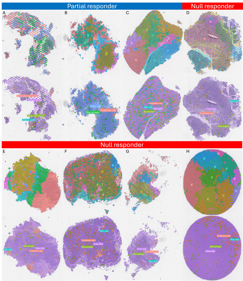
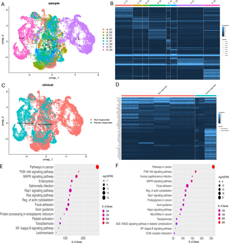
Clinically aggressive lactotroph pituitary neuroendocrine tumors (PitNET) are invasive tumors with an unusually rapid growth rate despite maximally tolerated doses of dopamine agonist (DA). We aimed to unravel the molecular heterogeneity of lactotroph PitNET and to identify biomarkers of aggressiveness and resistance to pharmacological treatment. A total of 13 patients harboring DA-resistant lactotroph PitNET were included in this study. Visium Spatial Transcriptomics (ST), whole transcriptome sequencing (WTS), and whole exome sequencing (WES) were performed in tumors from 4 of these patients; WTS and WES was carried out in 5; tumors from two patients underwent ST and WES and tumors from two other patients underwent only ST. Tumors were classified as null or partial responders according to their response to DA treatment. The eight PitNET analyzed by ST exhibited significant intratumoral heterogeneity, with clones showing alterations in PI3K/AKT and lipid metabolism pathways, particularly inositol phosphate, glycerophospholipid, and sphingolipid metabolism. The cell-cell communication analysis showed FGF-FGFR ligand receptor interaction whilst the transcription factors RXRA and CREM showed participation in both groups. A trajectory exploration was performed by including all PitNET together in a single analysis to determine whether there was a tendency or molecular pathway showing a differentiation pattern that would guide the transition from a partially responsive PitNET to a completely unresponsive one. We did not observe any such pattern. All of these findings were corroborated in the cohort of DA-resistant PitNETs in which only bulk WTS and WES were performed. The bulk WTS corroborated lipid metabolism and PI3K-AKT pathway alteration in PitNET, whereas the WES showed only SF3β1 and TP53 variants in one tumor each. Our work suggests that the PI3K/AKT pathway may constitute a molecular target at which to aim therapeutic strategies designed to treat aggressive and DA-resistant lactotroph PitNET.
Keywords: Aggressive PitNET; Cabergoline; Lactotroph PitNET; Pharmacological resistant; Prolactinoma; Resistant; Spatial transcriptomic.
© 2025. The Author(s).
Conflict of interest statement
Declarations. Compliance with ethical standards: The study protocol was approved by the Comisión Nacional de Ética e Investigación Científica del Instituto Mexicano del Seguro Social (approval: R-2022-3601-175, R-2022-3601-164 and R-2024-3601-239) and it has been carried out in accordance with the principles of the Helsinki declaration. All participating patients signed an informed consent. Competing interests: The authors declare no competing interests.
Figures


References
-
- Derwich A, Sykutera M, Bromińska B, Rubiś B, Ruchała M, Sawicka-Gutaj N (2023) The role of activation of PI3K/AKT/mTOR and RAF/MEK/ERK pathways in aggressive pituitary Adenomas—New potential therapeutic Approach—A systematic review. Int J Mol Sci 24(13):10952. 10.3390/ijms241310952 - DOI - PMC - PubMed
Publication types
MeSH terms
Substances
Grants and funding
LinkOut - more resources
Full Text Sources
Medical
Research Materials
Miscellaneous
