

Core-shell nanofiber dressings with zinc oxide nanoparticles and cell-free fat extract: boosting fibroblast activity and antibacterial efficacy
- PMID: 40390077
- PMCID: PMC12090510
- DOI: 10.1186/s13036-025-00511-1
Core-shell nanofiber dressings with zinc oxide nanoparticles and cell-free fat extract: boosting fibroblast activity and antibacterial efficacy
Abstract
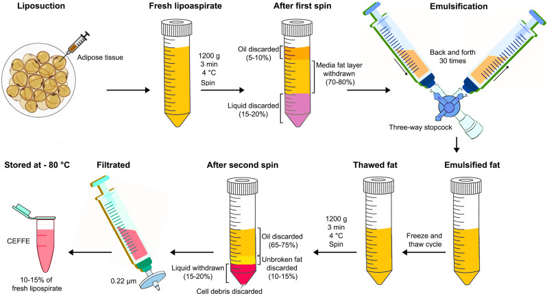
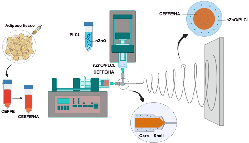
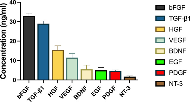
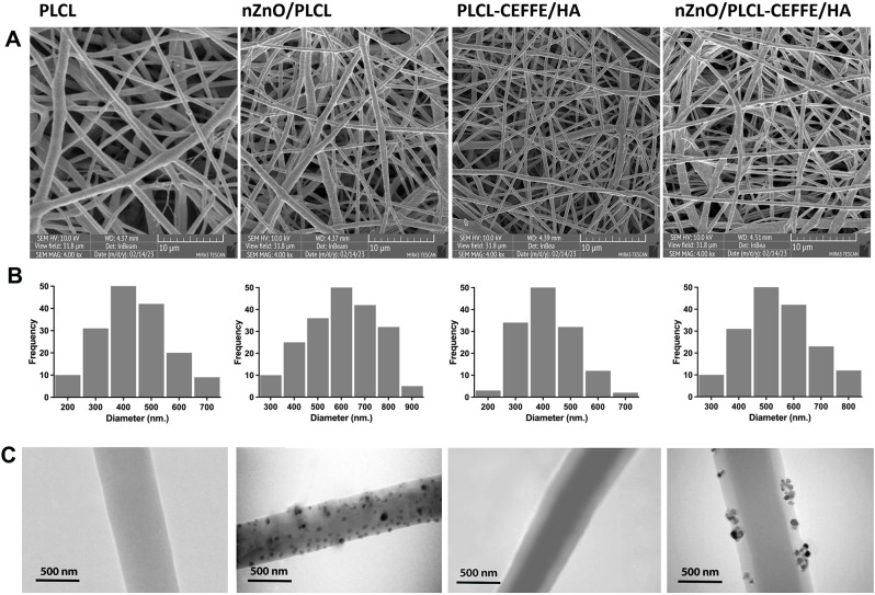
Background: This study presents the development and characterization of innovative core-shell nanofiber wound dressings incorporating zinc oxide nanoparticles (nZnO) and cell-free fat extract (CEFFE) to enhance fibroblast activity and antibacterial efficacy.
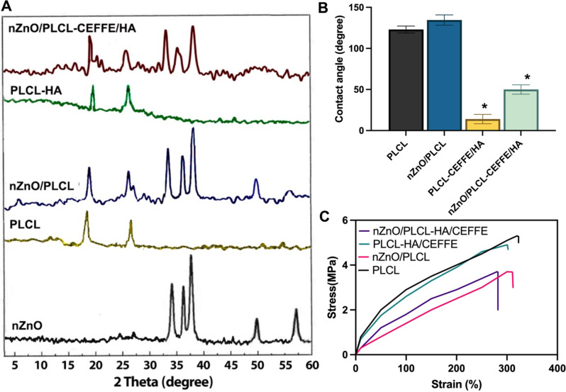
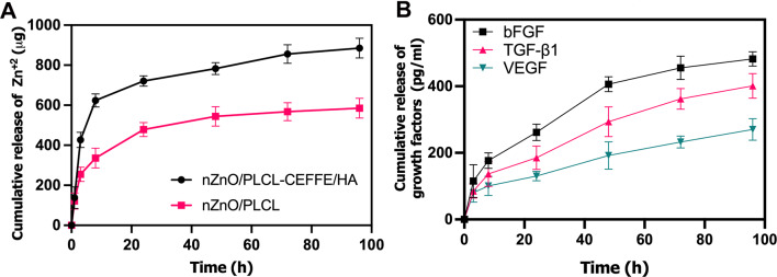
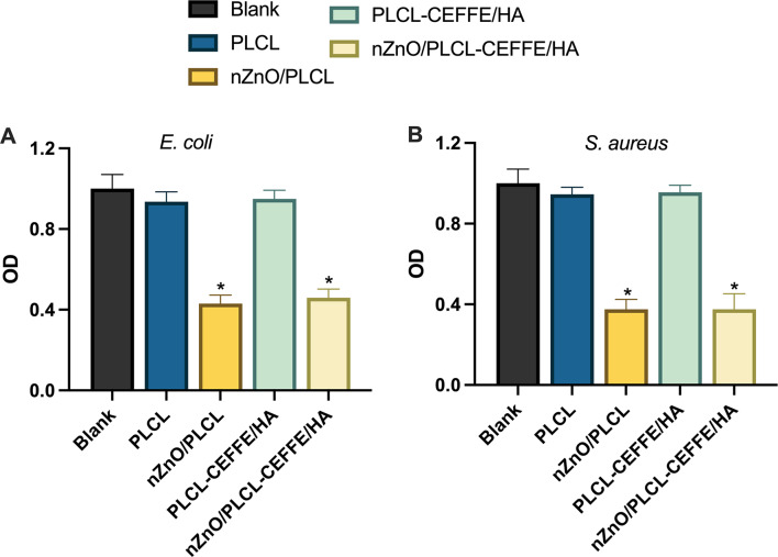
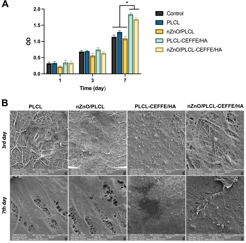
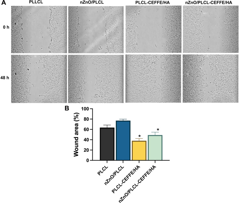
Results: CEFFE was prepared and analyzed, revealing high concentrations of essential growth factors, particularly bFGF and TGF-β1, supporting its therapeutic potential in tissue regeneration. The fabricated nanofibers (PLCL, nZnO/PLCL, PLCL-CEFFE/HA, and nZnO/PLCL-CEFFE/HA) were examined using FE-SEM and TEM, demonstrating successful encapsulation and morphological variations due to nZnO incorporation. XRD analysis confirmed the structural integrity and effective loading of nZnO and CEFFE. Hydrophilicity assessment via water contact angle measurements showed that CEFFE/HA significantly enhanced the hydrophilicity of PLCL membranes, crucial for wound exudate management. Mechanical tests indicated that CEFFE/HA addition maintained the scaffold's mechanical robustness, while nZnO slightly reduced mechanical properties. In vitro release studies revealed a biphasic release pattern of Zn²⁺ ions and growth factors from nZnO/PLCL-CEFFE/HA nanofibers, ensuring prolonged antibacterial activity and sustained therapeutic effects. Antibacterial assays demonstrated significant efficacy against E. coli and S. aureus, attributed to nZnO. MTT assays and FE-SEM analysis confirmed enhanced NIH-3T3 cell proliferation and adhesion on PLCL-CEFFE/HA nanofibers due to the controlled release of growth factors. The scratch assay showed superior cell migration and wound healing potential for PLCL-CEFFE/HA formulations.
Conclusions: These findings underscore the potential of nZnO/PLCL-CEFFE/HA core-shell nanofibers as multifunctional wound dressings, combining antibacterial properties with enhanced tissue regeneration capabilities. However, further studies are needed to assess long-term stability and in vivo performance, which represent key challenges for future research.
Keywords: Cell-free fat extract; Core-shell nanofiber; Wound dressing; Zinc oxide nanoparticles.
© 2025. The Author(s).
Conflict of interest statement
Declarations. Ethics approval and consent to participate: Not applicable. Consent for publication: Not applicable. Competing interests: The authors declare no competing interests.
Figures
Similar articles
-
Sodium alginate hydrogels co-encapsulated with cell free fat extract-loaded core-shell nanofibers and menstrual blood stem cells derived exosomes for acceleration of articular cartilage regeneration.Int J Biol Macromol. 2024 Sep 21;280(Pt 3):135851. doi: 10.1016/j.ijbiomac.2024.135851. Online ahead of print. Int J Biol Macromol. 2024. PMID: 39307503
-
Synergistic berberine chloride and Curcumin-Loaded nanofiber therapies against Methicillin-Resistant Staphylococcus aureus Infection: Augmented immune and inflammatory responses in zebrafish wound healing.Int Immunopharmacol. 2024 Oct 25;140:112856. doi: 10.1016/j.intimp.2024.112856. Epub 2024 Aug 8. Int Immunopharmacol. 2024. PMID: 39121609
-
In situ Fabrication of Nano ZnO/BCM Biocomposite Based on MA Modified Bacterial Cellulose Membrane for Antibacterial and Wound Healing.Int J Nanomedicine. 2020 Jan 6;15:1-15. doi: 10.2147/IJN.S231556. eCollection 2020. Int J Nanomedicine. 2020. PMID: 32021161 Free PMC article.
-
A focused review on hyaluronic acid contained nanofiber formulations for diabetic wound healing.Int J Biol Macromol. 2023 Dec 31;253(Pt 8):127607. doi: 10.1016/j.ijbiomac.2023.127607. Epub 2023 Oct 21. Int J Biol Macromol. 2023. PMID: 37871723 Review.
-
Wound Dressing with Electrospun Core-Shell Nanofibers: From Material Selection to Synthesis.Polymers (Basel). 2024 Sep 5;16(17):2526. doi: 10.3390/polym16172526. Polymers (Basel). 2024. PMID: 39274158 Free PMC article. Review.
References
-
- Hamed SH, Azooz EA, Al-Mulla EAJ. Nanoparticles-assisted wound healing: a review. Nano Biomed Eng. 2023;15(4):425–35.
-
- Pilehvar-Soltanahmadi Y, Dadashpour M, Mohajeri A, Fattahi A, Sheervalilou R, Zarghami N. An overview on application of natural substances incorporated with electrospun nanofibrous scaffolds to development of innovative wound dressings. Mini Rev Med Chem. 2018;18(5):414–27. - PubMed
-
- Wang X, Hu R, Han L, Lu X. Multifunctional hydrogels for chronic wounds repairing. Biosurface Biotribology. 2023;9(4):85–100.
LinkOut - more resources
Full Text Sources
