

A Comparative Study on Genotoxic and Oxidative DNA Damage in Oral Epithelial Cells of COVID-19 Suspected Patients
- PMID: 40390722
- PMCID: PMC12088234
- DOI: 10.7759/cureus.82593
A Comparative Study on Genotoxic and Oxidative DNA Damage in Oral Epithelial Cells of COVID-19 Suspected Patients
Abstract
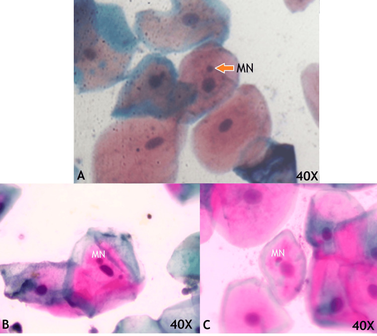
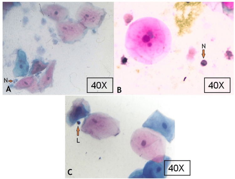
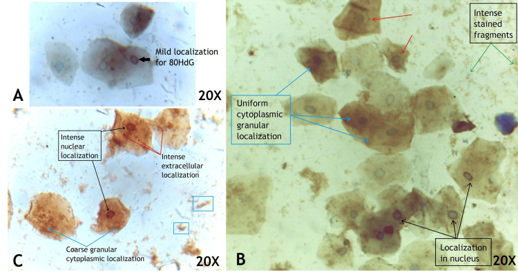
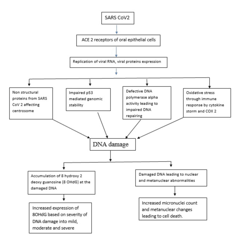
Introduction The SARS-CoV-2 virus causes COVID-19 by chiefly infecting the nasal and oral mucosal cavities, where its spike proteins bind to angiotensin-converting enzyme II (ACE2) receptors on epithelial cells. This study aimed to assess SARS-CoV-2-related genotoxicity in buccal mucosal cells through micronuclei counts and 8-Oxo-2'-deoxyguanosine (8-OHdG) expression. Methods This cross-sectional study was conducted in 86 COVID-19 suspected patients aged 18-45 years attending an outpatient screening area for reverse transcription polymerase chain reaction (RT-PCR) SARS-CoV-2 testing between December 2023 and February 2024. Salivary swabs were obtained and smeared on glass slides for each patient. These slides were stained to study and compare cellular toxicity by Papanicolaou (Pap) staining and oxidative DNA damage expression by immunohistochemistry. The categorical and continuous variables were assessed using the independent t-test and ANOVA. A p-value of less than 0.05 was considered statistically significant. Results The number of micronucleated cells, total micronuclei, neutrophil, lymphocyte and inflammatory cell count in COVID-19 RT-PCR positive patients were 11.32 ± 7.00, 17.32± 12.53, 1.93 ± 1.17, 3.98 ± 2.55 and 5.90 ± 3.15 respectively which is higher than COVID-19 RT-PCR negative individuals 6.09± 3.83, 9.04± 5.82, 0.18± 0.49, 2.13± 0.84 and 2.31±1.01 respectively. Moreover, buccal cells showed increased oxidative DNA damage with intense staining for 8-OHdG in COVID-19-positive patients. The epithelial-to-inflammatory cell ratio was very low in COVID-19-positive buccal smear patients. Conclusion This study concludes that SARS-CoV-2 has a genotoxic effect by increasing the micronuclei count and has oxidative DNA damage by increasing the 8-OHdG expression in the exfoliated buccal cells.
Keywords: 8 hydroxy deoxy guanosine; buccal cells; covid-19; oxidative dna damage; sars-cov2.
Copyright © 2025, Poojari et al.
Conflict of interest statement
Human subjects: Consent for treatment and open access publication was obtained or waived by all participants in this study. AIIMS/MG/IEC/2023-24/57 issued approval IEC/2023-24/57. Animal subjects: All authors have confirmed that this study did not involve animal subjects or tissue. Conflicts of interest: In compliance with the ICMJE uniform disclosure form, all authors declare the following: Payment/services info: All authors have declared that no financial support was received from any organization for the submitted work. Financial relationships: All authors have declared that they have no financial relationships at present or within the previous three years with any organizations that might have an interest in the submitted work. Other relationships: All authors have declared that there are no other relationships or activities that could appear to have influenced the submitted work.
Figures
References
-
- WHO coronavirus (COVID-19) dashboard. 2024. https://data.who.int/dashboards/covid19/deaths https://data.who.int/dashboards/covid19/deaths
LinkOut - more resources
Full Text Sources
Research Materials
Miscellaneous