Review
. 2025 May 19;4(2):e70010.
doi: 10.1002/smmd.70010. eCollection 2025 Jun.

The Application of Microfluidic Chips in Primary Urological Cancer: Recent Advances and Future Perspectives

Affiliations
Review

The Application of Microfluidic Chips in Primary Urological Cancer: Recent Advances and Future Perspectives

Jiafu Liu et al. Smart Med. .

Abstract

The research of primary urological cancers, including bladder cancer (BCa), prostate cancer (PCa), and renal cancer (RCa), has developed rapidly. Microfluidic technology provides a good variety of benefits compared to the heterogeneity of animal models and potential ethical issues of human study. Microfluidic technology and its application with cell culture (e.g., organ-on-a-chip, OOC) are extensively used in urological cancer studies in preclinical and clinical settings. The application has provided diagnostic and therapeutic benefits for patients with urological diseases, especially by evaluating biomarkers for urinary malignancies. In this review, we go through the applications of OOC in BCa, Pca and Rca, and discuss the prospects of reducing the cost and improving the repeatability and amicability of the intelligent integration of urinary system organ chips.

Keywords: in vitro model; microfluidic technology; organ‐on‐a‐chip; urological cancer.


Conflict of interest statement

The authors declare no conflicts of interest.

Figures

FIGURE 1
FIGURE 1
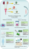
Application of microfluidics and OOCs in the detection, treatment, and prognosis of urinary system tumors.
FIGURE 2
FIGURE 2
Models for diseases of the urinary system. Reproduced with permission [53]. Copyright 2017, National Academy of Sciences. Reproduced with permission [133a]. Copyright 2009, Royal Society of Chemistry. Reproduced with permission [133b]. Copyright 2018, Journal of Visualized Experiments.
FIGURE 3
FIGURE 3
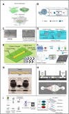
Application of microfluidic technology in bladder organs. (A) Microfluidic device for filtering ETCs by controlling pore size. Reproduced with permission [62]. Copyright 2018, Elsevier. (B) Microfluidic device with spiral channel filtration system. Reproduced under terms of the CC‐BY license [134]. Copyright 2019, The Authors, published by MDPI. (C) A microfluidic chip capable of concentrating fresh or frozen urine for dual biomarker immunoassays such as pan‐CK (epithelial biomarker), Sialyl‐Tn (tumor‐associated biomarker), and DAPI (nuclear staining). Reproduced under terms of the CC‐BY license [65]. Copyright 2020, The Authors, published by Frontiers Media S.A. (D) Dual‐filtration microfluidic device for capturing exosomes. Reproduced under terms of the CC‐BY license [74]. Copyright 2017, The Authors, published by Springer Nature. (E) Herringbone‐structured microfluidic chip for capturing CTCs. Reproduced with permission [77]. Copyright 2020, Taylor & Francis. (F) Impedance‐based microfluidic chip for identifying BCa cells grading BCa OOC. Reproduced with permission [79]. Copyright 2022, Royal Society of Chemistry. (G) BCa cultures in microfluidic devices. Reproduced under terms of the CC‐BY license [84]. Copyright 2017, The Authors, published by Springer Nature. (H) BCa organ‐on‐chip. Reproduced under terms of the CC‐BY license [85]. Copyright 2024, The Authors, published by John Wiley and Sons. (I) Structure of BCa OOC based on BCG vaccine. Reproduced under terms of the CC‐BY license [87]. Copyright 2021, The Authors, published by MDPI.
FIGURE 4
FIGURE 4
Application of microfluidic technology in prostate organ. (A) The α2,3‐Sia‐PSA assay system. MAA was included in the buffer to separate α2,3‐Sia‐PSA from α2,6‐Sia‐PSA by affinity electrophoresis. Fluorescence signals from laser‐induced fluorescence detection were analyzed, and the percentage of α2,3‐Sia‐PSA was calculated from their peak areas. Reproduced with permission [96]. Copyright 2022, The Authors, published by Springer Nature. (B) Exosome detection and capture using a microfluidic Raman chip. Reproduced with permission [102]. Copyright 2020, Royal Society of Chemistry. (C) Based on the analysis of different cancer cells. Reproduced under terms of the CC‐BY license [105]. Copyright 2019, The Authors, published by MDPI. (D) A vortex microfluidic chip for isolation of prostate CTCs. Reproduced under terms of the CC‐BY license [108]. Copyright 2017, The Authors, published by Springer Nature. (E) A lateral magnetophoretic microseparator for separation of CTCs for a deeper understanding of cancer characterization. Reproduced under terms of the CC‐BY license [110]. Copyright 2020, The Authors, published by MDPI. (F) A microfluidic device (GO chip) based on graphene oxide for circulating tumor cell counts and RNA extraction, respectively. Reproduced under terms of the CC‐BY license [112]. Copyright 2019, The Authors, published by John Wiley and Sons.
FIGURE 5
FIGURE 5
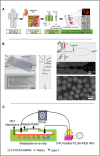
Application of microfluidics in kidney organs. (A) Microfluidics‐based in vitro model in which primary patient‐specific TEnCs and NEnCs are used to create patient‐specific biomimetic blood vasculature. Reproduced with permission [121]. Copyright 2019, The Authors, published by Elsevier. (B) Five‐entry On‐Chip Organoid structure diagram. Reproduced under terms of the CC‐BY license [127]. Copyright 2022, The Authors, published by MDPI. (C) Schematic of the tumor progression model based on metastasis‐on‐a‐chip. Reproduced under terms of the CC‐BY license [130]. Copyright 2020, The Authors, published by Ivyspring International Publisher.

Similar articles

References

    1. Sung H., Ferlay J., Siegel R. L., et al., “Global Cancer Statistics 2020: GLOBOCAN Estimates of Incidence and Mortality Worldwide for 36 Cancers in 185 Countries,” CA: A Cancer Journal for Clinicians 71 (2021): 209. - PubMed
    1. Kriegmair M. C., Rother J., Grychtol B., et al., “Multiparametric Cystoscopy for Detection of Bladder Cancer Using Real‐Time Multispectral Imaging,” European Urology 77 (2020): 251. - PubMed
    1. Siegel R., Ma J., Zou Z., and Jemal A., “Cancer Statistics, 2014,” CA: A Cancer Journal for Clinicians 64 (2014): 9. - PubMed
    1. Gandaglia G., Leni R., Bray F., et al., “Epidemiology and Prevention of Prostate Cancer,” European Urology Oncology 4 (2021): 877. - PubMed
    1. Suarez‐Ibarrola R., Sigle A., Eklund M., et al., “Artificial Intelligence in Magnetic Resonance Imaging–Based Prostate Cancer Diagnosis: Where Do We Stand in 2021,” European Urology Focus 8 (2022): 409. - PubMed

LinkOut - more resources