. 2025 Apr 23;9(6):ziaf063.
doi: 10.1093/jbmrpl/ziaf063. eCollection 2025 Jun.

Denosumab treatment of giant cell tumors in the spine induces woven bone formation

Affiliations

Denosumab treatment of giant cell tumors in the spine induces woven bone formation

Robyn Birch et al. JBMR Plus. .

Abstract

Giant cell tumors of bone (GCTB) are rare but aggressive, locally destructive tumors. They typically affect young people, significantly reducing their quality of life and increasing mortality rates. Giant cell tumors of bone are composed of osteoclast-like giant cells that respond to increased secretion of RANKL by stromal cells, triggering osteolysis. For over a decade, denosumab, a monoclonal antibody targeting this receptor activator, has been approved as a neo-adjuvant to facilitate surgical resection or in the setting of inoperable tumors. Denosumab treatment has shown rapid pain improvement and tumor size reduction in the spine. Although variable degrees of tumor mineralization have been observed in clinical applications of this drug, the nature of this newly formed mineralized tissue has yet to be determined. To characterize both mineralization and collagen organization in the newly formed bone, we conducted extensive analyses on 4 posttreatment giant cell tumor vertebral samples, involving quantitative backscattered imaging, electron probe microanalysis, and a novel method for determining the alignment of collagen fibrils using second harmonic generation. Additionally, biological mechanisms involved in bone mineralization and matrix formation were analyzed using histological staining and mass spectroscopy. Our results concluded that denosumab treatment after giant cell tumor of bone in the spine was associated with the formation of woven bone and increased mineral density in a matrix of disorganized collagen fibers characterized by increased collagen III content, with the response appearing to depend on patient age and extension of treatment. To our knowledge, this is the first comprehensive material-based study on the bone formed during denosumab treatment for GCTB, providing valuable information on how denosumab affects bone quality and how the reported methodology can be applied to similar studies.

Keywords: bone microscopy; bone mineralization; denosumab; giant cell tumor; spine; woven bone.


Conflict of interest statement

R.C.M has received research support from a 2024 AOSpine Knowledge Forum Associate Research Award, AOSpine, the Canadian Orthopaedic Foundation, a 2023 NASS translational award, a 2023 New Frontiers in Research Exploration Award, a 2023 Vancouver Coastal Health Research Institute Investigator Award, and a 2022 AOSpine DIA, they serve on the advisory board for Cerapedics Education Event as a co-chair, is co-chairing a Stryker educational event, and a CarboFix-joint solutions educational event. N.D. has been a consultant for Baxter, Cerapedics, and Stryker, owns stock in Medtronic, and has fellowship support paid to their institution from AOSpine, Medtronic, and Depuy Synthes. C.F. has been a consultant for Medtronic, is awarded royalties from Medtronic, and has fellowship support paid to their institution from AOSpine, Medtronic, and Depuy Synthes.

Figures

Graphical Abstract
Graphical Abstract
Figure 1
Figure 1
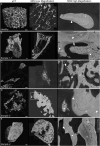
Micro-computed tomography 3D renderings of full-size cadaveric and surgically resected vertebrae (left column), followed by a stitched SEM low magnification image of the specimen (middle column), and a high magnification image of a selected ROI, outlined in white on the low magnification image (right column). (A) A vertebral matched healthy control demonstrating normal trabecular lamellar structure (open box) and ovoid lacunae (closed arrowhead) oriented along the lamellar major axis. (B) Sample 1, a mixed bone type with lytic-like disorganized bone formation inside large voids within original cortical and trabecular bone, with lamellar bone (open box) on the surfaces of woven bone (closed box) and irregular diffuse edges and porous erosive-like patterns (arrows, open and closed). The presence of ovoid lacunae (closed arrowheads) in lamellar bone is accompanied by round or irregular-shaped lacunae (open arrowheads) present in increased density in the woven bone. (C) Sample 2, a disorganized bone sample with lytic-like woven bone formation (closed box), porous indentations on the trabecular surfaces (closed arrows), and increased mineral deposits indicated by asterisks. (D) Sample 3, a lytic-like disorganized bone sample, with evidence of normal lamellar structure (open box), accompanied by adjacent woven bone (closed box), with ovoid (closed arrowhead), and nonovoid (open arrowhead) lacunae presence in adjacent bone types. (E) Sample 4, an osteoblastic sample characterized by thick trabeculae demonstrating an irregular, woven calcified matrix (closed box), with instances of lamellar bone forming on the surface (open box). Again, ovoid lacunae (closed arrowheads) are found in lamellar bone with round or irregular-shaped lacunae (open arrowheads) present in increased density in the woven bone. The scale of μCT renders = 1 mm, and all SEM high magnification images = 200 μm.
Figure 2
Figure 2
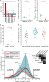
Analysis of bone morphological parameters acquired using μCT and mineral density of each sample measured using SEM-qBSE. (A) A waterfall plot of the bone volume/total volume (BV/TV) grouped by individual scans. The horizontal line indicates average healthy BV/TV, with the transparent box representing the minimum to maximum range. (B–D) Scatterplots of trabecular morphology quantification of the healthy and GCTB groups based on μCT evaluation. Statistical significance for each parameter was evaluated using a Mann-Whitney test. (E) Scatterplot of mineral content grouped by bone type. (F) A bone mineral density distribution using average histogram data for all controls and each GCTB sample. Statistical significance was evaluated between samples using a Kruskal–Wallis test. *p < .05, **p < .01, and ***p < .001, ns = not significant.
Figure 3
Figure 3
Lacunar morphology characterization scatterplots with median formula image IQR for healthy vertebrae and both disorganized and lamellar bone from GCTB samples. (A) Lacunae density, (B) lacunae number, (C) individual lacunae area, (D) distribution of major and minor axis length, (E) major axis to minor axis length ratio, and (F) measure of lacunae roundness. *p < .05, **p < .01, and ***p < .001, ns = not significant. Number of analyzed lacunae in healthy lamellar areas = 809, lacunae in GCTB lamellar areas = 1457, and in GCTB disorganized areas = 2108.
Figure 4
Figure 4
SHG collagen fiber alignment measurement. (A) A waterfall plot to illustrate the alignment present in the SHG images with grouping of bone types based on alignment ratio. The healthy treatment group, and the normal GCTB lamellar bone fall to the left side of the plot, indicating the fibers are more aligned—a result of pixels of the same intensity, directed in the same direction. The GCTB disorganized bone falls to the far right of the waterfall plot due to the decreased alignment of the fibers, a result of disorganized collagen fibers, seen by randomized intensities in different directions. (B) An ROI of sample 1 containing both disorganized and lamellar bone tissue, depicting a lamellar ROI, and a disorganized ROI. Branching trabeculae are visible in greyscale, with the collagen in the normal lamellae appearing brighter with linear striations. The collagen in the disorganized regions appears thinner, with variable intensity and random alignment. (C) A selected disorganized ROI, and (D) its resultant FFT illustrating minimal alignment of the fibers. (E) A selected lamellar ROI, and (F) its resultant FFT illustrating a preferred direction of alignment of the fibers. The resulting FFTs were rotated 90° for orientational analysis. Scale bar = 200 μm.
Figure 5
Figure 5
Visualization of protein clustering. (A) Dendrogram showing clustering of the sample groups with similar protein expression and clustering based on similar expression patterns. (B) A ranked order of the protein cluster plotted by the protein fold change.
Figure 6
Figure 6
Histological and immunohistochemical analysis showed differences in staining between the lamellar and disorganized bone in the GCTB samples. (A) H&E, (B) toluidine blue staining results in a staining pattern across the entire matrix, with differences in the stain intensity result in disorganized bone staining darker (closed arrowheads) than the lamellar regions (open arrowheads). The abnormal lacunae tend to stain darker along the lacunar edges, whereas lamellar bone lacunae tend to stain with a clean, smooth surface. (C) Collagen III, (D) osteopontin, (E) osteomodulin, and (F) decorin immunohistochemical staining, demonstrating increased staining in the disorganized matrix (closed arrowheads) compared to the lamellar matrix (open arrowheads). Images correspond to GCTB sample 4. Scale bar = 200 μm.

Similar articles

Cited by

References

    1. Xara-Leite F, Coutinho L, Fleming C, et al. Can denosumab cure giant cell tumors of the spine? A case report and literature review. Eur J Orthop Surg Traumatol. 2020;30(1):19–23. 10.1007/s00590-019-02554-9 - DOI - PubMed
    1. Goldschlager T, Dea N, Boyd M, et al. Giant cell tumors of the spine: has denosumab changed the treatment paradigm? J Neurosurg Spine. 2015;22(5):526–533. 10.3171/2014.10.SPINE13937 - DOI - PubMed
    1. Liede A, Bach BA, Stryker S, et al. Regional variation and challenges in estimating the incidence of giant cell tumor of bone. J Bone Joint Surg Am. 2014;96(23):1999–2007. 10.2106/JBJS.N.00367 - DOI - PubMed
    1. Amelio JM, Rockberg J, Hernandez RK, et al. Population-based study of giant cell tumor of bone in Sweden (1983–2011). Cancer Epidemiol. 2016;42:82–89. 10.1016/j.canep.2016.03.014 - DOI - PubMed
    1. Dahlin DC, Cupps RE, Johnson EW. Giant-cell tumor: a study of 195 cases. Cancer. 1970;25(5):1061–1070. 10.1002/1097-0142(197005)25:5<1061::AID-CNCR2820250509>3.0.CO;2-E - DOI - PubMed

LinkOut - more resources