. 2025 Apr 26:32:101805.
doi: 10.1016/j.mtbio.2025.101805. eCollection 2025 Jun.

Ultrasound-driven wireless piezoelectric hydrogel synergizes with cotransplantation of NSCs-hUCMSCs for structural and functional recovery in spinal cord injury

Affiliations

Ultrasound-driven wireless piezoelectric hydrogel synergizes with cotransplantation of NSCs-hUCMSCs for structural and functional recovery in spinal cord injury

Hao Zhong et al. Mater Today Bio. .

Abstract

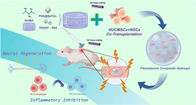
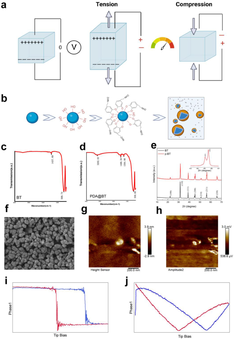
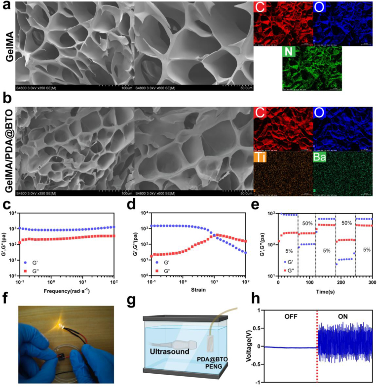
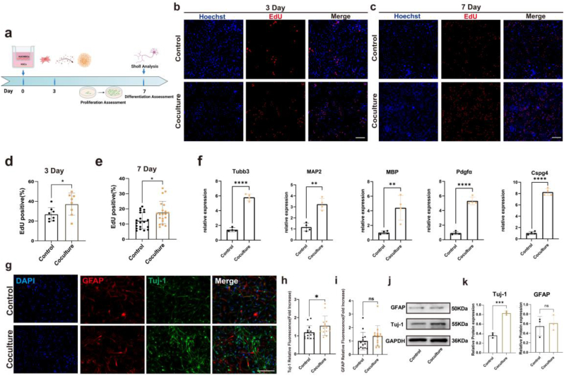
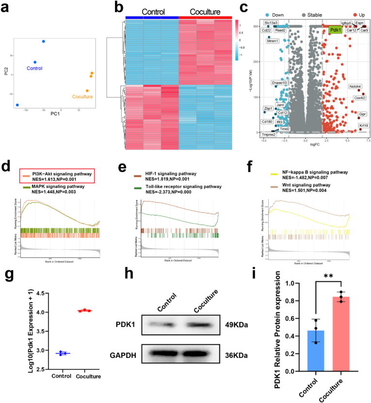
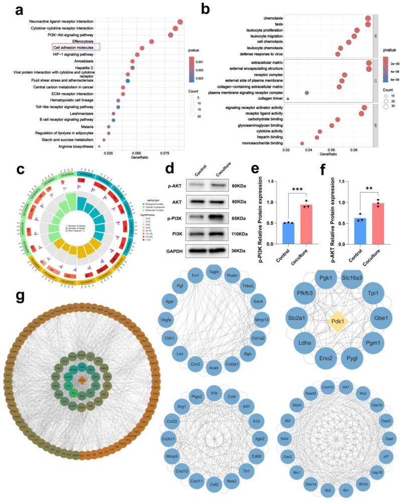
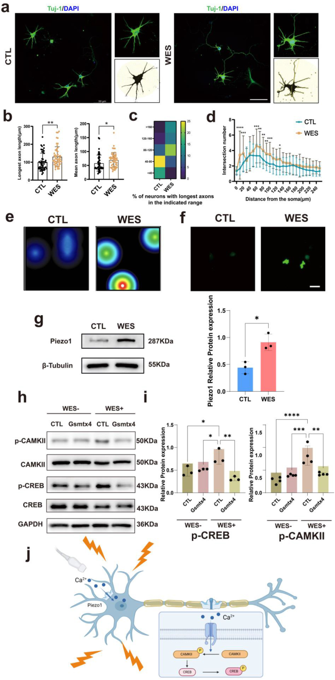
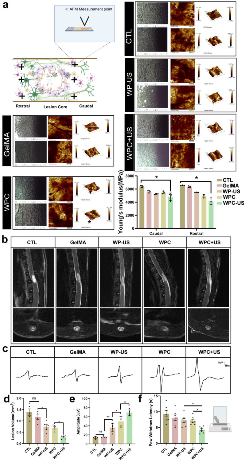
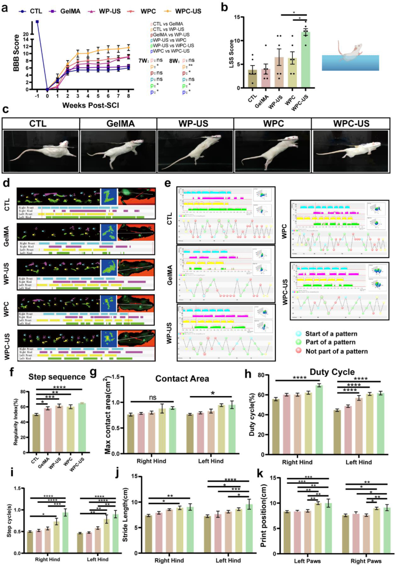
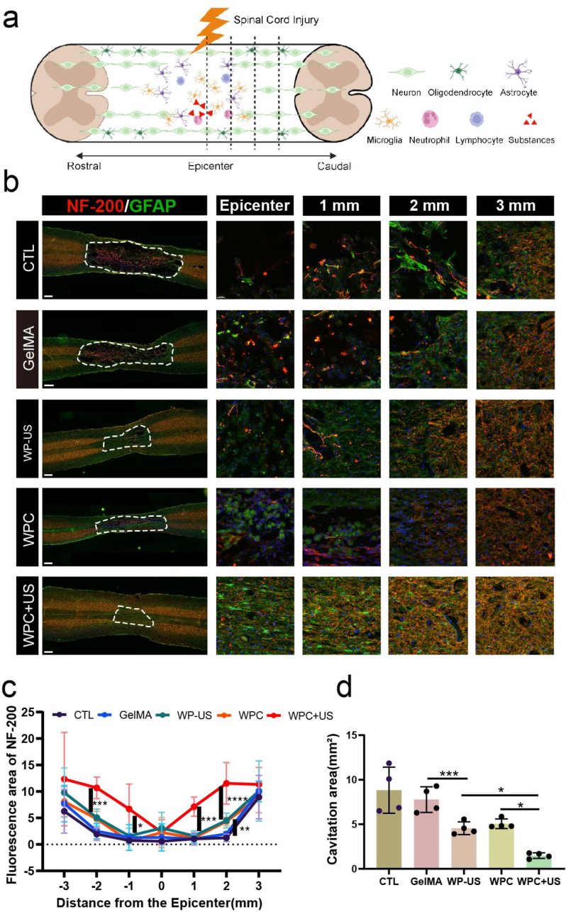
Spinal cord injury (SCI) is a devastating condition of the central nervous system, characterized by disrupted regulation of the immune microenvironment and the loss of electrical signaling, which poses significant challenges to repair. Neural stem cells (NSCs) have the potential to promote functional recovery after SCI; however, their therapeutic potential is limited by poor survival, restricted proliferation, and suboptimal differentiation. Human umbilical cord-derived mesenchymal stem cells (hUCMSCs) possess powerful paracrine and immunomodulatory properties, providing a supportive niche that improves the engraftment and function of NSCs. Recently, piezoelectric materials have attracted increasing attention for their ability to convert mechanical energy into electrical signals, thus providing a noninvasive, wireless alternative to traditional electrode-based therapies for neural regeneration. In this study, we investigated the synergistic effects of NSCs and hUCMSCs, focusing on how hUCMSCs direct NSC differentiation and the mechanisms underlying this action. We also introduce an ultrasound-driven wireless piezoelectric hydrogel, which generates electrical signals through the piezoelectric effect. In vitro, wireless electrical stimulation activated primary cortical neurons, stimulated axonal growth, and promoted neuronal plasticity through the Piezo1 channel and downstream CREB/CAMKII signaling pathways. In a rat SCI model, wireless piezoelectric hydrogel synergized with cotransplanting NSCs-hUCMSCs and modulated the immune microenvironment during the acute phase, thereby restructuring scar cavities during the chronic phase, suppressing scar formation, accelerating neurogenesis, and facilitating axonal regeneration. These results emphasize the potential of synergizing stem cell therapies with wireless piezoelectric stimulation as a promising strategy for SCI repair, providing novel insights into the clinical translation of regenerative treatments.

Keywords: Piezoelectric nanogenerator; Spinal cord injury; Ultrasound; Wireless electrical stimulation.


Conflict of interest statement

The authors declare that they have no known competing financial interests or personal relationships that could have appeared to influence the work reported in this paper.

Figures

Image 1
Graphical abstract
Fig. 1
Fig. 1
Schematic of the WPC hydrogel for recovery after SCI. The WPC hydrogel is activated by an external US to generate electrical cues. Created with BioRender.com.
Fig. 2
Fig. 2
Characterization and piezoelectric properties of PDA@BT NPs. a) Schematic of piezoelectric signal generation. b) Schematic of the preparation of PDA@BT NPs. c, d) FT-IR spectra of BT NPs and PDA@BT NPs. e) XRD curves of BT and PDA@BT, respectively. f) TEM images of PDA@BT NPs. Scale bar = 500 nm. g, h) Images of topography g) and amplitude h) of PDA@BT NPs observed via PFM. i, j) Piezoresponse phase curves i) and amplitude–voltage j) PDA@BT NPs.
Fig. 3
Fig. 3
Characterization and piezoelectric properties of the PEDOT/PDA@BT hydrogel. a, b). SEM images and EDX surface-scan element distribution of GelMA a) and WPC hydrogel b). (Scale bar = 100 and 50 μm, respectively.) c-e) Rheological properties of WPC hydrogel. f) Images visually demonstrate the conductivity of the PEDOT/PDA@BT hydrogel. g) Schematic of the electrical output of piezoelectric nanogenerator (PENG) made of WP hydrogel under US activation. h) The open-circuit voltage generated by the piezoelectric nanogenerator under US activation.
Fig. 4
Fig. 4
hUCMSCs induce NSCs proliferation and differentiation. a) Schematic of the evaluation timeline of coculture. Created with BioRender.com. b-e) Proliferation of NSCs determined using the EDU assay. Scale bars, 100 μm. f) RT-qPCR analysis of the expression of the neuron-specific genes Tuj-1, MAP2, MBP, PDGFα, and CSPG4 in NSCs cultured with hUCMSCs for 7 days. g) Representative images of Tuj-1/GFAP/nuclear immunostaining of NSCs. Scale bars, 100 μm. h, i) Quantification of the differentiation rates of NSCs. j) Representative western blotting images of Tuj-1 and GFAP of NSCs that received different treatments; GAPDH was used as the reference protein. k) Quantitative analysis of the expression level of Tuj-1 and GFAP. (ns indicates no significant, ∗p < 0.05, ∗∗p < 0.01, ∗∗∗p < 0.001, ∗∗∗∗p < 0.0001).
Fig. 5
Fig. 5
Identification of DEGs in the control and coculture groups. a) Principal component analysis (PCA) of transcriptome sequencing samples b) Heatmap of DEGs. c) Volcano plot of the identified DEGs. d-f) GSEA to show the enriched biological processes in cocultured NSCs. g) PDK1 expression in NSCs in gene level. h) Representative western blotting images of PDK1. i) Quantitative analysis of the expression level of PDK1. (∗∗p < 0.01).
Fig. 6
Fig. 6
The mechanism by which hUCMSCs regulate the cell fate of NSCs. a) KEGG pathway enrichment analysis depicted the major enriched pathway among upregulated genes in Cocultured NSCs. b) Enrichment of GO biological process based on differentially expressed genes between the control and coculture groups. c) Pie chart showing the enriched GO terms. d-f) Western blot images showing the activation of PI3K/AKT pathway proteins in NSCs after coculture with hUCMSCs. g) Visualization of PPI protein interaction network analysis. (∗∗p < 0.01, ∗∗∗p < 0.001, ∗∗∗∗p < 0.0001).
Fig. 7
Fig. 7
WPC hydrogel-facilitated wireless electrical stimulation(WES) activates Piezo1 channel regulating neuronal plasticity through CaMKII/CREB in neurons. a) Tuj-1 immunostaining of neurons cultured for 7 days. Scale bar = 50 μm. b-d) Sholl analysis of dendritic morphology in neurons. e) Heatmaps of spontaneous firing of neurons. f) Representative images of real-time calcium fluorescence image of neurons. Scale bar = 20 μm. g) Expression of Piezo1 in neurons. h, i) Representative western blot images and quantitative analysis of the expression level of p-CAMKII/CaMKII and p-CREB/CREB in neurons. j) Schematic of the mechanism of WES in regulating neuronal plasticity. Created with BioRender.com. (∗p < 0.05, ∗∗p < 0.01, ∗∗∗p < 0.001, ∗∗∗∗p < 0.0001).
Fig. 8
Fig. 8
WPC hydrogel inhibits inflammation and regulates macrophage/microglia polarization in the acute phase after SCI. a) Illustration of the process of hydrogel injection. b) ELISA analysis of the concentrations of TNF-α and IL-1β proteins in spinal cord tissues. c) Immunofluorescence image of iNOS expression in macrophage/microglia. Scale bar = 100 μm. d) Immunofluorescence images of Arg-1 expression in macrophages/microglia. Scale bar = 100 μm. e) Quantitative analysis of the mean fluorescence intensity of iNOS and Arg-1 protein expression. f, g) Representative western blot images and quantitative analysis of the expression level of iNOS and Arg-1. (∗p < 0.05, ∗∗p < 0.01, ∗∗∗p < 0.001, ∗∗∗∗p < 0.0001).
Fig. 9
Fig. 9
WPC hydrogel with wireless electrical stimulation reduces scar formation and lesion cavity. a) AFM stiffness characterization of scar tissue. b) Sagittal and axial T2-weighted images of rats in different groups. c) Representative electrophysiological images of Motor evoked potentials (MEP). d) Quantitative analysis of lesion cavity volume. e) Quantitative analysis MEP signals in each group (n = 5). f) Latency to withdrawal in the hot plate test. (ns indicates no significant, ∗p < 0.05, ∗∗p < 0.01).
Fig. 10
Fig. 10
WPC hydrogel with WES promotes locomotor function recovery after SCI. a) BBB scores from preinjury to 8 weeks postinjury (n = 6). b,c) LSS scores Photographs of swimming test at 56 days postinjury (n = 6). d, e) Representative images of footprint tracks and gait patterns. Left forelimb (yellow), LF; right forelimb (blue), RF; left hindlimb (green), LH; right hindlimb (purple), RH. f-k) Statistical analysis of six commonly used catwalk parameters (regularity index, max contact area, duty cycle, step cycle, stride length, and print position) (n = 6). (ns indicates no significant, ∗p < 0.05, ∗∗p < 0.01, ∗∗∗p < 0.001, ∗∗∗∗p < 0.0001). (For interpretation of the references to colour in this figure legend, the reader is referred to the Web version of this article.)
Fig. 11
Fig. 11
The WPC hydrogel with wireless ES facilitates axonal regeneration and remodeling of the neuronal network after SCI. a) Schematic of the measured area of spinal cord gray matter. (Created with BioRender.com.) b) Immunofluorescence staining of the injured area 28 days after SCI. Red represents NF200-positive, and green represents GFAP-positive. Scale bars: 500 μm. Scale bars of magnified image: 30 μm. c) Quantification o the fluorescence intensity at different distances to the epicenter. d) Cavitation area of lesion core. (∗p < 0.05, ∗∗p < 0.01, ∗∗∗p < 0.001, ∗∗∗∗p < 0.0001). (For interpretation of the references to colour in this figure legend, the reader is referred to the Web version of this article.)

Similar articles

References

    1. Hao D., Du J., Yan L., He B., Qi X., Yu S., Zhang J., Zheng W., Zhang R., Huang D.-G., et al. Trends of epidemiological characteristics of traumatic spinal cord injury in China, 2009-2018. Eur. Spine J. 2021;30(10):3115–3127. doi: 10.1007/s00586-021-06957-3. PubMed. - DOI - PubMed
    1. Zhou M., Xu Z., Zhong H., Ning G., Feng S. Spinal cord injury and inflammatory mediators: role in "fire barrier" formation and potential for neural regeneration. Neural Regen Res. 2025 doi: 10.4103/NRR.NRR-D-24-00792. PubMed. - DOI - PubMed
    1. Hu X., Xu W., Ren Y., Wang Z., He X., Huang R., Ma B., Zhao J., Zhu R., Cheng L. Spinal cord injury: molecular mechanisms and therapeutic interventions. Signal Transduct. Target. Ther. 2023;8(1):245. doi: 10.1038/s41392-023-01477-6. PubMed. - DOI - PMC - PubMed
    1. Zipser C.M., Cragg J.J., Guest J.D., Fehlings M.G., Jutzeler C.R., Anderson A.J., Curt A. Cell-based and stem-cell-based treatments for spinal cord injury: evidence from clinical trials. Lancet Neurol. 2022;21(7):659–670. doi: 10.1016/S1474-4422(21)00464-6. PubMed. - DOI - PubMed
    1. Chen S.-Y., Yang R.-L., Wu X.-C., Zhao D.-Z., Fu S.-P., Lin F.-Q., Li L.-Y., Yu L.-M., Zhang Q., Zhang T. Mesenchymal stem cell transplantation: neuroprotection and nerve regeneration after spinal cord injury. J. Inflamm. Res. 2023;16:4763–4776. doi: 10.2147/JIR.S428425. PubMed. - DOI - PMC - PubMed

LinkOut - more resources