. 2025 May 5:16:1527115.
doi: 10.3389/fimmu.2025.1527115. eCollection 2025.

Self and parasite-derived peptides selected upon DERAA-bearing HLA-DRB1 alleles activate CD4+ T cells from Chagas cardiomyopathy patients and are associated with ventricular dysfunction

Affiliations

Self and parasite-derived peptides selected upon DERAA-bearing HLA-DRB1 alleles activate CD4+ T cells from Chagas cardiomyopathy patients and are associated with ventricular dysfunction

Thaiany G Souza-Silva et al. Front Immunol. .

Abstract

Introduction: Human infection with the protozoan Trypanosoma cruzi causes Chagas disease, which may lead to a deadly dilated cardiomyopathy resulting from T-cell-mediated inflammation. We found that specific HLA-DRB1 alleles (*0103, *0402, *1301, and *1302) that display the DERAA motif are linked to this severe clinical manifestation of Chagas disease.

Methods: We employed computational analysis, in vitro functional assays, and single-cell RNA sequencing to determine the response of CD4+ T cells from indeterminate (IND) and cardiac (CCC) Chagas patients to peptides selected on DERAA-bearing alleles.

Results: We observed that these alleles display binding affinity towards host-derived peptides with sequence similarity to parasite-derived proteins. These peptides can activate and induce proliferation of CD4+ T-cells from CCC, but not IND. Importantly, the magnitude of this response correlated with the severity of ventricular dysfunction and increased production of soluble factors associated with myocardial fibrosis. Analysis of differentially expressed genes (DEGs) in activated CD4+ T-cells from individuals with the DERAA-DRB1 alleles demonstrated a high expression of cytotoxic, chemotactic and proapoptotic genes, linking these cells with pathogenic functions. Finally, we observed the upregulation of genes that code for the host proteins that contain the potentially pathogenic peptides in the cardiac tissue of CCC, suggesting their involvement in cardiomyopathy.

Discussion: Our findings highlight the ability of CD4+ T-cells from CCC patients to recognize and react to foreign and self-peptides, thereby emphasizing the importance of HLA-DRB1 alleles in the presentation of potentially pathogenic antigens and in the amplification of CCC pathology.

Keywords: Chagas disease; HLA-DRB1; T-cells; cardiomyopathy; cross-reactivity.


Conflict of interest statement

The authors declare that the research was conducted in the absence of any commercial or financial relationships that could be construed as a potential conflict of interest. The author(s) declared that they were an editorial board member of Frontiers, at the time of submission. This had no impact on the peer review process and the final decision.

Figures
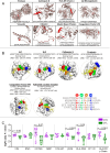
Figure 1
Figure 1
Three-dimensional visualization and interaction of antigens potentially recognized by the different classes of DERAA-bearing HLA-DRB1. (A) The 3D structures of human-proteins potentially recognized by HLA-DRB1 *0103, *0401, *1301 and *1302 alleles visualized using the PyMOL software (version 1.2r3pre). The location of the peptide recognized by HLA-DRB1 is highlighted in red. (B) Peptides bounds to the HLA-DRB1 interface visualized using as template structure PBD 1aqd to *13 allele and 1j8h to *04 allele. The HLA-DRB1 molecule is shown in white surface. The alignment of the peptide sequences in the core region is shown. The amino acids that interact with the protein pockets are colored in red (P1), green (P4), orange (P6) and blue (P9). The amino acids residues colored in yellow are the amino acids residues that bridge TCR contact. (C) Differently expressed genes in the Chagas heart tissue. Cardiac tissue transcripts from CCC patients (10 end-stage heart failure patients) and health subjects (7 heart tissues), available in Gene Expression Omnibus (accession number GEO 84796), were measured as described in Material and Methods. The expression of heart transcripts was expressed as logFC (adj. p value). Comparative analysis between CCC (chronic Chagas cardiomyopathy) and CTL (Control) was carried out by Student’s t-test according to data normality distribution. Significant differences with p < 0.05 are demonstrated. Ig1, Immunoglobulin 1; Ig2, Immunoglobulin 2; CTSS, Cathepsin S; MBP, Myelin Basic Protein; COL2A1, Collagen α-1; β2-M, β2-microglobulin; CF VIII, Coagulation factor VIII.
Figure 2
Figure 2
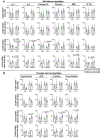
Frequency of CD4+CD69+ total lymphocytes, small and blast cells after stimulation with self-derived and T. cruzi-derived peptides. (A) Frequency (%) of CD4+CD69+ T lymphocytes, small and blast cells proliferating (R2) from indeterminate (IND, n=5) and chronic Chagas cardiomyopathy (CCC, n=5) patients stimulated with medium and anti-CD28 or self-peptides and anti-CD28. (B) Frequency (%) of CD4+CD69+ T lymphocytes, small and blast cells proliferating (R2) from indeterminate (IND, n=5) and chronic Chagas cardiomyopathy (CCC, n=5) patients stimulated with medium and anti-CD28 or parasite-derived peptides and anti-CD28. The analysis was performed as described in the materials and methods for comparison between the groups. Significant differences with p < 0.05 are demonstrated.
Figure 3
Figure 3
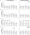
Proliferation index of CD4+CD69+ cells stimulated with self and non-self-peptides. (A) Proliferating index of CD4+CD69+ T lymphocytes, (B) small and (C) blast cells from indeterminate (IND, n=5) and chronic Chagas cardiomyopathy (CCC, n=5) patients stimulated with self-derived and parasite-derived peptides plus anti-CD28. The analysis was performed as described in the materials and methods for comparison between the groups. (D) Proliferating index of CD4+CD69+ blast cells indicating DERAA-HLA-DRB1 bearing patients as orange dots and non-DERAA as blue dots. The statistical analysis was performed as described in the material and methods for comparisons between the groups. Significant differences with p < 0.05 are demonstrated. Ig1, Immunoglobulin 1; Ig2, Immunoglobulin 2; CTSS, Cathepsin S; VIM, Vimentin; MBP, Myelin basic protein; CF VIII, Coagulation factor VIII; Trypan, Trypanothione; ACR, Adenylate cyclase receptor; Cruzip., Cruzipain; Trans-Sial., Trans-Sialidase.
Figure 4
Figure 4
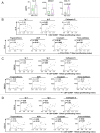
Plasma level of soluble mediators and correlation analysis between proliferation index of CD4+CD69+ blast cells and cardiac function markers. (A) levels of IL-4, CXCL9 and CCL11 in plasma from indeterminate (IND) and chronic Chagas cardiomyopathy (CCC) patients. (B) Correlation between the proliferation index of CD4+CD69+ blast cells from indeterminate and chronic Chagas cardiomyopathy patients stimulated with self-derived peptides (immunoglobulins 1 and 2 and cathepsin S), parasite-derived peptides (Trypanothione, Adenylate cyclase receptor (ACR), Cruzipain and Trans-Sialidase) and IL-4. (C) Correlation between the proliferation index of CD4+CD69+ blast cells from indeterminate and chronic Chagas cardiomyopathy patients stimulated with self-derived peptides (immunoglobulins 1 and 2 and cathepsin S), parasite-derived peptides (Trypanothione, ACR (Adenylate cyclase receptor), Cruzipain and Trans-Sialidase) and CXCL9. (D) Correlation between the proliferation index of CD4+CD69+ blast cells from indeterminate and chronic Chagas cardiomyopathy patients stimulated with self-derived peptides (immunoglobulins 1 and 2 and cathepsin S), parasite-derived peptides (Trypanothione, ACR (Adenylate cyclase receptor), Cruzipain and Trans-Sialidase) and CCL11. Parametric data were analyzed using Pearson’s correlation test and non-parametric data were analyzed using Spearman’s test. Significant differences with p < 0.05 are demonstrated.
Figure 5
Figure 5
Correlation between clinical parameters of cardiac dysfunction and proliferative response to self- and parasite-derived DERAA-selected peptides. (A) Correlation between the proliferation index of CD4+CD69+ blast cells from indeterminate and chronic Chagas cardiomyopathy patients stimulated with self-derived peptides (immunoglobulins 1 and 2 and cathepsin S) and left ventricular ejection fraction (LVEF) or left systolic diameter (LVDD). (B) Correlation between the proliferation index of CD4+CD69+ blast cells from indeterminate and chronic Chagas cardiomyopathy patients stimulated with parasite-derived peptides (Trypanothione, Adenylate cyclase receptor (ACR), Cruzipain and Trans-Sialidase) and left ventricular ejection fraction (LVEF) or left ventricular systolic diameter (LVDD). Parametric data were analyzed using Pearson’s correlation test and non-parametric data were analyzed using Spearman’s test. Significant differences with p < 0.05 are demonstrated.
Figure 6
Figure 6
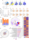
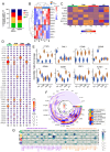
Differentially expressed genes in activated CD4+ T-cells from individuals with indeterminate and chronic Chagas cardiomyopathy. (A) Density of CD3, CD4, CD69 and CD8 gene expression in populations of CD4+CD3+CD69+ T cells formed by unsupervised analysis employing the tSNE algorithm. CD4+CD3+CD69+ T cells from non-DERAA donors are indicated in blue, while cells from donors expressing DRB1-DERAA HLA (*0103, *0402, *1301 and *1302 alleles) are indicated in orange. (B) Volcano Plot of patients with chronic Chagas cardiomyopathy compared to those with indeterminate form. Upregulated genes (Log2 Fold-change > 1.5 and Bonferroni-adjusted p <0.05) are shown in red, and downregulated genes (Log2 Fold-change < -1.5 and Bonferroni-adjusted p <0.05) are shown in blue. Genes without significant differential expression (Log2 Fold-Change between 1.5 and -1.5 and Bonferroni-adjusted p >0.05) are shown in grey. (C) Distributions of CTSW, GZMs (Granzyme A, B, H and K), LAMP1, PRF1 and KLRB1 gene expression in CD4+CD69+ T cells from non-DERAA and DERAA donors. (D) Bar graph demonstrating the expression of genes involved in cytotoxic processes in activated CD4+ T cells from non-DERAA (blue) and DRB1-DERAA (orange) donors. (E) Heatmap demonstrating the gene expression of cytokines and cytokine receptors, and (F) chemokines and chemokine receptors in activated CD4+ T cells from non-DERAA and DRB1-DERAA donors. (G) Radar graph of transcription factor gene expression in activated CD4+ T cells from non-DERAA (blue) and DRB1-DERAA (orange) donors. (H) Signaling pathways enriched in activated CD4+ T cells from donors expressing the HLA-DRB1 alleles. This analysis was performed considering only upregulated and downregulated DEGs. *Indicate differentially expressed genes (DEGs) upregulated (Log2 Fold-change > 1.5) and downregulated DEGs (Log2 Fold-change < -1.5) and Bonferroni-adjusted p value <0.05 are considered significant.
Figure 7
Figure 7
Differentially expressed genes in memory CD4+ T cells from patients with Chagas asymptomatic and chronic Chagas cardiomyopathy. (A) Graph showing the frequency of T naïve (TN, CCR7+CD45RA+), T central memory (TCM, CCR7+CD45RA-), T effector memory (TEM, CCR7-CD45RA-) and T effector (CCR7-CD45RA+) among CD4+CD69+ T cells from non-DERAA and DERAA donors. (B) Heatmap displaying transcription factor gene expression from non-DERAA (blue, n=8) and DERAA (orange, n=9) donors. (C) genes involved in apoptosis pathway in CD4+CD69+ T cell subpopulations from non-DERAA (ND) and DERAA (D) donors. (D) Dot plot of the mean expression of chemokines and chemokine receptors, in CD4+CD69+ T cell subpopulations from non-DERAA (ND) and DERAA (D) donors. (E) Violin plots show the expression of cathepsin W (CTSW), granulysin (GNLY), Granzyme (GZM) A, GZMB, GZMH, GZMK, perforin (PRF1) and KLRB1 in memory CD4+CD69+ T cells from non-DERAA (blue) and DERAA (orange) donors. (F) Heatmap of the mean expression of cytokines and cytokine receptors in CD4+CD69+ T cell subpopulations from non-DERAA (orange) and DERAA (blue) donors. (G) Heatmap of the mean expression of adhesion and migration (purple), proliferation and differentiation (orange) markers in CD4+CD69+ T cell subpopulations from non-DERAA (ND) and DERAA (D) donors. *Upregulated genes (Log2 Fold-change > 1.5) and downregulated genes (Log2 Fold-change < -1.5) and Bonferroni-adjusted p value <0.05 are considered significant.
Figure 8
Figure 8
Schematic representation of differentially expressed genes in memory CD4+ T cells from donors expressing HLA-DRB1 alleles. Illustration of dichotomous gene expression pattern in distinct CD4+CD69+ T cell subsets, including T naïve (TN, CCR7+CD45RA+), T central memory (TCM, CCR7+CD45RA-), T effector memory (TEM, CCR7-CD45RA-) and T effector (CCR7-CD45RA+) cells from Non-DERAA (Blue panel) and DERAA donors (Orange panel). TN CD4+CD69+ from non-DERAA donors exhibit upregulation of TNF-α and IFN-γ cytokines. Additionally, there is an upregulation of IL4R, STAT3, genes inhibiting T cell receptor (TCR) signaling pathway (TIGIT), suggesting potential regulatory roles, and chemotactic genes (CCL20, CCL3, CCL4, CXCL2 and CXCL8). In contrast, TN CD4+CD69+ cells from DERAA donors demonstrate upregulation of genes contributing to the TCR signaling pathway (TRAT1, PI3K), costimulatory molecule (CD28), and genes crucial for cellular differentiation (LTB, STAT1). TCM CD4+CD69+ cells from non-DERAA donors upregulate the expression of IL2RB, IL15RA and GZMB. These cells also show upregulate the expression of IL4R and gene inhibitors of the TCR signaling pathway (TIGIT and PD1). On the other hand, TCM CD4+CD69+ cells from DERAA donors upregulate characteristic genes of Th1 cells (STAT1, STAT4), cytotoxic markers (CD161, granzyme A) and crucial genes for TCR signaling (PI3K, AKT, ZAP70, and LAT). TEM and EC CD4+CD69 cells from non-DERAA donors express regulatory transcription factors (FOXP3, STAT3, STAT6, and IRF4). Both subsets also upregulate the expression of IL4R, KLRC3, BCL2, and BCL2A1, potentially contributing to regulatory and antiapoptotic response. Conversely, TEM and EC cells from DERAA donors downregulate IL4R and upregulate CTLA4, TBX21, and genes involved in cytotoxic processes (GZMA, GZMK, PRF1). In DERAA donors, TEM cells express CD27, CD28, PD1 and NF-кB, while EC cells upregulate FAS, BTLA, and IL18R.

Similar articles

References

    1. Kubinak JL, Ruff JS, Hyzer CW, Slev PR, Potts WK. Experimental viral evolution to specific host MHC genotypes reveals fitness and virulence trade-offs in alternative MHC types. Proc Natl Acad Sci U S A. (2012) 109:3422–7. doi: 10.1073/pnas.1112633109 - DOI - PMC - PubMed
    1. La Gruta NL, Gras S, Daley SR, Thomas PG, Rossjohn J. Understanding the drivers of MHC restriction of T cell receptors. Nat Rev Immunol. (2018) 18:467–78. doi: 10.1038/s41577-018-0007-5 - DOI - PubMed
    1. El Bissati K, Chentoufi AA, Krishack PA, Zhou Y, Woods S, Dubey JP, et al. . Adjuvanted multi-epitope vaccines protect HLA-A*11:01 transgenic mice against Toxoplasma gondii. JCI Insight. (2016) 1:1–18. doi: 10.1172/jci.insight.85955 - DOI - PMC - PubMed
    1. Heide J, Vaughan KC, Sette A, Jacobs T, Zur Wiesch JS. Comprehensive review of human Plasmodium falciparum-specific CD8+ T cell epitopes. Front Immunol. (2019) 10:1–23. doi: 10.3389/fimmu.2019.00397 - DOI - PMC - PubMed
    1. Hamrouni S, Bras-Gonçalves R, Kidar A, Aoun K, Chamakh-Ayari R, Petitdidierid E, et al. . Design of multi-epitope peptides containing hla class-i and class-ii-restricted epitopes derived from immunogenic Leishmania proteins, and evaluation of CD4+ and CD8+ T cell responses induced in cured cutaneous leishmaniasis subjects. PloS Negl Trop Dis. (2020) 14:1–31. doi: 10.1371/journal.pntd.0008093 - DOI - PMC - PubMed

LinkOut - more resources