. 2025 Apr 17;36(2):102540.
doi: 10.1016/j.omtn.2025.102540. eCollection 2025 Jun 10.

Fighting RNA viruses with a gold nanoparticle Cas13d gene-editing armor

Affiliations

Fighting RNA viruses with a gold nanoparticle Cas13d gene-editing armor

Alessandro De Carli et al. Mol Ther Nucleic Acids. .

Abstract

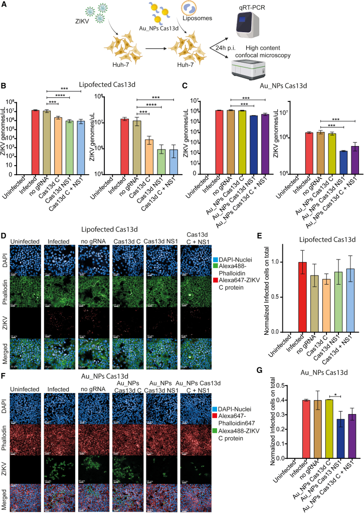
A novel Cas13d-based gene-editing approach has been developed to target viral RNAs in infected cells, reducing the replication of severe acute respiratory syndrome coronavirus 2 (SARS-CoV-2) and Zika virus (ZIKV) by up to 90% compared with controls. Despite its potential, the use of Cas13d as an antiviral faces several challenges that limit its effectiveness before reaching target cells. This study presents a proof-of-concept strategy for constructing Cas13d with gold nanoparticles (Au_NPs) to destroy SARS-CoV-2 and ZIKV genomes into cells. The Au_NPs Cas13d complexes were administered to Huh-7 cells infected with either virus, in single or multiple doses. The study demonstrated that Au_NPs Cas13d cuts target RNAs with comparable efficiency as lipofected ribonucleoprotein (RNP). Additionally, we found that Au_NPs Cas13d can spontaneously enter cells by endocytosis or diffusion, before the first 4 h of treatment. Au_NPs Cas13d co-localized with SARS-CoV-2 virions in early endosomes and reduced SARS-CoV-2 replication after a single administration, unlike RNPs, which showed no antiviral activity. However, Au_NPs Cas13d was less efficient at reducing ZIKV replication compared with lipofected Cas13d-RNPs, likely due to different intracellular localization. These results suggest that Au_NPs can be adapted as a new antiviral strategy, highlighting an innovative delivery method of Cas13d against viruses without the need for transfecting, providing a new gene-editing-based approach against emerging RNA viruses.

Keywords: Au_NPs Cas13d; Cas13d; MT: RNA/DNA Editing; RNA viruses; SARS-CoV-2; Zika virus; emerging viruses; gold nanoparticles.


Conflict of interest statement

The authors declare that they have no competing interests.

Figures

None
Graphical abstract
Figure 1
Figure 1
Assessing the best gRNAs against SARS-CoV-2 and ZIKV (A) Representative illustration of the experimental workflow. Briefly, eight lentiviral vectors were produced and used to obtain stable cell lines expressing a single gRNA and Cas13. Transduced cells were then infected with SARS-CoV-2 or ZIKV and analyzed by RT-qPCR or HCS. (B) On the left, RT-qPCR quantification of ZIKV genomes released in supernatants. On the right, RT-qPCR quantification on ZIKV viral genomes detected in cell lysates. Data are expressed as mean ± SD and analyzed by one-way ANOVA (∗∗∗∗p < 0.0001; multiple comparison Dunnett’s test: ∗p < 0.05, ∗∗p < 0.01, ∗∗∗p < 0.001, and ∗∗∗∗p < 0.0001). (C) On the left, RT-qPCR quantification of SARS-CoV-2 genomes released in supernatants. On the right, RT-qPCR quantification of SARS-CoV-2 viral genomes detected in cell lysates. Data are expressed as mean ± SD and analyzed by one-way ANOVA (∗∗∗∗p < 0.0001; multiple comparison Dunnett’s test: ∗p < 0.05, ∗∗p < 0.01, ∗∗∗p < 0.001, and ∗∗∗∗p < 0.0001). (D) Representative images of HCS of ZIKV-infected cells, transduced or not with the 4 gRNAs selected. Cells were stained for nuclei (DAPI, blue), F-actin (Phalloidin 488, green), and ZIKV (NS1, red). (E) Automated count of ZIKV infected cells on samples shown in (D). Data are expressed as mean ± SD and analyzed by one-way ANOVA (∗∗p < 0.01; multiple comparison Dunnett’s test: ∗p < 0.05, ∗∗p < 0.01, ∗∗∗p < 0.001, and ∗∗∗∗p < 0.0001). (F) Representative images of HCS on SARS-CoV-2-infected cells, transduced or not with the four gRNAs selected. Cells were stained for Nuclei (DAPI, blue), F-actin (Phalloidin 488, green), and SARS-CoV-2 (N protein, red). (F) Automated count of SARS-CoV-2-infected cells on samples shown in (G). Data are expressed as mean ± SD and analyzed by one-way ANOVA (∗∗p < 0.01; multiple comparison Dunnett’s test: ∗p < 0.05, ∗∗p < 0.01, ∗∗∗p < 0.001, and ∗∗∗∗p < 0.0001).
Figure 2
Figure 2
Au_NPs Cas13d co-localize with early endosomes in the cytoplasm (A) Representative scheme of the high-content confocal 3D acquisition. Briefly, Huh-7-mGFP cells treated or not with Au_NPs Cas13d marked in orange (Alexa 546) were stained for nuclei (DAPI, blue) and early endosomes (EEA1-Alexa 647). Then cells were 3D reconstructed taking 1 image every 0.5 μm, for 30 μm. (B) Representative images of 3D reconstructed Huh-7 cells described in (A). (C) Statistical analysis of Au_NPs Cas13d and EEA1 co-localization. Data are expressed as mean ± SD and analyzed by one-way ANOVA (∗∗∗p < 0.001; multiple comparison Dunnett’s test: ∗p < 0.05, ∗∗p < 0.01, ∗∗∗p < 0.001, and ∗∗∗∗p <0.0001). (D, top) Representative image of the experimental workflow. The live imaging acquisition was performed for 18 h, taking a picture every 30 min. (Bottom) Representative images of the experiment described in (C). (E) Statistical analysis of Au_NPs Cas13d localization over time. Data are expressed as mean ± SD and analyzed by one-way ANOVA (∗∗∗∗p < 0.01; multiple comparison Tukey’s test: ∗p < 0.05, ∗∗p < 0.01, ∗∗∗p < 0.001, and ∗∗∗∗p < 0.0001).
Figure 3
Figure 3
Au_NPs Cas13d co-localization with SARS-CoV-2 during the entry phase (A) Representative scheme of TEM experiment. Briefly, Huh-7 cells were first infected with SARS-CoV-2 at 100 MOI, in the presence or not of Au_NPs Cas13d in a 1:100 ratio. Then cells were fixed and analyzed by TEM 2 h after infection. (B) Representative images taken by TEM, black circles highlight SARS-CoV-2 virions, Au_NPs Cas13d appear as black spots, co-localizing in the same vesicles containing SARS-CoV-2 virions.
Figure 4
Figure 4
Au_NPs Cas13d do not co-localize with ZIKV virions during replication and egress (A) Representative scheme of the TEM experiment. Briefly, Huh-7 cells were first infected with ZIKV at 100 MOI, in the presence or absence of Au_NPS Cas13d at a 1:100 ratio. Cells were then fixed and probed for TEM 2 h post infection. In parallel, to assess the co-localization during the replication-egress phase, we administered Au_NPs Cas13d 20 h post infection, fixing cells 24 h post treatment. (B) Representative images taken by TEM. Black circles highlight ZIKV virions while Au_NPs Cas13d appear as black spots.
Figure 5
Figure 5
Au_NPs Cas13d reduce SARS-CoV-2 replication (A) Representative scheme of the experimental workflow. Briefly, cells were infected with SARS-CoV-2 at 1 MOI, then treated or not with Au_NPs or RNP Cas13d. Cell supernatants were then analyzed for virions released by RT-qPCR on or immunostaining, with the aim to automatically count the number of infected cells out of the total. (B) Statistical analysis of SARS-CoV-2 virion release after a single administration of Cas13d (RNP). Data are expressed as mean ± SD and analyzed by One-Way ANOVA: NS > 0.05. (C) Statistical analysis of SARS-CoV-2 virion release after a single administration of Au_NPs Cas13d. Data are expressed as mean ± SD and analyzed by One-Way ANOVA: NS > 0.05; Multiple comparison Dunnett’s test: ∗p < 0.05, ∗∗p < 0.01, ∗∗∗p < 0.001, and ∗∗∗∗p < 0.0001. (D) Representative images of HCS performed on Huh-7 cells treated or not with lipofected Cas13d. Cells were then infected with SARS-CoV-2 and stained for nuclei (DAPI, blue), F-actin (Phalloidin 488, green), and SARS-CoV-2 nucleocapsid protein (N protein 647, red). Then SARS-CoV-2+ Huh-7 cells were counted using the following building blocks: Find Nuclei>Find Cytoplasm (488+)>Calculate Intensity Properties> Select Population (infected cells)> formula output infected cells/total. (E) Automated count of SARS-CoV-2 infected cells treated with Cas13d RNP. Data are expressed as mean ± SD and analyzed by one-way ANOVA (∗p < 0.05; multiple comparison Dunnett’s test: ∗p < 0.05, ∗∗p < 0.01, ∗∗∗p < 0.001, and ∗∗∗∗p < 0.0001). (F) Representative images of HCS performed on Huh-7 cells treated or not with Au_NPs Cas13d as described in D. (G) Automated count of SARS-CoV-2 infected cells treated or not with Cas13d delivered by Au_NPs.
Figure 6
Figure 6
Au_NPs Cas13d administration reduces ZIKV replication (A) Representative illustration of the experimental workflow. (B) RT-qPCR quantification of ZIKV genomes released in supernatants after Cas13d lipofection. One-Way ANOVA: ∗∗∗p < 0.001; multiple comparison Dunnett’s test: ∗p < 0.05, ∗∗p < 0.01, ∗∗∗p < 0.001, and ∗∗∗∗p < 0.0001. (C) RT-qPCR quantification on ZIKV genomes released in supernatant after Cas13d administration delivered by Au_NPs. One-way ANOVA: ∗∗∗p < 0.001; multiple comparison Dunnett’s test: ∗p < 0.05, ∗∗p < 0.01, ∗∗∗p < 0.001, and ∗∗∗∗p < 0.0001. (D) Representative images of HCS on ZIKV-infected cells, treated with Cas13d delivered by lipofection. Cells were stained for nuclei (DAPI, blue), F-actin (Phalloidin 488, green), and ZIKV (C protein1, red). (E) Automated count of ZIKV-infected cells on samples shown in (D). Data are expressed as mean ± SD and analyzed by one-way ANOVA (p < NS; multiple comparison Dunnett’s test: ∗p < 0.05, ∗∗p < 0.01, ∗∗∗p < 0.001, and ∗∗∗∗p < 0.0001). (F) Representative images of HCS on ZIKV-infected cells, treated with Cas13d delivered by Au_NPs. Cells were stained for nuclei (DAPI, blue), F-actin (Phalloidin 647, red), and ZIKV (C protein1, green). (G) Automated count of ZIKV infected cells on samples shown in (D). Data are expressed as mean ± SD and analyzed by one-way ANOVA (∗p < 0.05; multiple comparison Dunnett’s test: ∗p < 0.05, ∗∗p < 0.01, ∗∗∗p < 0.001, and ∗∗∗∗p < 0.0001).
Figure 7
Figure 7
Single Au_NPs administration slows down but does not abolish ZIKV replication (A) Representative illustration of the experimental workflow. (B) RT-qPCR quantification on ZIKV genomes detected in cell lysates collected at different time points after single administration via lipofection of Cas13d. Data are expressed as mean ± SD and analyzed by one-way ANOVA: ∗∗∗∗p < 0.001; Multiple comparison Dunnett’s test: ∗p < 0.05, ∗∗p < 0.01, ∗∗∗p < 0.001, and ∗∗∗∗p < 0.0001. (C) RT-qPCR quantification on ZIKV genomes detected in cell lysates collected at different time points after single administration via Au_NPs of Cas13d. Data are expressed as mean ± SD and analyzed by one-way ANOVA: ∗∗∗p < 0.001; multiple comparison Dunnett’s test: ∗p < 0.05, ∗∗p < 0.01, ∗∗∗p < 0.001, and ∗∗∗∗p < 0.0001.
Figure 8
Figure 8
Multiple administrations of Cas13d blocks ZIKV replication (A) Representative scheme of the experimental workflow. (B) RT-qPCR quantification of virions released at different time points. (C) RT-qPCR quantification of virions released after 48 h from ZIKV-infected cells after 3 administrations of Cas13d delivered by lipofection. Data are expressed as mean ± SD and analyzed by one-way ANOVA: ∗∗∗p < 0.001; multiple comparison Dunnett’s test: ∗p < 0.05, ∗∗p < 0.01, ∗∗∗p < 0.001, and ∗∗∗∗p < 0.0001. (D) RT-qPCR quantification of virions released at different time points. (E) RT-qPCR quantification of virions released after 48 h from ZIKV infected cells after three administrations of Cas13d delivered by Au_NPs. Data are expressed as mean ± SD and analyzed by one-way ANOVA: ∗p < 0.001; multiple comparison Dunnett’s test: ∗p < 0.05, ∗∗p < 0.01, ∗∗∗p < 0.001, and ∗∗∗∗p <0.0001.

Similar articles

Cited by

References

    1. Yuan Y., Jiao B., Qu L., Yang D., Liu R. The development of COVID-19 treatment. Front. Immunol. 2023;14 doi: 10.3389/fimmu.2023.1125246. - DOI - PMC - PubMed
    1. Chinta S., Rodriguez-Guerra M., Shaban M., Pandey N., Jaquez-Duran M., Vittorio T.J. COVID-19 therapy and vaccination: a clinical narrative review. Drugs Context. 2023;12:1–11. doi: 10.7573/dic.2022-7-2. - DOI - PMC - PubMed
    1. Fiolet T., Kherabi Y., MacDonald C.J., Ghosn J., Peiffer-Smadja N. Comparing COVID-19 vaccines for their characteristics, efficacy and effectiveness against SARS-CoV-2 and variants of concern: a narrative review. Clin. Microbiol. Infect. 2022;28:202–221. doi: 10.1016/j.cmi.2021.10.005. - DOI - PMC - PubMed
    1. Wilder-Smith A., Ooi E.E., Horstick O., Wills B. Dengue. Lancet. 2019;393:350–363. doi: 10.1016/S0140-6736(18)32560-1. - DOI - PubMed
    1. Huang Z., Zhang Y., Li H., Zhu J., Song W., Chen K., Zhang Y., Lou Y. Vaccine development for mosquito-borne viral diseases. Front. Immunol. 2023;14 doi: 10.3389/fimmu.2023.1161149. - DOI - PMC - PubMed

LinkOut - more resources