. 2025 May 31;39(10):e70651.
doi: 10.1096/fj.202402703R.

SMYD2 Promotes Renal Tubular Cell Apoptosis and Chronic Kidney Disease Following Cisplatin Nephrotoxicity

Affiliations

SMYD2 Promotes Renal Tubular Cell Apoptosis and Chronic Kidney Disease Following Cisplatin Nephrotoxicity

Siyang Zuo et al. FASEB J. .

Abstract

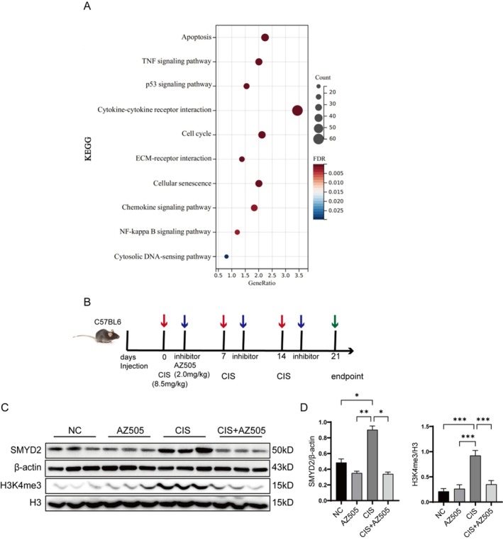
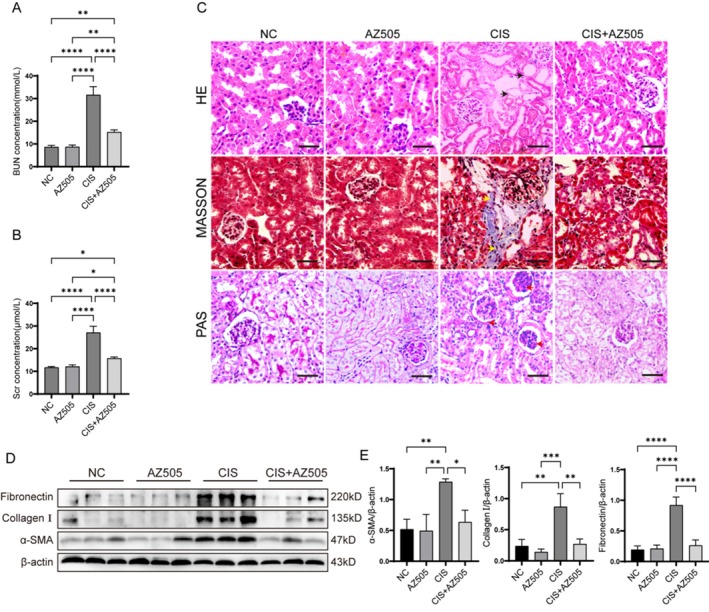
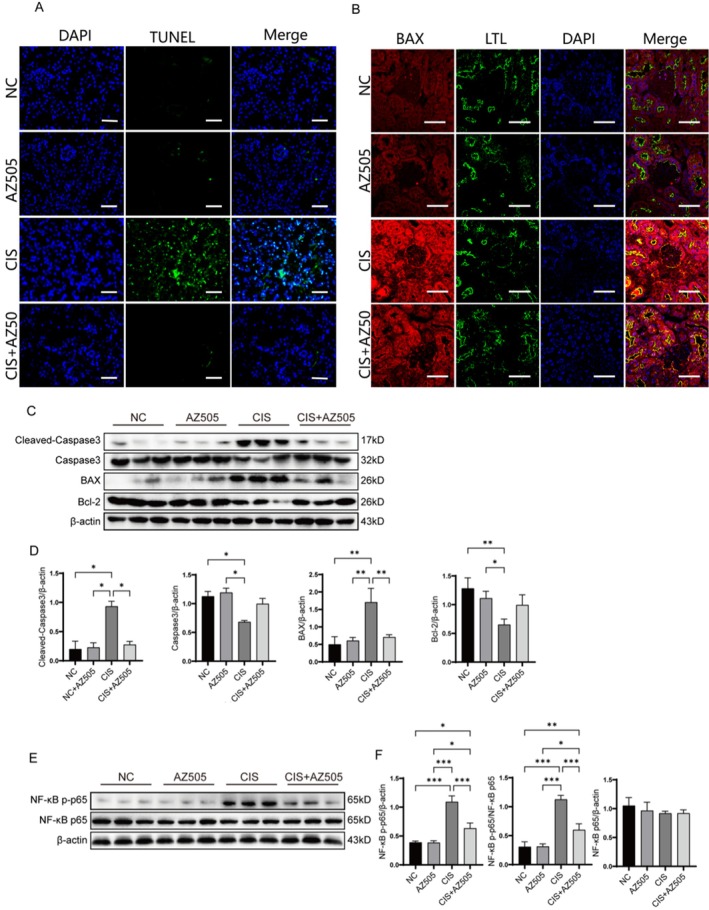
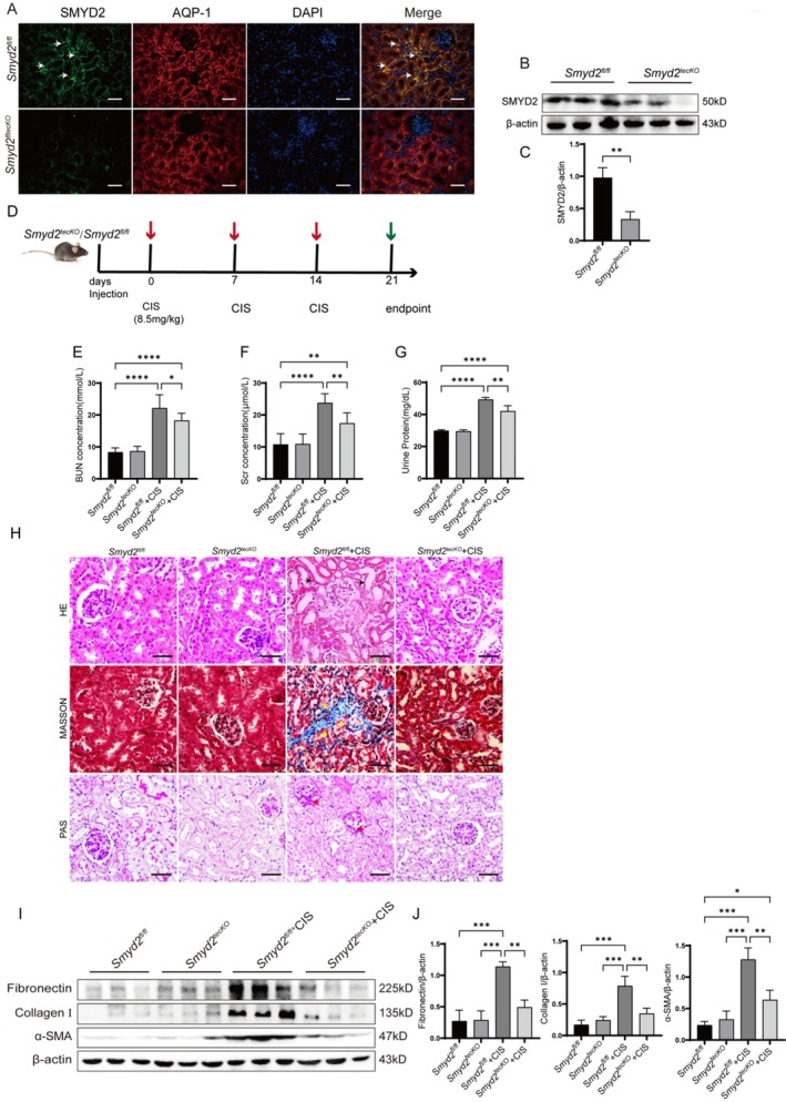
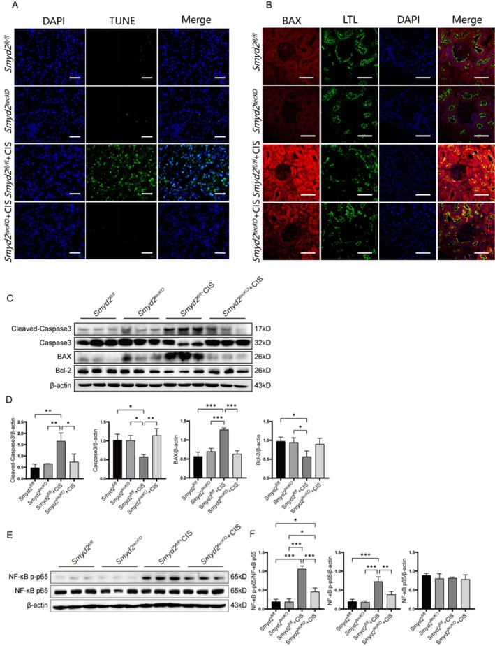
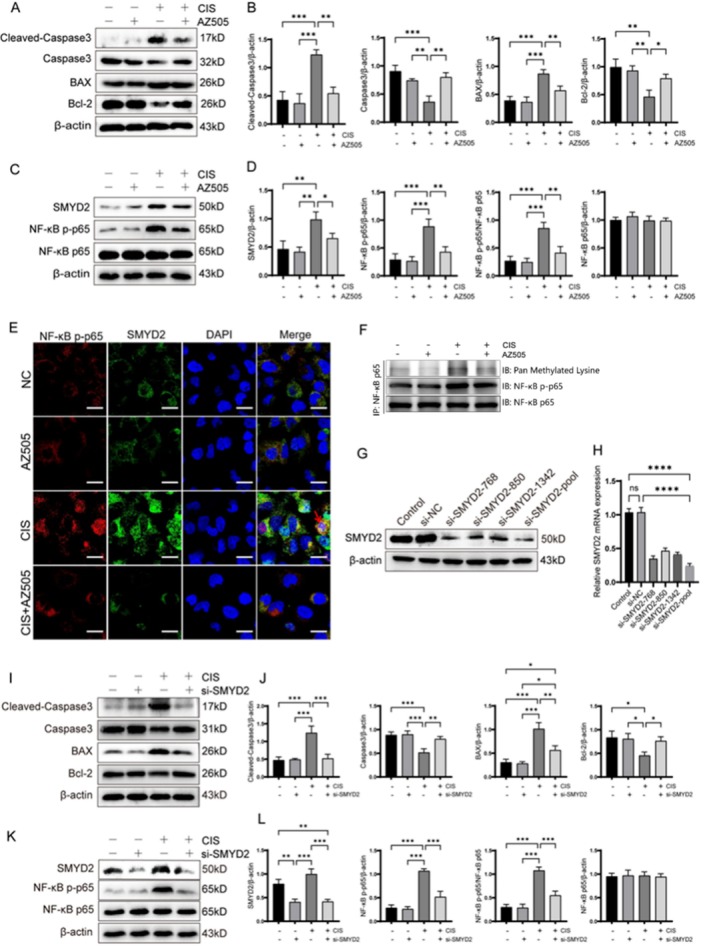
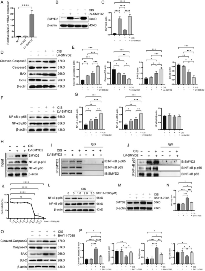
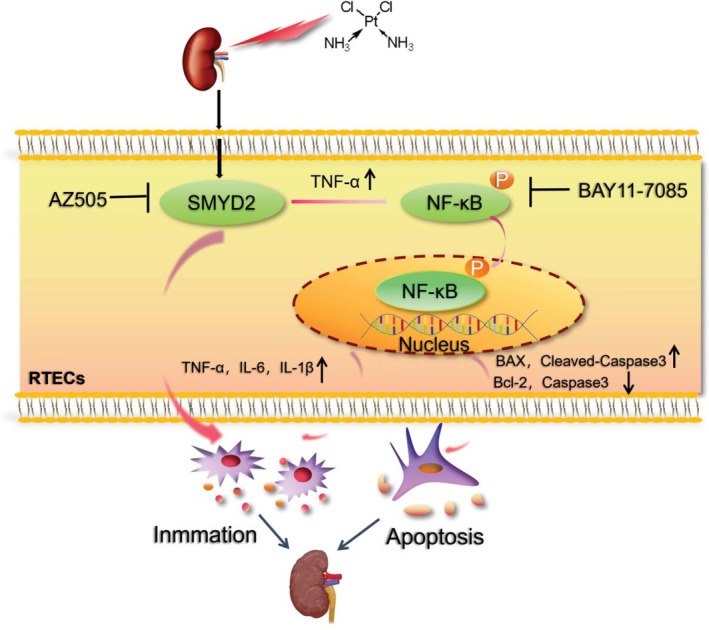
The protein lysine methyltransferase 2 (SMYD2) can affect cell proliferation, differentiation, and survival through methylation of its histone and non-histone substrates. SMYD2 has been shown to act as an oncogene to promote disease progression in a variety of cancer diseases, but its role in chronic kidney diseases (CKD) pathogenesis has not been fully elucidated. This study aims to investigate the effect of SMYD2 on cisplatin-induced CKD and its underlying mechanisms. In this study, we found that cisplatin caused severe renal injury in mice, which was accompanied by the up-regulation of SMYD2 expression. AZ505 treatment significantly down-regulated cisplatin-induced renal injury and fibrosis. It also alleviated renal apoptosis and inhibited the phosphorylation level of NF-κB p65. Conditional knockdown of Smyd2 achieved similar effects as AZ505. In renal tubular epithelial cells, inhibition or silencing of SMYD2 down-regulated cisplatin-induced apoptotic response, while overexpression of SMYD2 induced apoptotic response and activated NF-κB in response to the up-regulation of SMYD2 expression. Up-regulation of SMYD2 induced interaction and phosphorylation of SMYD2 and NF-κB p65, and inhibition of NF-κB activation further suppressed cisplatin-induced NF-κB activation and apoptosis. The present study suggests that up-regulation of SMYD2 expression in cisplatin-induced CKD may promote apoptosis of renal tubular epithelial cells and accelerate the process of renal injury through NF-κB activation. SMYD2 may serve as a potential target for effective CKD treatment.

Keywords: NF‐κB signal path way; apoptosis; renal tubular epithelial cells; the protein lysine methyltransferase 2.


Conflict of interest statement

The authors declare no conflicts of interest.

Figures

FIGURE 1
FIGURE 1
Changes in SMYD2 expression in renal tissues of mice with cisplatin‐induced CKD. (A) Differential expression of genes associated with cisplatin‐induced CKD was analyzed in renal tissues using KEGG enrichment analysis through the Cluster Profiler R package. (B) A CKD mouse model was established via cisplatin treatment in conjunction with AZ505. (C, D) Western blot analysis was conducted to evaluate the expression levels of SMYD2 and H3K4me3 in the kidneys of all groups of mice (n = 3). *p < 0.05, **p < 0.01, ***p < 0.001.
FIGURE 2
FIGURE 2
AZ505 mitigates renal injury and suppresses the upregulation of ECM proteins in the renal tissue of mice with cisplatin‐induced CKD. (A, B) Serum blood urea nitrogen (BUN) and serum creatinine (Scr) levels in AZ505‐treated and untreated cisplatin‐induced CKD mice (n = 5). (C) Hematoxylin and eosin (H&E), Masson's trichrome, and periodic acid‐Schiff (PAS) staining of renal tissue sections from AZ505‐treated and untreated cisplatin‐induced CKD mice. Scale bar: 10 μm. (D, E) Western blot analysis of ECM protein expression in renal tissues from AZ505‐treated and untreated cisplatin‐induced CKD mice (n = 3). *p < 0.05, **p < 0.01, ***p < 0.001, ****p < 0.0001.
FIGURE 3
FIGURE 3
Effects of SMYD2 inhibition on apoptosis and phosphorylated NF‐κB p65 levels in renal tubular epithelial cells from mice with cisplatin‐induced CKD. (A) TUNEL staining of renal tissues from AZ505‐treated and control groups. Scale bar: 50 μm. (B) Immunofluorescence analysis of BAX expression and localization in kidney tissues. Scale bar: 50 μm. (C, D) Western blot analysis of BAX, Bcl‐2, cleaved caspase‐3, and caspase‐3 protein expression in kidney tissues (n = 3). (E, F) Western blot analysis of total NF‐κB p65 and phosphorylated NF‐κB p65 (p‐p65) protein levels (n = 3). *p < 0.05, **p < 0.01, ***p < 0.001.
FIGURE 4
FIGURE 4
Smyd2 tecKO Mitigates kidney injury in cisplatin‐induced CKD mice and suppresses ECM protein upregulation. (A–C) Immunofluorescence and Western blot analysis of SMYD2 expression and localization in renal tissues of Smyd2 tecKO mice, with β‐Actin as a loading control (scale bar: 50 μm; N = 3). (D) Schematic of cisplatin‐induced CKD model in Smyd2 tecKO mice. (E–G) Blood urea nitrogen (BUN), serum creatinine (Scr), and proteinuria levels in cisplatin‐treated Smyd2 tecKO mice and Smyd2 fl/fl mice (n = 5). (H) H&E, Masson, and PAS staining of kidney sections. Scale bar: 10 μm. (I, J) Western blot analysis of renal ECM proteins (α‐SMA, fibronectin, collagen I) in cisplatin‐treated groups (n = 3). *p < 0.05, **p < 0.01, ***p < 0.001.
FIGURE 5
FIGURE 5
Effects of Smyd2 tecKO on apoptosis and phosphorylated p65 levels in renal tubular epithelial cells of cisplatin‐induced CKD mice. (A) TUNEL staining of renal tissues in Smyd2 tecKO and Smyd2 fl/fl mice. Scale bar: 50 μm. (B) Immunofluorescence analysis of BAX expression and localization in renal tissues. Scale bar: 50 μm. (C, D) Western blotting analysis of renal protein expression levels of BAX, Bcl‐2, cleaved caspase‐3, and caspase‐3 (n = 3). (E, F) Western blotting analysis of renal protein expression levels of NF‐κB p65 and phosphorylated p65 (n = 3). *p < 0.05, **p < 0.01, ***p < 0.001.
FIGURE 6
FIGURE 6
Effects of SMYD2 Inhibition or Silencing on Apoptosis and Phosphorylated p65 Levels in HK‐2 Cells. (A, B) Western blot analysis of BAX, Bcl‐2, Cleaved‐Caspase 3, and Caspase 3 protein expression in cisplatin‐induced HK‐2 cells treated with AZ505 (n = 3). (C, D) Western blot analysis of SMYD2, NF‐κB p65, and NF‐κB p‐p65 (n = 3). (E) Immunofluorescence detection of SMYD2 and NF‐κB p‐p65 expression and localization. Scale bar: 20 μm. (F) Immunoprecipitation detection of lysine methylation levels in NF‐κB p65. (G, H) Western blot and qPCR analysis of SMYD2 protein and mRNA expression in SMYD2 si‐RNA transfected HK‐2 cells. (I, J) Western blot analysis of BAX, Bcl‐2, Cleaved‐Caspase 3, and Caspase 3 protein expression in SMYD2 si‐RNA transfected HK‐2 cells (n = 3). (K, L) Western blot analysis of SMYD2, NF‐κB p65, and NF‐κB p‐p65 expression in SMYD2 si‐RNA transfected HK‐2 cells (n = 3). *p < 0.05, **p < 0.01, ***p < 0.001, ****p < 0.0001.
FIGURE 7
FIGURE 7
SMYD2 promotes cisplatin‐induced apoptosis in HK‐2 cells via NF‐κB pathway activation. (A–C) Western blot and qRT‐PCR analysis of SMYD2 protein and mRNA levels in LV‐SMYD2‐transfected HK‐2 cells (n = 3). (D, E) Western blot analysis of BAX, Bcl‐2, Cleaved‐Caspase3, and Caspase3 (n = 3). (F, G) Western blot analysis of NF‐κB p65 and NF‐κB p p65 n = 3). (H–J) Co‐IP assay of SMYD2 and NF‐κB p65. (K) CCK‐8 viability assay of HK‐2 cells treated with BAY11‐7085 (n = 5). (L) Western blot analysis of p65 phosphorylation in cisplatin‐treated HK‐2 cells following BAY11‐7085 treatment at indicated concentrations. (M, N) Western blot analysis of SMYD2 protein levels in cisplatin‐treated HK‐2 cells after BAY11‐7085 (2 μM) treatment (n = 3). (O, P) Western blot analysis of BAX, Bcl‐2, Cleaved‐Caspase3, and Caspase3 protein levels in cisplatin‐treated SMYD2‐overexpressing HK‐2 cells after BAY11‐7085 (2 μM) treatment (n = 3). *p < 0.05, **p < 0.01, ***p < 0.001, ****p < 0.0001.
FIGURE 8
FIGURE 8
Cisplatin stimulation induces the upregulation of SMYD2 expression in renal tubular epithelial cells, which interacts with NF‐κB and promotes its phosphorylation. This, in turn, enhances the release of inflammatory mediators from the cells and induces apoptosis.

Similar articles

Cited by

References

    1. Li Y., Ning Y., Shen B., et al., “Temporal Trends in Prevalence and Mortality for Chronic Kidney Disease in China From 1990 to 2019: An Analysis of the Global Burden of Disease Study 2019,” Clinical Kidney Journal 16, no. 2 (2022): 312–321. - PMC - PubMed
    1. Gregg L. P., Bossola M., Ostrosky‐Frid M., and Hedayati S. S., “Fatigue in CKD: Epidemiology, Pathophysiology, and Treatment,” Clinical Journal of the American Society of Nephrology 16, no. 9 (2021): 1445–1455. - PMC - PubMed
    1. Duan Z., Cai G., Li J., and Chen X., “Cisplatin‐Induced Renal Toxicity in Elderly People,” Therapeutic Advances in Medical Oncology 12 (2020): 1758835920923430. - PMC - PubMed
    1. Sharfuddin A. A. and Molitoris B. A., “Pathophysiology of Ischemic Acute Kidney Injury,” Nature Reviews. Nephrology 7, no. 4 (2011): 189–200. - PubMed
    1. Fu Y., Tang C., Cai J., Chen G., Zhang D., and Dong Z., “Rodent Models of AKI‐CKD Transition,” American Journal of Physiology. Renal Physiology 315, no. 4 (2018): F1098–F1106. - PMC - PubMed

MeSH terms

LinkOut - more resources