. 2025 May-Jun;73(5-6):197-221.
doi: 10.1369/00221554251338066. Epub 2025 May 20.

The Human Mechanosensory Corpuscles: A New Schwann Cell Localization of the Wilms' Tumor Protein WT1

Affiliations

The Human Mechanosensory Corpuscles: A New Schwann Cell Localization of the Wilms' Tumor Protein WT1

Alfonso Cepeda-Emiliani et al. J Histochem Cytochem. 2025 May-Jun.

Abstract

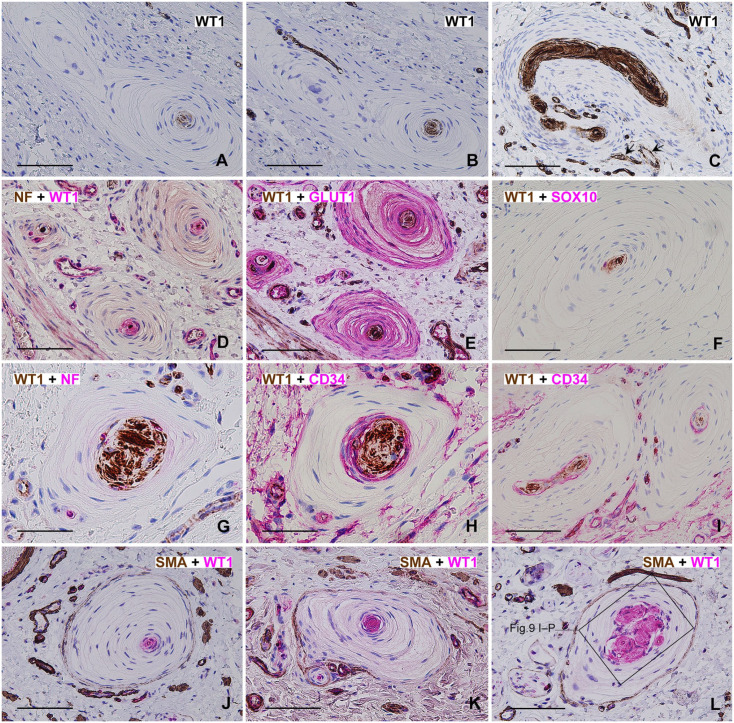
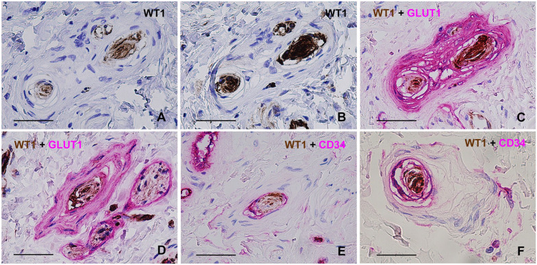
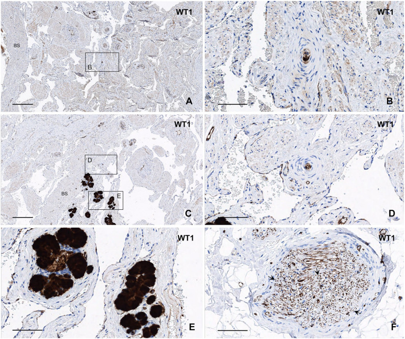
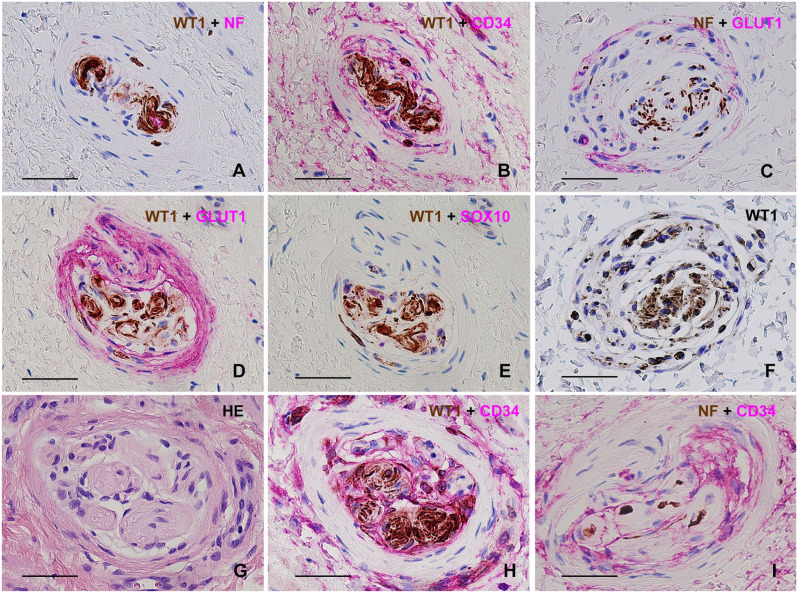
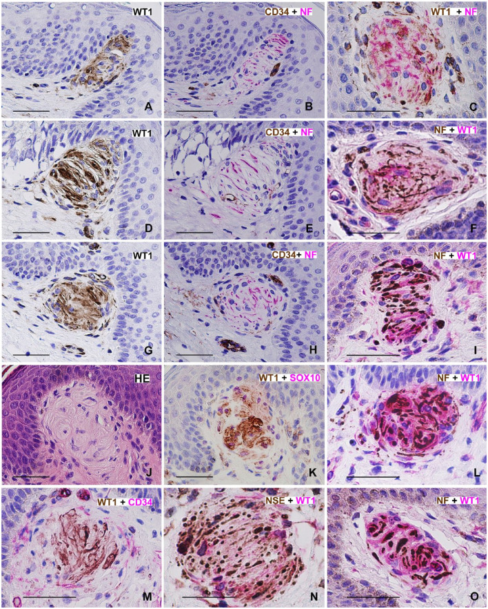
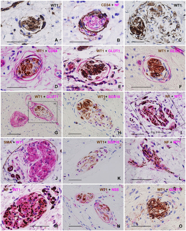
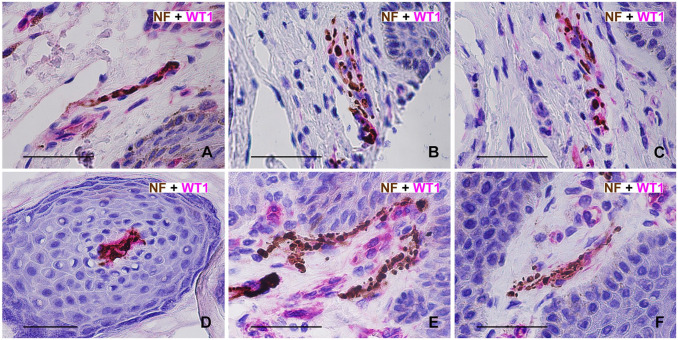
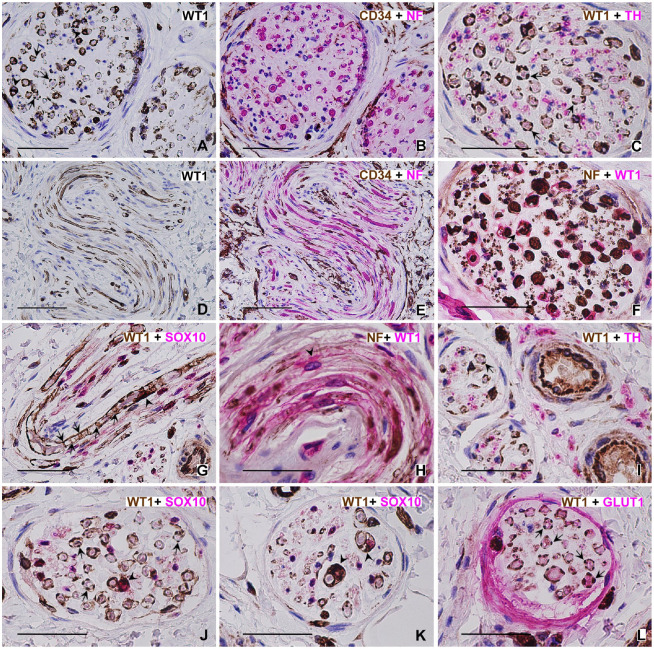
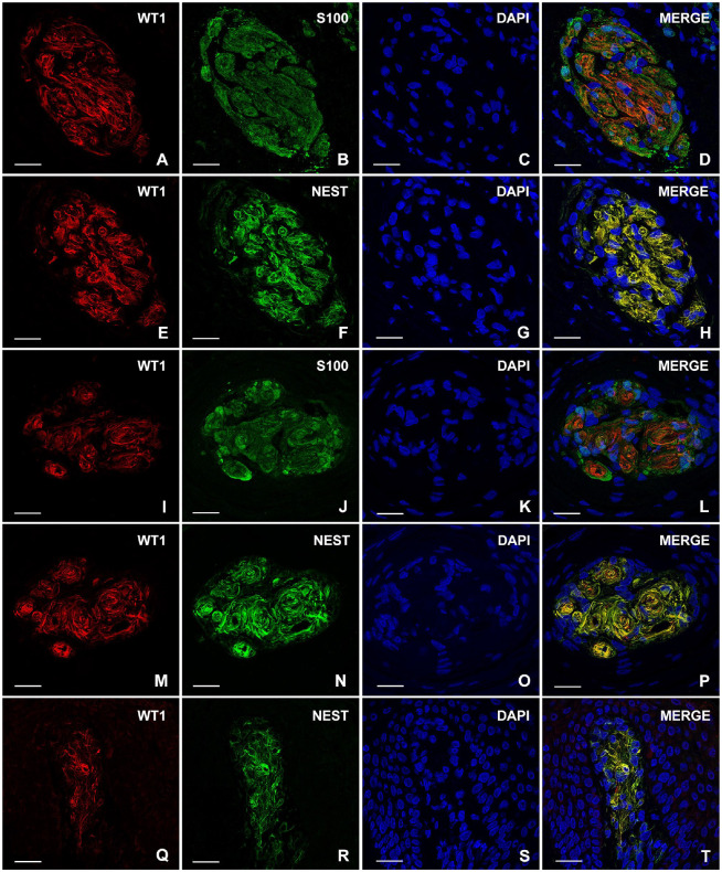
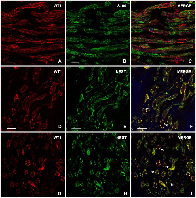
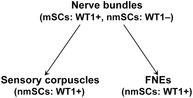
SummaryThe Wilms' Tumor protein WT1 is a zinc-finger transcription factor with crucial roles in organogenesis, cell differentiation, tissue homeostasis, and oncogenesis. While its expression has been extensively studied in various tissues, its presence in the nervous system, particularly in peripheral glial cells, remains largely unexplored. In this study, we examined WT1 expression in the Schwann cells of mechanosensory corpuscles, nerve bundles, and free nerve endings (FNEs) within human penile tissues. Using single and double immunohistology, we analyzed WT1 coexpression with Schwann cell markers (S100, nestin, SOX10) and its association with axonal (neurofilaments, neuron-specific enolase, tyrosine hydroxylase) and perineurial/endoneurial markers (Glut-1, α-SMA, CD34). We found consistent WT1 cytoplasmic expression in the Schwann cells of Pacinian, Meissner, Krause, genital, Golgi-Mazzoni, and Ruffini-like corpuscles, with variable staining intensity. Confocal microscopy revealed WT1 colocalized with nestin but not S100, suggesting involvement in cytoskeletal organization. In addition, we documented WT1 in myelinating Schwann cells of nerve bundles, with distinct staining patterns in Cajal bands and Schmidt-Lanterman incisures, as well as in non-myelinating Schwann cells of FNEs. This is the first study to describe WT1 expression in sensory corpuscles, implicating it in Schwann cell development, maintenance, or plasticity, with potential relevance for peripheral nerve biology, pathology, and mechanosensation.

Keywords: cytoskeleton; foreskin; genitalia; immunohistochemistry; mechanoreceptors; nerve endings; neuroglia; penis; peripheral nervous system; urogenital system.


Conflict of interest statement

Competing InterestsThe author(s) declared no potential conflicts of interest with respect to the research, authorship, and/or publication of this article.

Figures

Figure 1.
Figure 1.
Preputial Pacinian corpuscles with WT1 expression in the inner core Schwann cells, demonstrated through single and double immunohistochemistry. (A, B) Serially sectioned Pacinian showing weak WT1 immunoreactivity without linker in A and stronger WT1 expression with signal amplification via linker in the consecutive section (B). WT1 signal amplification was applied in all subsequent WT1 microphotographs in this study unless otherwise specified. (C) Pacinian corpuscle showing strong WT1 staining in the inner core. Note the adjacent WT1+ endothelial staining (arrows). (D, E) Serially sectioned Pacinians: (D) NF+ punctate axonal profiles in the inner cores (brown) and WT1+ inner core Schwann cells (magenta); (E) WT1 in the inner cores (brown) and Glut-1 in the outer cores (magenta). (F) Pacinian corpuscle with WT1+ inner core (brown) and SOX10+ nuclear immunoreactivity (magenta) in inner core Schwann cells. Some SOX10+ nuclei are obscured due to chromogen overlap with strong WT1 staining, though a clearly visible SOX10+ nucleus is present in the peripheral inner core. (G, H) Serially sectioned small Pacinian corpuscle: (G) WT1+ lamellar cells in the inner core (brown) with NF+ axonal profiles (magenta). An additional axonal profile is visible in the lower portion of the image, within the peripheral part of the outer core; (H) WT1+ lamellar cells in the inner core (brown) and CD34+ cells in the intermediate layer. CD34+ fibroblast-like cells or dermal dendrocytes (magenta) surround the Pacinian. (I) Pacinian corpuscles with WT1 expression in the inner cores (brown) and CD34 in the intermediate layers (magenta). (J–L) Pacinian corpuscles showing WT1 expression in the inner core (magenta) and SMA immunoreactivity in the capsule (brown). Surrounding the corpuscles are small smooth muscle bundles and blood vessels. In the upper left corner of J, note a blood vessel with WT1+ endothelium (magenta) and an SMA+ tunica media (brown). Adjacent SMA+ smooth muscle bundles and WT1+ endothelial cells are also visible. In L note multiple WT1+ Schwann lamellar cell systems within the enlarged Pacinian inner core (magenta), potentially representing a transitional form between the Pacinian and Golgi-Mazzoni corpuscles. Scale bars = 100 µm (A–F, I, J–L), 50 µm (G, H).
Figure 2.
Figure 2.
Glans Pacinian and Paciniform corpuscles with WT1 expression in the inner core Schwann cells, demonstrated through single and double immunohistochemistry. (A–C) Serially sectioned small Pacinian corpuscle with double inner core and a common perineurial outer core: (A) Faint WT1 immunoreactivity in the inner cores without signal amplification; (B) strong WT1 expression in the inner cores following signal amplification; (C) WT1 in the inner cores (brown) and strong Glut-1 expression in the common outer core (magenta). (D, E) Serially sectioned small Pacinian corpuscle: (D) WT1 expression in the inner core with signal amplification (brown) and Glut-1 in the outer core (magenta). Note adjacent WT1+ nerve bundles encased by Glut-1+ perineurium; (E) WT1 expression in the inner core without signal amplification (brown) and CD34 in the intermediate layer (magenta). (F) Paciniform corpuscle displaying WT1 in the inner core (brown) and CD34 in the intermediate layer (magenta). Scale bars = 50 µm (A–E), 25 µm (F).
Figure 3.
Figure 3.
Bulbar Pacinian and Paciniform corpuscles with WT1 expression in the inner core Schwann cells and WT1 immunoreactivity in related structures, all observed without signal amplification. (A) Low-magnification image of the bulbar region of the corpus spongiosum, with the bulbar septum (BS) visible on the left. (B) High-magnification view of the outlined area in A, showing a small Pacinian corpuscle with WT1 expression in the inner core, located within a bulbar trabecula. (C) Low-magnification image of the bulbar region of the corpus spongiosum, again showing the bulbar septum (BS) labeled. (D) High-magnification view of the outlined area in C, depicting a Paciniform corpuscle with WT1+ Schwann cells in the inner core. (E) Bulbar urethral Littré glands exhibiting intense WT1+ staining. (F) Nerve bundle within the penile suspensory apparatus displaying WT1 immunoreactivity in the Cajal bands of Schwann cells (arrows), surrounded by adipose tissue. Scale bars = 400 µm (A, C), 100 µm (B, D), 50 µm (E, F).
Figure 4.
Figure 4.
Preputial Golgi-Mazzoni corpuscles with WT1 expression in the inner core Schwann cells, demonstrated through single and double immunohistochemistry. (A, B) Serially sectioned Golgi-Mazzoni corpuscle: (A) WT1+ Schwann cells in the inner core (brown) and NF+ axons in the inner core (magenta); (B) WT1 expression in the inner core (brown) and CD34+ cells positioned between the inner and outer core, extending into the inner core (magenta). Unlike Pacinian corpuscles (Figs. 1H and I and 2E and F), a well-defined CD34+ intermediate layer is absent. (C) Golgi-Mazzoni corpuscle displaying numerous NF+ axonal profiles in the inner core (brown) and Glut-1 immunoreactivity in the outer core/capsule (magenta). The capsular layers appear loosely arranged, in contrast to the more compact capsules observed in the other Golgi-Mazzoni corpuscles in this figure. (D, E) Serially sectioned Golgi-Mazzoni: (D) WT1+ Schwann lamellar cells in the inner core (brown) with strong Glut-1 staining in the compact outer core (magenta); (E) WT1+ Schwann cells in the inner core (brown) and SOX10 nuclear staining in the same cells (magenta). (F) Consecutive serial section of the Golgi-Mazzoni corpuscle shown in C, exhibiting WT1+ cells in both the inner and outer core, an uncommon finding. (G, H) Serially sectioned Golgi-Mazzoni: (G) HE staining; (H) WT1 expression in the lobulated inner core (brown) and CD34+ fibroblast-like cells (possible telocytes) with long, slender processes extending between the inner and outer core and into the inner core (magenta), compartmentalizing the lamellar cells. As observed previously, a well-defined CD34+ intermediate layer, characteristic of Pacinian corpuscles, is absent. (I) Another typical Golgi-Mazzoni corpuscle, showing NF+ axonal profiles in the inner core (brown) and CD34+ cells (magenta) situated between the inner and outer core, as well as within the inner core itself, resembling the distribution patterns seen in B and H. Scale bars = 50 µm (A–I).
Figure 5.
Figure 5.
Preputial papillary corpuscles with WT1 expression in Schwann cells, demonstrated through single and double immunohistochemistry. (A, B) Serially sectioned Meissner corpuscle: (A) WT1 immunoreactivity is prominent in the Schwann cell cytoplasm, with no staining in the nuclei; (B) consecutive section showing NF+ axons (magenta) transversely oriented relative to the corpuscle’s long axis. Note the absence of CD34+ capsule. (C) Papillary corpuscle displaying faint WT1 immunoreactivity without signal amplification (brown) and a dense network of glomerularly arranged NF+ axons (magenta). (D, E) Serially sectioned Meissner corpuscle: (D) WT1 strong expression is restricted to the Schwann cell cytoplasm, with unstained nuclei; (E) consecutive section showing transversely arranged NF+ axons (magenta) and no CD34+ capsule. (F) Papillary corpuscle with a dense NF+ axonal network (brown) and weak to strong WT1 immunoreactivity in the lamellar cells (magenta). A Schwann cell at the center of the corpuscle exhibits a strong perinuclear WT1+ immunostaining pattern (magenta). (G, H) Serially sectioned rounded Meissner corpuscle: (G) WT1 staining is confined to the Schwann cell cytoplasm, with no expression in the nuclei; (H) consecutive section showing NF+ axons and absent CD34+ capsule. (I) Meissner corpuscle with NF+ axons aligned transversely to its long axis and WT1+ Schwann cells displaying cytoplasmic immunoreactivity (magenta). (J, K) Serially sectioned Meissner corpuscle: (J) HE staining; (K) WT1+ Schwann cells (brown) with SOX10+ nuclear expression in the same cells (magenta); (L) rounded subepithelial corpuscle with dense glomerular NF+ axons (brown) and WT1+ Schwann cells (magenta). (M) Papillary corpuscle with WT1+ lamellar cells (brown) and CD34+ adjacent dermal fibroblast-like cells (magenta). No distinct capsule is observed. (N) Meissner corpuscle with densely packed NSE+ axons (brown) and faint WT1 staining in the lamellar cells (magenta). (O) Small Meissner corpuscle with NF+ axons (brown) enclosed by WT1+ lamellar cells showing strong immunoreactivity (magenta). Scale bars = 50 µm (A–O).
Figure 6.
Figure 6.
Preputial deep dermal Krause, genital, and Ruffini-like corpuscles with WT1 expression in Schwann cells. (A, B) Serially sectioned encapsulated Krause corpuscle: (A) WT1+ Schwann cells within the corpuscle, with some capsular cells exhibiting perinuclear and cytoplasmic WT1 expression, an uncommon finding; (B) well-defined CD34+ capsule (brown) and NF+ axons (magenta) inside the corpuscle. (C) Large encapsulated genital corpuscle with WT1+ lamellar Schwann cells compartmentalized into lobules. (D) Small encapsulated Krause corpuscle displaying WT1+ Schwann cells (brown) and a CD34+ capsule (magenta). (E, F) Two small encapsulated Krause corpuscle with WT1+ Schwann cells (brown) and Glut-1+ capsules (magenta). (G, H) Serially sectioned genital corpuscle: (G) WT1+ Schwann cells in the inner core (brown) and a Glut-1+ capsule (magenta). The capsule exhibits Glut-1+ perineurial continuity with an adjacent small nerve bundle supplying the corpuscle. On the left, a small Pacinian or Paciniform corpuscle, likely sectioned through its Glut-1+ capsule (magenta), is visible, with its inner core out of the plane of section; (H) higher magnification of the outlined region in G, highlighting WT1+ Schwann cells (brown) and SOX10+ Schwann cell nuclei (magenta). (I) Large genital corpuscle with dense NF+ axonal profiles (brown) surrounded by WT1+ Schwann cell cytoplasm (magenta). (J) Large genital corpuscle with an SMA+ capsule (brown) and WT1+ lamellar Schwann cells organized in lobules (magenta). (K) Ruffini-like fusiform corpuscle displaying WT1+ Schwann cell cytoplasm (brown) and SOX10+ Schwann cell nuclei (magenta). (L) Ruffini-like fusiform corpuscle with dense NF+ axons (brown) enveloped by WT1+ Schwann cell cytoplasm (magenta). Note the axons supplying the corpuscle on the left part of the image, also associated with WT1+ Schwann cells (magenta). (M) Large genital corpuscle containing densely packed NF+ axons (brown), surrounded by WT1+ Schwann cell cytoplasm (magenta). (N) Pair of Krause or genital corpuscles showing WT1+ Schwann cells (brown) and NSE+ axonal profiles (magenta). (O) Probable Krause corpuscle featuring WT1+ Schwann cells (brown). No Glut-1+ perineurial capsule is observed. Scale bars = 50 µm (A–F, H–O), 100 µm (G).
Figure 7.
Figure 7.
Preputial free nerve endings (FNEs) with WT1 expression in non-myelinating Schwann cells, as shown by double immunohistochemistry. (A–F) FNEs in the papillary dermis, with NF+ axons (brown) associated with WT1+ Schwann cell cytoplasm (magenta). The staining intensity is weak to strong. Scale bars = 50 µm (A–F).
Figure 8.
Figure 8.
Preputial nerve bundles with WT1 expression in Schwann cells, demonstrated through single and double immunohistochemistry. (A, B) Serially sectioned nerve bundles with axons in cross-section: (A) WT1 exhibits a punctate or dot-like expression pattern characteristic of Cajal bands along the outermost (abaxonal) Schwann cell compartment of numerous myelinated axons (arrows). In some axons, WT1 is restricted to the perinuclear Schwann cell cytoplasm (arrowhead), whereas in others, staining appears circumferential around the internodal myelin sheath; (B) CD34 highlights endoneurial fibroblasts and surrounding dermal fibroblasts (brown). Myelinated and unmyelinated axons within the nerve bundles both display strong NF immunostaining (magenta). (C) Nerve bundle with WT1+ Schwann cells exhibiting punctate periaxonal Cajal band-like expression (brown, arrows). TH+ unmyelinated axons (magenta) are present but unassociated with WT1+ Schwann cells. (D, E) Serially sectioned S-shaped nerve bundle: (D) WT1 immunostaining highlights the longitudinal bands of Schwann cell cytoplasm surrounding longitudinally sectioned axons; (E) CD34 labels endoneurial fibroblasts and surrounding dermal dendrocytes (brown). NF+ myelinated and unmyelinated axons are visible within the bundle (magenta). (F) Consecutive serial section of the same nerve bundle shown in C, with axons in cross-section showing strong NF immunostaining in both myelinated and unmyelinated axons (brown). WT1 expression (magenta) is restricted to Schwann cells of myelinated axons, with staining patterns ranging from circumferential internodal myelin to the characteristic punctate pattern associated with Cajal bands (arrow). (G) Nerve bundle predominantly containing longitudinally sectioned axons. WT1 immunopositivity is observed in Schwann cell cytoplasmic channels along the myelinated axons, including perinuclear staining (arrowhead) and Schmidt–Lanterman incisures (arrows). Schwann cell nuclei are SOX10+ (magenta). (H) Nerve bundle with longitudinally sectioned NF+ axons (brown). WT1 expression (magenta) is observed in the perinuclear (arrowhead) and internodal Schwann cell cytoplasm. (I) Two small nerve bundles with axons in cross-section. WT1 (brown) exhibits a characteristic punctate Cajal band-like staining pattern in the Schwann cell cytoplasm (arrow). TH immunoreactivity is selectively present in unmyelinated axons (magenta), which are unassociated with WT1+ Schwann cells. Two small blood vessels in the field also show WT1+ endothelial cells (brown) and TH+ autonomic innervation (magenta). (J, K) Nerve bundles displaying WT1+ Schwann cells (brown) with perinuclear (arrowheads) and internodal Cajal band-like staining (arrows), along with SOX10+ Schwann cell nuclei (magenta). (L) Nerve bundle exhibiting the typical dot-like WT1+ (brown) Schwann cell staining pattern associated with Cajal bands (arrows) and a well-defined Glut-1+ perineurium (magenta). Scale bars = 50 µm (A–C, F–L), 100 µm (D, E).
Figure 9.
Figure 9.
Double immunofluorescence analysis of WT1, nestin, and S100 in Schwann cells of preputial genital, Pacinian, and Meissner corpuscles. (A–D) Genital corpuscle corresponding to the corpuscle in Fig. 6I, in a consecutive serial section: (A) WT1 staining in the lamellar cell cytoplasm (red); (B) S100 staining in the same cell compartment (green); (C) DAPI nuclear staining (blue); (D) although WT1 and S100 are coexpressed in these Schwann cells with a typical cytoplasmic staining pattern, the merged image shows no colocalization, as indicated by the absence of yellow. (E–H) The same genital corpuscle shown in A–D, in the next consecutive section: (E) WT1 staining in the lamellar cell cytoplasm (red); (F) nestin staining in the same cell compartment (green); (G) DAPI nuclear staining (blue); (H) merged image showing colocalization of WT1 and nestin in the Schwann cell cytoplasm (yellow). (I–L) Inner core of a Pacinian corpuscle corresponding to the Pacinian in Fig. 1L, in a consecutive serial section: (I) WT1 staining in the lamellar cell cytoplasm (red); (J) S100 staining in the same cell compartment (green); (K) DAPI nuclear staining (blue); (L) merged image showing no colocalization of WT1 and S100, as indicated by the absence of yellow. (M–P) Inner core of the same Pacinian corpuscle shown in I–L, in the next consecutive section: (M) WT1 staining in the lamellar cell cytoplasm (red); (N) nestin staining in the same cell compartment (green); (O) DAPI nuclear staining (blue); (P) merged image demonstrating colocalization of WT1 and nestin in the Schwann cell cytoplasm (yellow). (Q–T) Meissner corpuscle: (Q) WT1 expression in Schwann or lamellar cells (red); (R) nestin expression in the same cells (green); (S) DAPI nuclear staining (blue); (T) merged image showing colocalization of WT1 and nestin in the Schwann cell cytoplasm (yellow). Scale bars = 25 µm (A–T).
Figure 10.
Figure 10.
Double immunofluorescence analysis of WT1, nestin, and S100 in Schwann cells of preputial nerve bundles. (A–C) Longitudinally sectioned nerve bundle: (A) WT1 staining of Schwann cell cytoplasmic channels (red); (B) S100 staining in the same compartment (green); (C) although WT1 and S100 are coexpressed in these Schwann cells, the merged image reveals no colocalization of WT1 and S100, as indicated by the absence of yellow. (D–F) Longitudinally sectioned nerve bundle: (D) WT1 staining of Schwann cell cytoplasmic channels (red); (E) nestin staining in the same cell compartment (green); (F) merged image demonstrating colocalization of WT1 and nestin (yellow). Cajal band-like staining is observed in transversely sectioned axons (arrow), along with a more linear pattern resembling the appositional regions flanking Cajal bands (arrowhead). In addition, a staining pattern similar to that of Schmidt–Lanterman incisures is present (green arrowhead). (G–I) Transversely sectioned nerve bundle: (G) WT1 expression in Schwann cell cytoplasmic channels (red); (H) nestin staining of the same cell compartment (green); (I) merged image showing colocalization of WT1 and nestin (yellow). Note the Cajal band-like staining in transversely sectioned axons (arrows), along with a linear staining pattern that more closely resembles the appositional regions flanking Cajal bands (arrowhead). Scale bars = 12 µm (A–C, G–I), 16 µm (D–F).
Figure 11.
Figure 11.
Summary of WT1 immunohistochemical expression in Schwann cell subtypes identified in this study. Peripheral nerve bundles contained a variable mixture of myelinated and unmyelinated axons, associated with WT1+ myelinating Schwann cells (mSCs) and WT1 non-myelinating Schwann cells (nmSCs), respectively. These nerve bundles gave rise to (i) sensory corpuscles containing modified WT1+ nmSCs; (ii) free nerve endings (FNEs) associated with WT1+ nmSCs. Abbreviations: mSCs, myelinating Schwann cells; nmSCs, non-myelinating Schwann cells; FNEs, free nerve endings.

Similar articles

References

    1. Zelená J. Nerves and mechanoreceptors: the role of innervation in the development and maintenance of mammalian mechanoreceptors. London: Chapman & Hall; 1994.
    1. Malinovský L, Pác L. Morphology of sensory corpuscles in mammals. Brno: Acta Facultatis Medicae Universitatis Brunensis; 1982.
    1. Krause W. Die nervenendigungen innerhalb der terminalen korperchen [The nerve endings within the terminal corpuscles]. Arch Mikr Anat Entwgsch. 1881;19:53–136.
    1. Chouchkov C. Cutaneous receptors. New York: Springer-Verlag; 1978.
    1. Cobo R, García-Piqueras J, Cobo J, Vega JA. The human cutaneous sensory corpuscles: an update. J Clin Med. 2021;10(2):227. - PMC - PubMed

LinkOut - more resources