. 2025 May;42(5):1509-1523.
doi: 10.1007/s10815-025-03497-3. Epub 2025 May 20.

Three-dimensional culture in a bioengineered matrix and somatic cell complementation to improve growth and survival of bovine preantral follicles

Affiliations

Three-dimensional culture in a bioengineered matrix and somatic cell complementation to improve growth and survival of bovine preantral follicles

Juliana I Candelaria et al. J Assist Reprod Genet. 2025 May.

Abstract

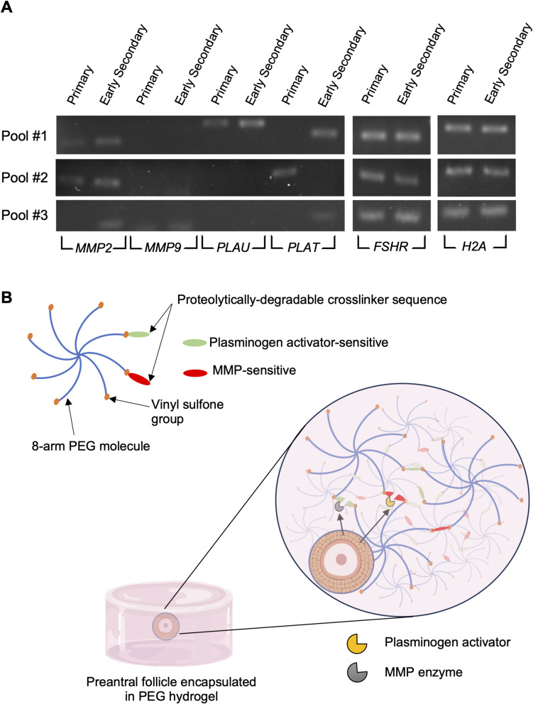
Purpose: Here, we explored poly(ethylene glycol) (PEG) bioengineered hydrogels for bovine preantral follicle culture with or without ovarian cell co-culture and examined the potential for differentiation of bovine embryonic stem cells (bESCs) towards gonadal somatic cells to develop a system better mimicking the ovarian microenvironment.

Methods: Bovine preantral follicles were first cultured in two-dimensional (2D) control or within PEG hydrogels (3D) and then co-cultured within PEG hydrogels with bovine ovarian cells (BOCs) to determine growth and viability. Finally, we tested conditions to drive differentiation of bESCs towards the intermediate mesoderm and bipotential gonad fate.

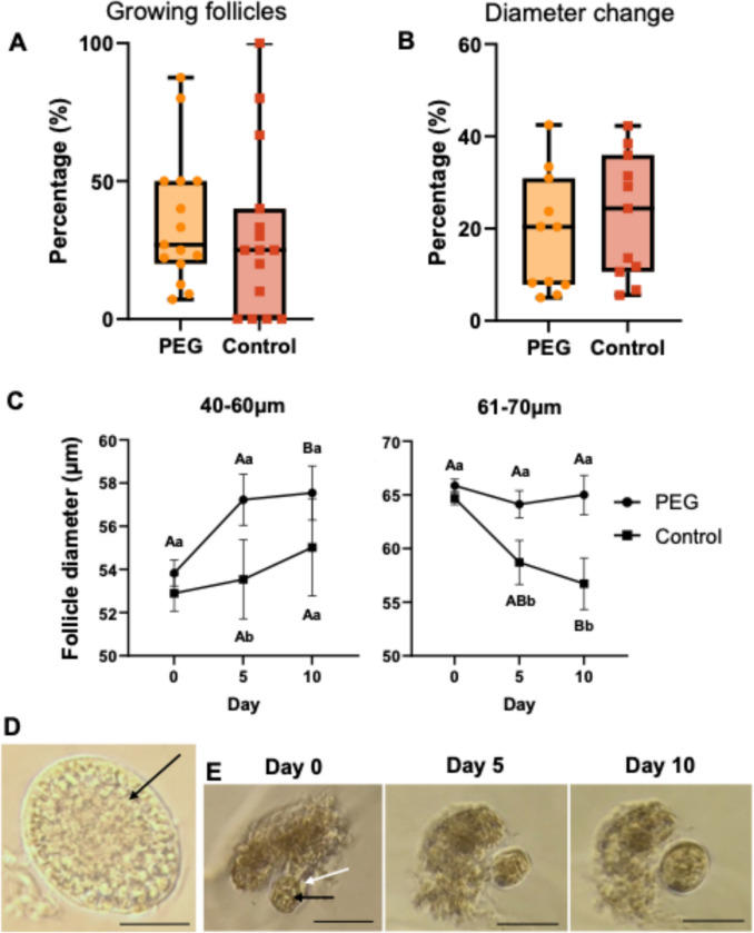
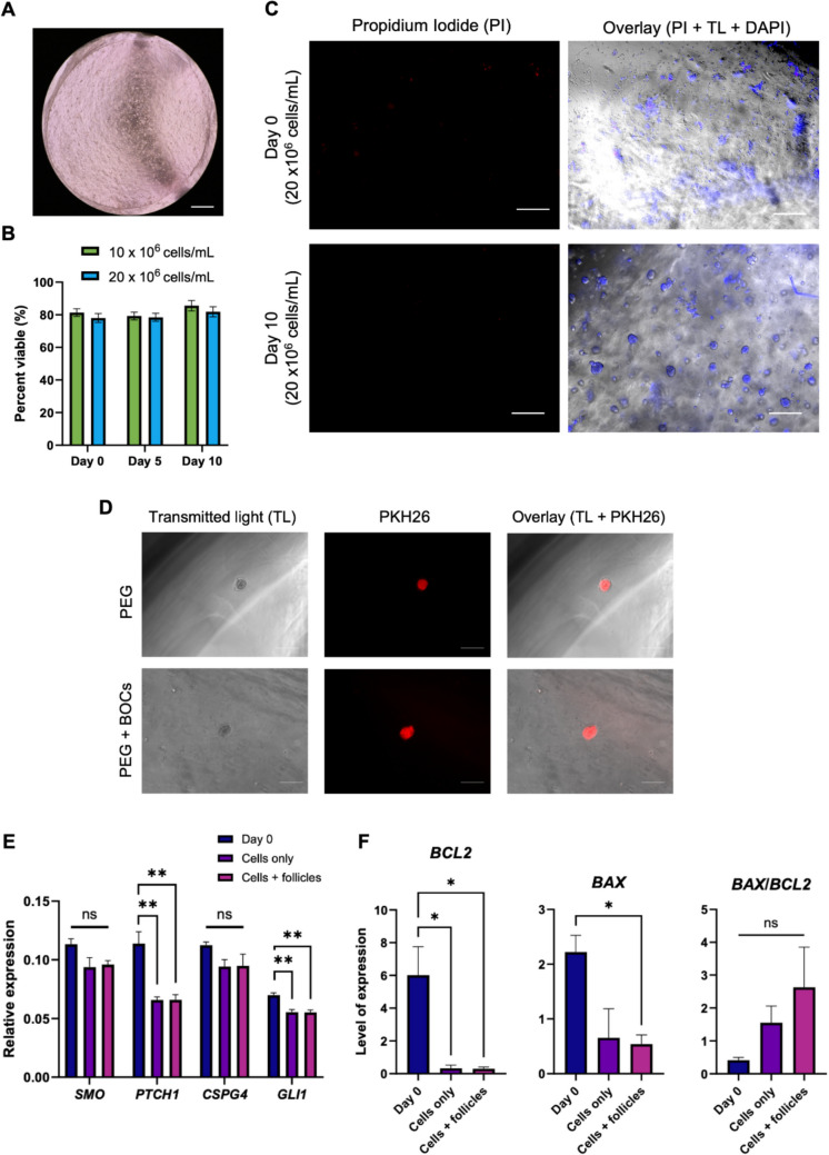
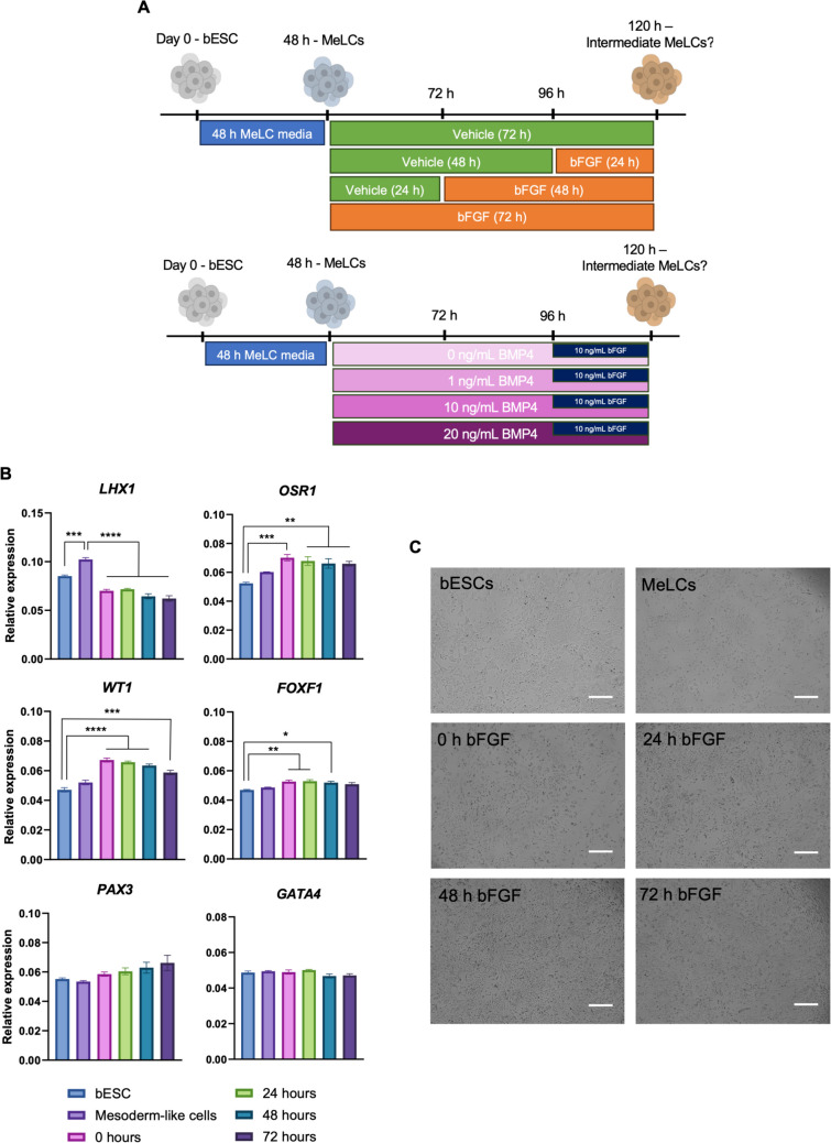
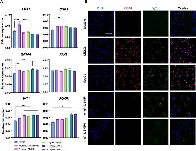
Results: Primary follicles grew over the 10-day culture period in PEG hydrogels compared to 2D control. Early secondary follicles maintained a similar diameter within the PEG while control follicles decreased in size. Follicles lost viability after co-encapsulation with BOCs; BOCs lost stromal cell signature over the culture period within hydrogels. Induction of bESCs towards gonadal somatic fate under WNT signaling was sufficient to upregulate intermediate mesoderm (LHX1) and early coelomic epithelium/bipotential gonad markers (OSR1, GATA4, WT1). Higher BMP4 concentrations upregulated the lateral plate mesoderm marker FOXF1. PAX3 expression was not induced, indicating absence of the paraxial mesoderm lineage.

Conclusions: Culture of primary stage preantral follicles in PEG hydrogels promoted growth compared to controls; BOCs did not maintain identity in the PEG hydrogels. Collectively, we demonstrate that PEG hydrogels can be a potential culture system for early preantral follicles pending refinements, which could include addition of ESC-derived ovarian somatic cells using the protocol described here.

Keywords: Biomimetic; Bovine; Embryonic stem cells; Ovary; Preantral follicle; Three-dimensional culture.


Conflict of interest statement

Declarations. Competing interests: The authors declare no competing interests.

Figures

Fig. 1
Fig. 1
Bovine preantral follicles express extracellular matrix-degrading enzymes, allowing the design of a PEG hydrogel containing target peptides for controlled degradation. A Agarose gel with products of RT-PCR of primary and early secondary follicles expressing mRNA for MMP2, MMP9, PLAU, PLAT, FSHR, and H2A. Each pool of primary and early secondary follicles (n = 3 pools per stage) contained 46–56 and 34–40 follicles, respectively. B Schematic of PEG hydrogel for follicle encapsulation. PEG, poly(ethylene glycol); MMP, matrix metalloproteinase. Figure made with Biorender
Fig. 2
Fig. 2
Bovine preantral follicle in vitro growth using PEG or 2D control culture systems. A Percentage of preantral follicles that grew (n = 236) and B percent change in diameter from follicles that grew (n = 171). C Bovine preantral follicle diameter change (from all follicles cultured; n = 236) during in vitro culture at indicated starting diameters. Lowercases letters (a, b) that differ = significant difference (P < 0.05) between treatment groups in the same day. Uppercase letters (A, B) that differ = significant difference (P < 0.05) within the same treatment group but compared to day 0. Data are presented as mean ± SEM. D Representative image of a freshly isolated follicle with a visible oocyte. Scale bar = 50 µm. E Representative images of a follicle grown in a PEG hydrogel over the 10-day culture period. White arrow shows the follicle attached to ovarian tissue; black arrows indicate the oocyte. Scale bar = 100 µm
Fig. 3
Fig. 3
Co-culture of bovine preantral follicles with bovine ovarian cells (BOCs) in PEG hydrogels. A Representative image of BOCs in PEG hydrogel (scale bar = 500 µm). B Viability of BOCs encapsulated in PEG hydrogels during in vitro culture. C Propidium iodine (PI) staining in BOCs encapsulated in PEG hydrogels at day 0 and day 10 of culture. Counterstained with DAPI (TL, transluminescent; scale bar = 100 µm). D Unviable preantral follicle encapsulated in PEG hydrogel with or without BOCs using PKH26 to label follicles at day 10 of culture (scale bar = 100 µm). E Gene expression of pre-theca cell markers in BOCs (relative expression = 1/ΔCT). F Anti- and pro-apoptotic gene marker expression in BOCs (level of expression = absolute quantification from standard curve). Data are presented as mean ± SEM
Fig. 4
Fig. 4
In vitro differentiation of bovine embryonic stem cells (bESCs) towards progenitors of somatic bipotential gonad-like cells. A Schematic diagram of experimental design for bFGF and BMP4 experiments. B Gene expression of intermediate mesoderm, bipotential gonad, paraxial and lateral plate mesoderm markers before and after bESC differentiation using bFGF at various days of exposure. C Representative images of cells from each treatment group. Data are presented as mean ± SEM. Significant differences are noted by *P < 0.05, **P < 0.01, ***P < 0.001, ****P < 0.0001. Figure made with Biorender
Fig. 5
Fig. 5
In vitro differentiation of bovine embryonic stem cells (bESCs) towards progenitors of somatic bipotential gonad-like cells testing BMP4 concentrations. A Gene expression of intermediate mesoderm, bipotential gonad, paraxial and lateral plate mesoderm markers before and after bESC differentiation. B Representative images of cells from each treatment group. Data are presented as mean ± SEM. Significant differences are noted by *P < 0.05, **P < 0.01, ***P < 0.001, ****P < 0.0001

Update of

Similar articles

References

    1. Bus A, Langbeen A, Martin B, Leroy JLMR, Bols PEJ. Is the pre-antral ovarian follicle the ‘holy grail’ for female fertility preservation? Anim Reprod Sci. 2019;207:119–30. 10.1016/j.anireprosci.2019.05.017. - PubMed
    1. Gutierrez CG, Ralph JH, Telfer EE, Wilmut I, Webb R. Growth and antrum formation of bovine preantral follicles in long-term culture in vitro1. Biol Reprod. 2000;62:1322–8. 10.1095/biolreprod62.5.1322. - PubMed
    1. Araújo VR, Gastal MO, Wischral A, Figueiredo JR, Gastal EL. Long-term in vitro culture of bovine preantral follicles: Effect of base medium and medium replacement methods. Anim Reprod Sci. 2015;161:23–31. 10.1016/j.anireprosci.2015.07.006. - PubMed
    1. Rossetto R, Saraiva MVA, Bernuci MP, Silva GM, Brito IR, Alves A, et al. Impact of insulin concentration and mode of FSH addition on the in vitro survival and development of isolated bovine preantral follicles. Theriogenology. 2016;86:1137–45. 10.1016/j.theriogenology.2016.04.003. - PubMed
    1. Gomes JE, Correia SC, Gouveia-Oliveira A, Cidadão AJ, Plancha CE. Three-dimensional environments preserve extracellular matrix compartments of ovarian follicles and increase FSH-dependent growth. Mol Reprod Dev. 1999;54:163–72. 10.1002/(SICI)1098-2795(199910)54:2<163::AID-MRD8>3.0.CO;2-4. - PubMed

LinkOut - more resources