Review
. 2025 Aug;48(4):921-941.
doi: 10.1007/s13402-025-01073-6. Epub 2025 May 20.

Pericytes in hematogenous metastasis: mechanistic insights and therapeutic approaches

Affiliations
Review

Pericytes in hematogenous metastasis: mechanistic insights and therapeutic approaches

Shuo Xu et al. Cell Oncol (Dordr). 2025 Aug.

Abstract

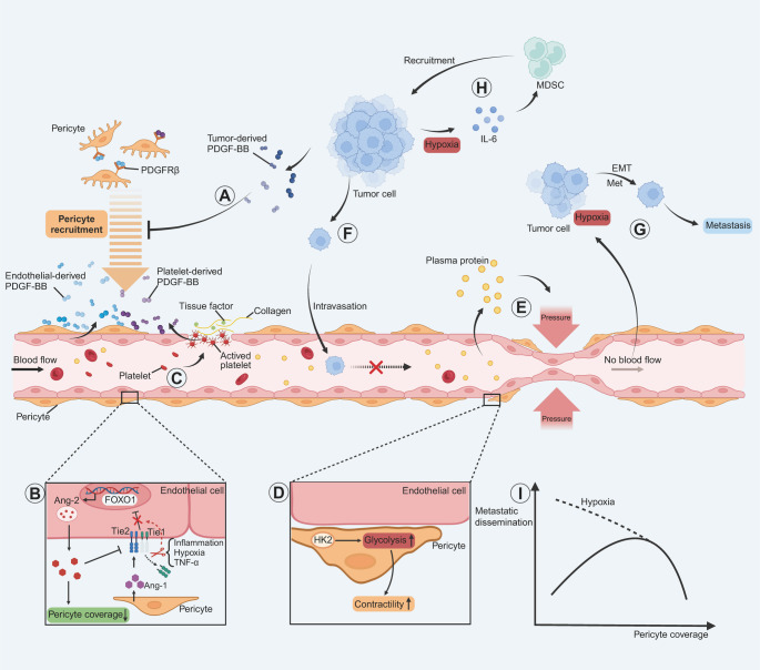
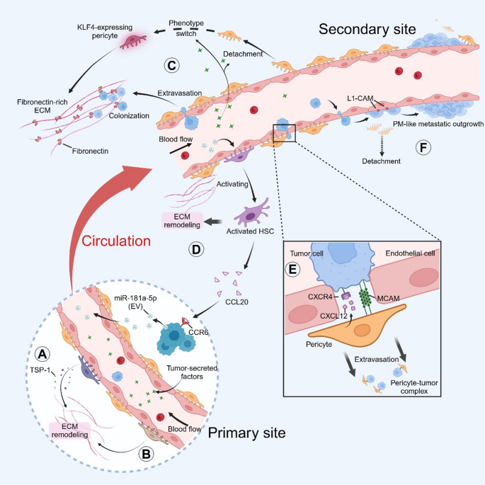
Metastasis, the leading cause of cancer-related deaths, underscores the critical need to understand its regulatory mechanisms to improve prevention and treatment strategies for late-stage tumors. Hematogenous dissemination is a key route of metastasis. However, as the gatekeeper of vessels, the role of pericytes in hematogenous metastasis remains largely unknown. In this review, we comprehensively explore the contributions of pericytes throughout the metastatic cascade, particularly their functions that extend beyond influencing tumor angiogenesis. Pericytes should not be perceived as passive bystanders, but rather as active participants in various stages of the metastatic cascade. Pericytes-targeted therapy may provide novel insights for preventing and treating advanced-stage tumor.

Keywords: Hematogenous metastasis; Pericytes; Plasticity; Tumor microenvironment.


Conflict of interest statement

Declarations. Ethics approval and consent to participate: Not applicable. Consent for publication: Not applicable. Competing interests: The authors declare no competing interests.

Figures

Fig. 1
Fig. 1
Relationship between pericyte coverage and intravasation. (A) In the PDGF-BB/PDGFRβ pathway, endothelial cell-derived PDGF-BB establishes a chemotactic gradient for PDGFRβ⁺ pericyte recruitment, whereas tumor cells with high PDGF-BB expression disrupt this gradient, leading to insufficient pericyte coverage. (B) Under physiological conditions, pericyte-derived Ang-1 activates ECs Tie2 via Tie1-dependent binding, triggering the PI3K/AKT/FOXO1 axis to suppress Ang-2 production and stabilize vasculature. In TME, TNF-α induces proteolytic shedding of Tie1’s extracellular domain, impairing Ang-1/Tie2 signaling and triggering Ang-2 release. This shifts the Ang-1/Ang-2 balance toward Ang-2 dominance, perpetuating Tie2 inhibition and driving vascular destabilization with pericyte shedding. (C) Aberrant expression of tissue factors in tumors and their discontinuous vascular endothelium result in sustained platelet activation, degranulation, and PDGF-BB release, which recruit pericytes and promote tumor vascular system stability. (D) Pericytes upregulate HK2-driven glycolysis and induce Rho-associated coiled-coil containing protein kinase 2 (ROCK2)-myosin light chain 2 (MLC2)-mediated contractility, which negatively affects their vascular support effects (EF) The deficiency of pericytes leads to plasma protein extravasation and increased interstitial pressure around blood vessels, subsequently compressing the vessels and causing vascular collapse. This impedes effective blood circulation, thereby hindering the intravasation and dissemination of tumor cells. (GH) The low coverage of pericytes leads to vascular collapse and results in circulatory obstruction and subsequent hypoxia within the tumor tissue. This condition triggers the upregulation of epithelial-to-mesenchymal transition (EMT) and Met pathways, facilitating metastasis. Concurrently, it stimulates the secretion of IL-6 by tumor cells, recruiting MDSCs and consequently promoting immune evasion and facilitating metastasis. (I) Schematic diagram depicting the possible relationship between pericyte coverage and metastatic dissemination
Fig. 2
Fig. 2
Pericyte-related cellular plasticity and metastatic dissemination. (A) Tumor cells can be recruited to blood vessels and establish adhesion junctions with endothelial cells by upregulating the expression of PDGFRβ and N-cadherin through pericyte-like transition. (B) Endothelial cells secrete CXCL12, attract CXCR4-expressing GSCs, and develop pericyte-like transition under the induction of TGF-β. (C) Pericyte-like transition enables CD44+ CSCs in the lungs to undergo trans-endothelial migration (TEM) and distant metastasis in the form of pericytes. (D) Tumor cells can replace normal pericytes via pericytic mimicry(PM) and migrate to distant sites via extravascular migratory metastasis (EVMM). (E) PDGF-BB derived from tumor cells induces the detachment of pericytes from the endothelial surface and triggers PFT. The generated CAFs then lead to an increase in the number of CTCs and distant metastasis. (F) Exosomes originating from gastric cancer cells contain BMP2, stimulating the PI3K/AKT and mitogen-activated protein kinase (MAPK) pathways. This process triggers PFT, thereby promoting the proliferation and migration of gastric cancer cells. (G) Tumor matrix stiffness may alter Yes-associated protein (YAP) signalling in pericytes and regulate the PFT process
Fig. 3
Fig. 3
Remodelling of the perivascular matrix by pericytes facilitates tumor dissemination and metastatic colonisation. (A) Pericytes secrete TSP-1, which independently regulates collagen expression and matrix remodelling at the primary tumor site. (B) TCF21+pericytes subpopulation can increase matrix stiffness, remodel collagen fibres, degrade basement membrane components, and promote the metastatic dissemination of primary tumors. (C) Tumor cell-secreted factors drive the detachment of pericytes from the endothelium at secondary sites, leading to an upregulation of KLF4 expression. Consequently, this leads to the deposition of fibronectin-rich ECM, shaping the premetastatic niche and facilitating integrin β1-mediated tumor cell colonisation and metastasis. (D) Colorectal cancer cells secrete EVs abundant in miR-181a-5p, thereby inducing the activation of HSCs at metastatic sites (liver). Activated HSCs facilitate ECM remodelling, consequently promoting the liver metastasis of colorectal cancer cells. Moreover, activated HSCs release CCL20 and interact with CCR6 receptors on colorectal cancer cells, enhancing miR-181a-5p expression and establishing a positive feedback loop. (E) Pericytes in bone metastatic sites attract tumor cells through the CXCL12/CXCR4 axis and form pericyte-MCAM-tumor complexes via homotypic interactions mediated by MCAM, facilitating efficient tumor cell extravasation. (F) After extravasation, tumor cells employ L1CAM to replace pericytes. They upregulate integrin β1/integrin-linked kinase (ILK) signalling for YAP activation, rapidly spreading through the capillaries in a PM-like manner and eventually forming clinically detectable metastases
Fig. 4
Fig. 4
Crosstalk between pericytes and the TME regulates metastasis. (A) Pericytes interact with tumor cells through direct contact and non-contact methods. CD248 may enhance the TEM ability of tumor cells through the formation of tumor-pericyte complexes. In glioblastoma, Cdc42 regulates the formation of invadopodia and mediates vascular co-option in tumors by transmitting Cdc42 and CD44 to pericytes at contact sites. The invadopodia also trigger a burst of reactive oxygen species (ROS) in glioblastoma cells, thereby inducing an immunosuppressive phenotype in pericytes and enhancing LAMP-2 A expression and chaperone-mediated autophagy (CMA) activity in pericytes, facilitating metastasis. An autocrine activation loop of TGF-β1 is formed between pericytes and colorectal cancer cells, stimulating pericytes to secrete IGFBP3, upregulating EMT and the CSC-like traits of tumor cells. TCAF2 inhibits the TRPM8 channel in pericytes, enhancing the secretion of Wnt5a and subsequent activation of STAT3 in tumor cells, thereby promoting EMT and facilitating liver metastasis. (B) pericytes possess immunoregulatory functions, inhibiting T cell activity and cytotoxic T lymphocyte (CTL) infiltration. (C) Impaired pericyte-endothelial interactions can lead to increased vascular leakage and metastasis. ARHGEF37 disrupts the pericyte-endothelial cell interaction mediated by N-cadherin and Cx43 through invadopodia formation induced by activation of Cdc42. The action of PGE2 on pericytes occurs via the activation of the receptors EP1 and EP4, resulting in the disruption of pericyte-EC junctions. Moreover, the level of glycolysis can also affect pericyte-EC interactions. (D) The regulation of metastasis involves a close interaction between pericytes and TAMs. PDGF-BB stimulates pericytes to secrete IL-33, which acts on the ST2 receptor on TAMs, leading to the recruitment and M2 polarisation of TAMs. Pericyte-derived CXCL12 can trigger the EGF/CSF-1 paracrine invasion loop to mediate the co-migration of TAMs and tumor cells. In nasopharyngeal carcinoma, pericytes secrete CXCL14 through the FGF-2/FGFR1/aryl hydrocarbon receptor (AHR) axis, causing tumor-associated macrophage (TAM) recruitment and M2 polarisation, promoting tumor cell intravasation and lung metastasis; pericytes influence macrophage M2 polarisation through MFG-E8 secretion

Similar articles

References

    1. G.P. Gupta, J. Massagué, Cancer metastasis: Building a framework. Cell. 127, 679–695 (2006) - PubMed
    1. P.S. Steeg, Tumor metastasis: mechanistic insights and clinical challenges. Nat. Med. 12, 895–904 (2006) - PubMed
    1. J. Massagué, K. Ganesh, Metastasis-Initiating cells and ecosystems. Cancer Discov. 11, 971–994 (2021) - PMC - PubMed
    1. A.W. Lambert, D.R. Pattabiraman, R.A. Weinberg, Emerging biological principles of metastasis. Cell. 168, 670–691 (2017) - PMC - PubMed
    1. J. Massagué, A.C. Obenauf, Metastatic colonization by Circulating tumour cells. Nature. 529, 298–306 (2016) - PMC - PubMed

LinkOut - more resources