Assessing Commercial Tissue-Based Assays for Autoimmune Neurologic Disorders (II): Antibodies to Surface Antigens
- PMID: 40393020
- PMCID: PMC12153941
- DOI: 10.1212/NXI.0000000000200406
Assessing Commercial Tissue-Based Assays for Autoimmune Neurologic Disorders (II): Antibodies to Surface Antigens
Abstract
Background and objectives: Detecting neural surface antibodies (NSAbs) is essential for diagnosing autoimmune encephalitis. The recommended diagnostic strategy involves initial screening with tissue-based assays (TBAs), followed by confirmation with cell-based assays (CBAs). While specialized centers use in-house TBAs, many clinical laboratories depend on commercial TBAs, whose accuracy is yet to be fully assessed.
Methods: We selected 92 CSF and 99 serum samples from patients with autoimmune encephalitis and NSAbs confirmed by in-house TBAs and CBAs (20 samples each for AMPAR, GABAAR, GABABR, IgLON5, LGI1, NMDAR, and CASPR2; 19 for mGluR5; 17 for DPPX; and 15 for mGluR1 antibodies), along with 50 CSF and 50 serum samples from negative controls. We assessed the performance of a commercial indirect immunofluorescence (IIF)-TBA (EUROIMMUN). Slides were evaluated as "positive" or "negative" by 2 experienced investigators and 2 less experienced raters. Discordant results were re-evaluated through interrater discussion and assessed using Cohen's kappa.
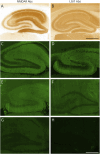
Results: The experienced raters agreed on 94% (133/142) of CSF and 88% (131/149) of serum classifications (Cohen's kappa = 0.87 and 0.75, respectively, p < 0.001). Among CSF samples, 75% (106/142) were correctly identified while 19% (27/142) were misclassified (13 false positives, 14 false negatives). Among serum samples, 66% (98/149) were correctly identified while 22% (33/149) were misclassified (11 false positives, 22 false negatives). The poorest performance was seen in detecting NMDAR, GABAAR, and mGluR5 Abs, which were not identified in 5 of 10, 6 of 10, and 5 of 9 serum samples and in 4 of 10, 5 of 10, and 5 of 10 CSF samples, respectively. The overall sensitivity of the commercial IIF-TBA was 84% for CSF and 76% for serum while the specificity was 72% for CSF and 73% for serum. Less experienced raters correctly identified 69% (98/142) of CSF samples and 73% (109/149) of serum samples and misclassified 13% (18/142) of CSF samples and 11% (16/149) of serum samples, and 18% (26/142) of CSF samples and 16% (24/149) of serum samples remained discordant.
Discussion: The diagnostic performance of EUROIMMUN IIF-TBA in detecting NSAbs in autoimmune encephalitis is suboptimal. NMDAR antibodies, among the most common NSAbs, can be missed in 50% of cases. This commercial TBA should not be used alone as a screening method nor as a confirmatory technique for NSAbs.
Conflict of interest statement
C. Papi receives research support from Spanish National Health Institute Carlos III (FIS grant PI23/01366) and 2023 EAN Research Training Fellowship. C. Milano receives research support from Spanish National Health Institute Carlos III co-funded by the European Union (Rio-Hortega grant CM24/00055). L. Marmolejo receives research support from Spanish National Health Institute Carlos III (predoctoral research grant FI24/00021). M. Spatola receives research support from La Caixa Foundation (Junior Leader) and Spanish National Health Institute Carlos III (ISCIII) and co-funded by the European Union (FIS grant PI23/01366). J. Dalmau receives research support from CaixaResearch Health 2022 (HR22-00221), Spanish National Health Institute Carlos III (ISCIII) and co-funded by the European Union (FIS grant PI23/00858), Cellex Foundation, Fundació Clínic per a la Recerca Biomèdica (FCRB) Programa Multidisciplinar de Recerca, Generalitat de Catalunya Department of Health (SLT028/23/000071), Edmond J. Safra Foundation. He receives royalties from Euroimmun for the use of NMDA as an antibody test. He received a licensing fee from Euroimmun for the use of GABAB receptor, GABAA receptor, DPPX and IgLON5 as autoantibody tests. He has received a research grant from Sage Therapeutics. All the other authors report no disclosures relevant to the manuscript. Go to
Figures


Comment in
-
Beyond the Lab Slip: Why Laboratory Expertise Matters in Neuronal Antibody Testing and Why Clinicians Should Care.Neurol Neuroimmunol Neuroinflamm. 2025 Jul;12(4):e200425. doi: 10.1212/NXI.0000000000200425. Epub 2025 May 20. Neurol Neuroimmunol Neuroinflamm. 2025. PMID: 40393019 Free PMC article. No abstract available.
-
Assessing Commercial Tissue-Based Assays for Autoimmune Neurologic Disorders (I): Antibodies to Intracellular Antigens.Neurol Neuroimmunol Neuroinflamm. 2025 Jul;12(4):e200410. doi: 10.1212/NXI.0000000000200410. Epub 2025 May 20. Neurol Neuroimmunol Neuroinflamm. 2025. PMID: 40393022 Free PMC article.
References
MeSH terms
Substances
Supplementary concepts
LinkOut - more resources
Full Text Sources
Medical
