. 2025 Jul;12(4):e200407.
doi: 10.1212/NXI.0000000000200407. Epub 2025 May 20.

Pro-Inflammatory Molecules Implicated in Multiple Sclerosis Divert the Development of Human Oligodendrocyte Lineage Cells

Affiliations

Pro-Inflammatory Molecules Implicated in Multiple Sclerosis Divert the Development of Human Oligodendrocyte Lineage Cells

Gabriela J Blaszczyk et al. Neurol Neuroimmunol Neuroinflamm. 2025 Jul.

Abstract

Background and objectives: Oligodendrocytes (OLs) and their myelin-forming processes are lost during the disease course of multiple sclerosis (MS), targeted by infiltrating leukocytes and their effector cytokines. Myelin repair is considered to be dependent on recruitment and differentiation of oligodendrocyte progenitor cells (OPCs). The basis of remyelination failure during the disease course of MS remains to be defined. The aim of this study was to determine the impact of the proinflammatory molecules tumor necrosis factor-⍺ (TNF⍺) and interferon-γ (IFNγ) on the differentiation of human OPCs.

Methods: We generated human OPCs from induced pluripotent stem cells with a reporter gene under the OL-specific transcription factor SOX10. We treated the cells in vitro with TNF⍺ or IFNγ and evaluated effects regarding cell viability, expression of OL lineage markers, and coexpression of astrocyte markers. To relate our findings to the molecular properties of OPCs as found in the MS brain, we reanalyzed publicly available single-nuclear RNA sequencing (RNAseq) datasets.

Results: Our analysis indicated that both TNF⍺ and IFNγ decreased the proportion of cells differentiating into the OL lineage, consistent with previous reports. Uniquely, we now observe that the TNF⍺ effect is linked to aberrant OPC differentiation in that a subset of O4+, reporter-positive cells coexpressing the astrocytic marker aquaporin-4. At the transcriptomic level, the cells acquire an astrocyte-like signature alongside a conserved reactive phenotype while downregulating OL lineage genes. Analysis of single-nuclear RNAseq datasets from the human MS brain revealed a subset of OPCs expressing an astrocytic signature.

Discussion: In the context of MS, these results imply that OPCs are present but inhibited from differentiating along the OL lineage, with a subset acquiring a reactive and stem cell-like phenotype, reducing their capacity to contribute toward repair. These findings help define a potential basis for the impaired myelin repair in MS and provide a prospective route for regenerative treatment.


Conflict of interest statement

The authors report no relevant disclosures. Full disclosure form information provided by the authors is available with the full text of this article at Neurology.org/NN.

Figures

Figure 1
Figure 1. Proinflammatory Molecules Block Human OPC Differentiation
(A) iPSC OPCs generated from our SOX10mO reporter line, subset based on fluorescence intensity before and after 21-day differentiation, showing an increase in SOX10mO with not all cells being terminally mature (mOrange high). Median percentage value of n = 4 passages displayed in heatmap. (B) Percentage of O4+ OPCs (normalized) as counted after immunofluorescence staining and treatment for 4 days, n = 4 passages. *p < 0.05, **p < 0.01. (C) Proportion of O4+ PI+ dead OPCs as measured by immunofluorescence staining after treatment for 4 days, n = 4 passages. ***p < 0.001. NG = no glucose, positive control. (D) Proportion of AQP4+ astrocytes in culture as measured by flow cytometry after treatment for 4–6 days, n = 3 passages. ****p < 0.0001. OPC = oligodendrocyte progenitor cell.
Figure 2
Figure 2. TNF⍺ Increases the Proportion of OPCs Coexpressing AQP4
(A) Representative gating scheme of OPC marker O4 (PE) coexpressing astrocyte marker AQP4 (AF488). (B) Percentage of O4+ OPCs also expressing AQP4 as measured by flow cytometry. n = 3, treatment between 4–6 days, **p < 0.01. (C) Histogram of SOX10mO fluorescence intensity of O4+AQP4+ cells (orange) against a nonreporter control (black). Fluorescence intensity values normalized to mode. AQP-4 = aquaporin-4; OPC = oligodendrocyte progenitor cell; TNF⍺ = tumor necrosis factor-⍺.
Figure 3
Figure 3. Transcriptome Changes in iPSC-Derived SOX10-Med Cells on Treatment With TNF⍺
(A) Principal component analysis (PCA) using all detected genes reveals a clear effect of TNF⍺ treatment in PC2. Control samples are shown in blue and treated samples in red. Normalized read counts from all detected genes were used. (B) Hierarchical clustering was performed on significantly differentially regulated genes (adjusted p value <0.05 and log2 fold change >1). Normalized read counts were used for clustering, and row normalization was applied to visualize the heatmap. Top genes (log2FC) include EXOC3L4, CSF2RB, CXCL10, ANO9, and CXCL13. (C) Gene Ontology (GO) analysis of significantly upregulated genes was conducted using g:Profiler, and GO terms were summarized and visualized with the Revigo tool. (D) 214 of 241 genes related to injury response in glial cells as published by Kopourtidou et al. (2024) were detected in our bulk RNAseq dataset. These genes were more enriched in TNF⍺-treated cells compared with control. 214 genes are visualized using log2 fold change after single-sample Gene Set Enrichment Analysis. TNF⍺ = tumor necrosis factor-⍺.
Figure 4
Figure 4. TNF⍺ Treatment of iPSC-Derived SOX10-Med Cells Deviates Glial Differentiation Program and Includes a Neural Progenitor Cell Proliferation Signature
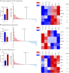
(A) Single-sample Gene Set Enrichment Analysis (ssGSEA) of the NPC proliferation signature shows significant enrichment in TNF⍺-treated cells compared with control cells (left panel). Log2 fold changes indicate a higher number of genes with increased expression in this signature on TNF⍺ treatment (middle panel). Examples of significantly upregulated NPC proliferation genes are visualized in a heatmap, with hierarchical clustering performed using normalized read counts, and DESeq2-calculated p values are provided for each gene (right panel). (B) ssGSEA of oligodendrocyte development genes using normalized read counts shows reduced enrichment of this signature in TNF⍺-treated cells (left panel). Log2 fold change analysis reveals a higher proportion of genes with elevated expression in control cells compared with TNF⍺-treated cells (middle panel). (C) ssGSEA of astrocyte differentiation genes indicates greater enrichment of this signature under TNF⍺ treatment conditions (left panel) and a higher proportion of genes with increased expression in this signature (middle panel). Examples of significantly upregulated genes involved in astrocyte differentiation are shown in the heatmap. Statistical significance is denoted as follows: ns = not significant, * = p < 0.01, ** = p < 0.001, and *** = p < 0.0001 (DESeq2 results). NPC = neural progenitor cell; TNF⍺ = tumor necrosis factor-⍺; ssGSEA = single-sample Gene Set Enrichment Analysis.
Figure 5
Figure 5. Identification and Characterization of Oligodendrocyte Progenitor Cells With Astrocytic Signatures in Human Single-Nuclear RNA Sequencing DataSets
(A) Analysis, clustering, and annotation of human snRNA-seq datasets derived from the studies by Jakel et al. (left panel) and Absinta et al. (right panel). OPC populations were identified using specific marker genes, notably PDGFRA and CSPG4/NG2. (B) A subset of OPCs was found to express AQP4, a marker typically associated with astrocytes. AQP4-positive cells are indicated in red, whereas cells lacking AQP4 expression are depicted in blue. (C) Bar graph depicting ratio of AQP4+ to AQP4- OPCs in different regions of the MS brain from the dataset by Absinta et al. Highest ratio of AQP4+ OPCs found within the core of an MS lesion. (D) Comparative analysis of AQP4-positive OPCs vs a gene signature based on top 100 genes in the astrocyte cluster by Jakel et al. (2019), with the log2 fold change (log2FC) depicted in bar plots. Top panel presents data from the study by Jakel et al., 2019, and bottom panel from the study by Absinta et al., 2021, with upregulated genes shown in red and downregulated genes in blue. AQP-4 = aquaporin-4; OPC = oligodendrocyte progenitor cell; snRNA-Seq = single-nuclear RNA sequencing.

References

    1. Kuhlmann T, Miron V, Cui Q, Wegner C, Antel J, Brück W. Differentiation block of oligodendroglial progenitor cells as a cause for remyelination failure in chronic multiple sclerosis. Brain. 2008;131(Pt 7):1749-1758.doi:10.1093/brain/awn096 - DOI - PubMed
    1. Jamann H, Cui QL, Desu HL, et al. . Contact-dependent granzyme B-mediated cytotoxicity of Th17-polarized cells toward human oligodendrocytes. Front Immunol. 2022;13:850616.doi:10.3389/fimmu.2022.850616 - DOI - PMC - PubMed
    1. Rodriguez M, Scheithauer B. Ultrastructure of multiple sclerosis. Ultrastruct Pathol. 1994;18(1-2):3-13.doi:10.3109/01913129409016267 - DOI - PubMed
    1. Wolswijk G. Oligodendrocyte survival, loss and birth in lesions of chronic-stage multiple sclerosis. Brain. 2000;123 ( Pt 1)(1):105-115.doi:10.1093/brain/123.1.105 - DOI - PubMed
    1. Lassmann H, van Horssen J. The molecular basis of neurodegeneration in multiple sclerosis. FEBS Lett. 2011;585(23):3715-3723.doi:10.1016/j.febslet.2011.08.004 - DOI - PubMed

MeSH terms

Substances