. 2025 Aug;75(4):100831.
doi: 10.1016/j.identj.2025.04.011. Epub 2025 May 19.

Artesunate Promotes Bone Remodeling Through TRAF6-Mediated NF-κB Signaling Under Orthodontic Stress in Diabetic Rats

Affiliations

Artesunate Promotes Bone Remodeling Through TRAF6-Mediated NF-κB Signaling Under Orthodontic Stress in Diabetic Rats

Yuxiang Zhan et al. Int Dent J. 2025 Aug.

Abstract

Objectives: To determine the effects of artesunate (ART) on bone remodeling in vivo under orthodontic stress in diabetic rats and explore the underlying mechanisms in vitro.

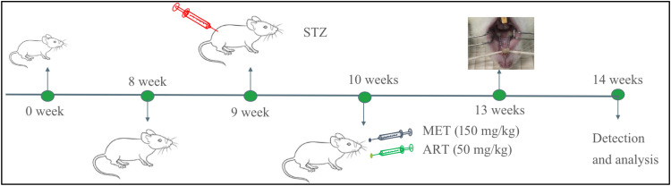
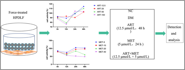
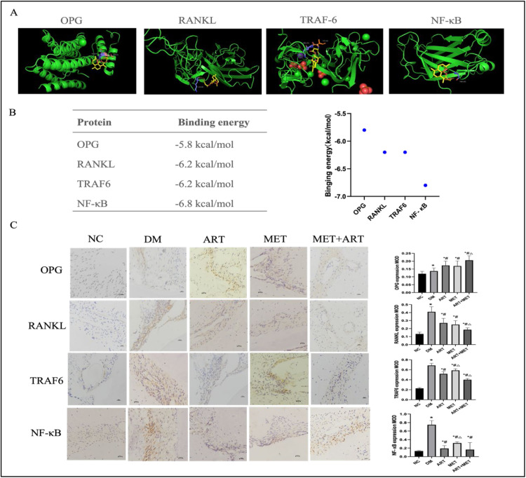
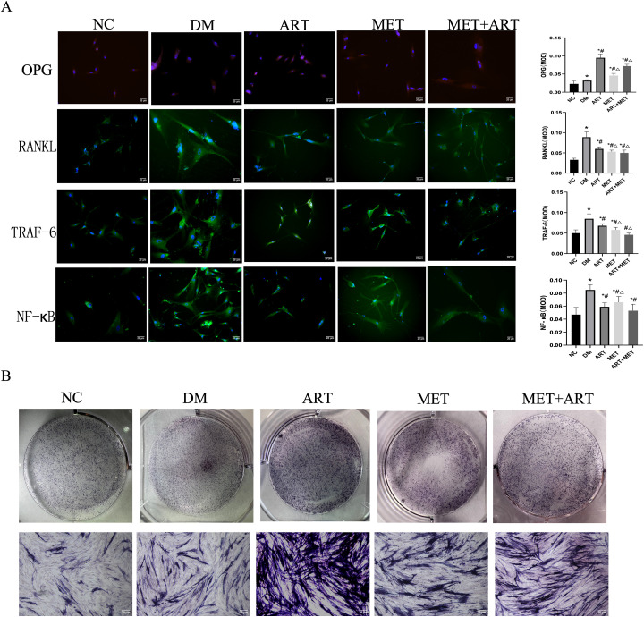
Materials and methods: A rat model of type 2 diabetes mellitus with orthodontic tooth movement was established. The rats received ART and/or metformin (Met) orally. The effects of ART and Met on periodontium changes were evaluated using tartrate-resistant acid phosphatase and immunohistochemical staining. Molecular docking analyses were employed to investigate the mechanisms of ART action. In vitro, the effects of ART on osteogenic and osteoclastic activity were explored by examining TRAF6 and NF-κB expression under hyperglycemic and static pressure conditions via immunofluorescence and Western blotting.

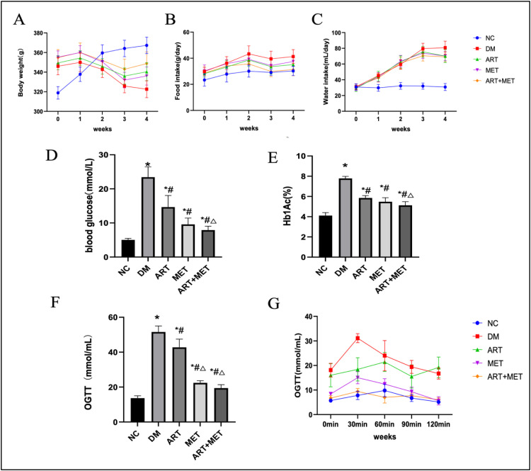
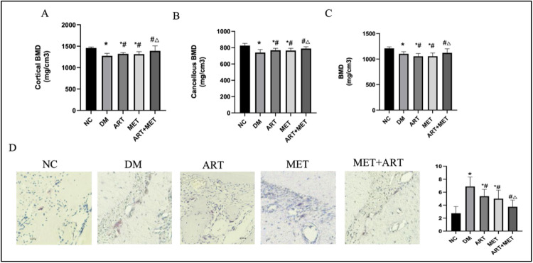
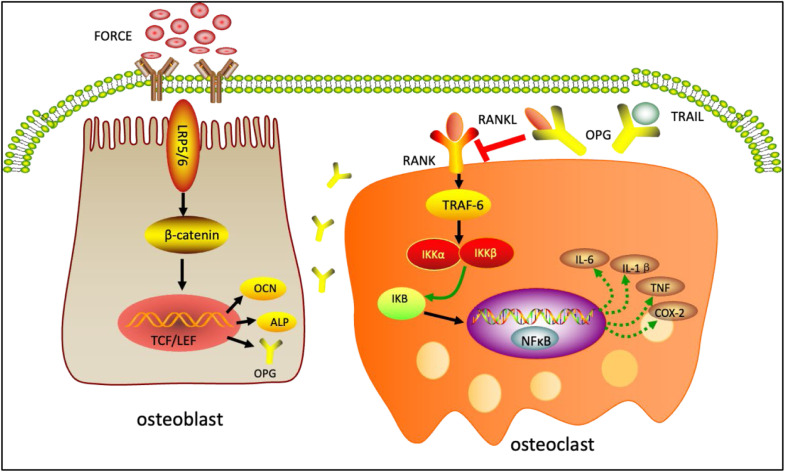
Results: ART enhanced bone metabolism despite hyperglycemia, though mechanical stress still induced bone resorption. Treatment with ART alone or in combination with Met promoted osteogenesis. TRAF6, NF-κB and the OPG/RANKL/RANK signaling pathways have been identified as key mediators of these effects. The expression of the osteogenesis-associated factor OPG increased after ART and Met treatment, while that of TRAF6 and the osteoclast-associated factors RANKL and NF-κB decreased.

Conclusions: Increased bone resorption and decreased bone formation are characteristics of type 2 diabetes, impacting orthodontic tooth movement. ART administration alone promotes bone remodeling under static pressure and hyperglycemic conditions. These effects are mediated by lowering blood sugar levels, inhibiting osteoclast function, and improving osteogenesis through mechanisms closely associated with the OPG/TRAF6/NF-κB signaling pathway.

Keywords: Artesunate; Bone reconstruction; NF-κB; Orthodontic force; TRAF6; Type 2 diabetes.


Conflict of interest statement

Conflict of interest The authors declared no potential conflicts of interest with respect to the research, authorship, and/or publication of this article.

Figures

Fig 1
Fig. 1
Experimental design. STZ was intraperitoneally injected at 8 weeks after HFD feeding. Rats with HFD/STZ treatment respectively received intragastric administration of ART, Met, and ART/Met combination for 3 consecutive weeks at 9 weeks after feeding. A rat orthodontic tooth movement model was then established, and the drug intervention was continued for 1 week.
Fig 2
Fig. 2
Cell compressive force equipment. The cck8 method was used to measure the cell viability of HPDLF after the intervention of different concentrations of ART or Met for 6, 12, 24 and 48 hours, respectively. The data are expressed as the mean ± standard deviation of 3 independent experiments.
Fig 3
Fig. 3
ART or MET alone and ART/ MET combination alleviate typical T2DM characteristics and reduce diabetes-related markers in T2DM rats. (A) Body weight, (B) food intake, (C) water intake, (D)FBG, (E) HbA1c, and (F) OGTT of rats in each group, (G) the blood glucose changes at different time points for different groups of OGTT (8 rats per group). Data are expressed as the mean ± SD. *P < .05 vs NC group. #P < .05 vs DM group. P < .05 vs ART group. NC, Normal control group; DM, Untreated diabetic group; ART, ART-treated diabetic group; MET, MET-treated diabetic group; ART+MET, ART/ MET co-treated diabetic group.
Figure 4
Fig. 4
ART or MET alone and ART/ MET combination relieve alveolar bone absorption in T2DM rats under orthodontic stress. (A) Cortical BMD, (B) Cancellous BMD, and (C) BMD, A-D: A Maxillary molar alveolar bone micro-CT scan of T2DM rats under orthodontic stress. (D) TRAP staining of the rats in each group (8 rats per group). Tartrate-resistant acid phosphatase–positive (TRAP+) odontoclasts decreased with drug treatment. scale bar 20 μm (400 ×). Data are expressed as the mean ± SD. *P < .05 vs NC group. #P < .05 vs DM group. P < .05 vs ART group. NC, Normal control group; DM, Untreated diabetic group; ART, ART-treated diabetic group; MET, MET-treated diabetic group; ART+MET, ART/ MET co-treated diabetic group.
Figure 5
Fig. 5
Molecular docking and the expressions of OPG, RANKL, TRAF6 and NF-κB. (A) Molecular docking of ART (yellow part of the pictures) to OPG, RANKL, TRAF6 and NF-κB protein, (B) The binding energy of each protein to ART, (C) Immunohistochemistry analysis of OPG, RANKL, TRAF6 and NF-κB in the periodontal membrane of rats in each group. scale bar 20 μm (400 ×). Data are expressed as the mean ± SD. *P < .05 vs NC group. #P < .05 vs DM group. P < .05 vs ART group. NC, Normal control group; DM, Untreated diabetic group; ART, ART-treated diabetic group; MET, MET-treated diabetic group; ART+MET, ART/ MET co-treated diabetic group.
Fig 6
Fig. 6
ART or MET on static pressure protection of HPDLF through TRAF6/NF-κB pathway. (A) Immunofluorescence analysis of OPG, RANKL, TRAF6 and NF-κB of HPDLF in each group, scale bar 20 μm (400 ×). (B) Alkaline phosphatase staining (ALP) of HPDLF in each group, Data are expressed as the mean ± SD. *P < .05 vs NC group. #P < .05 vs DM group. P < .05 vs ART group. NC, Normal control group; DM, Untreated diabetic group; ART, ART-treated diabetic group; MET, MET-treated diabetic group; ART+MET, ART/ MET co-treated diabetic group.
Fig 7
Fig. 7
OPG, RANKL, TRAF6 and NF-κB expression in HPDLF. (A) The messenger RNA expression of OPG, RANKL, TRAF6 and NF-κB of HPDLF, (B) Western blot analysis of OPG, RANKL, TRAF6 and NF-κB . Protein expression is normalized to GAPDH. The samples derive from the same experiment and the gels/blots were processed in parallel. Data are expressed as the mean ± SD. *P < .05 vs NC group. #P < .05 vs DM group. P < .05 vs ART group. NC, Normal control group; DM, Untreated diabetic group; ART, ART-treated diabetic group; MET, MET-treated diabetic group; ART+MET, ART/ MET co-treated diabetic group.
Figure 8
Fig. 8
Mechanism of the effect of ART on bone remodeling during orthodontic movement in type 2 diabetes. Under the action of orthodontic stress, osteoblast secretion of OPG increases, and the increased OPG can competitively bind RANK to RANKL secreted by osteoclasts, thereby reducing the activation of TRAF6/NF-κB axis and inhibiting osteoclast. On the 1 hand, ART regulates bone metabolism by regulating the OPG/RANKL/RANK pathway, and on the other hand, ART can also alleviate diabetes-induced bone resorption by directly regulating the TRAF6/NF-κB pathway.

Similar articles

References

    1. Khan R M M, Chua Z J Y, Tan J C, et al. From pre-diabetes to diabetes: diagnosis, treatments and translational research. Medicina (Kaunas) 2019;55(9):546. - PMC - PubMed
    1. Landon R., Gueguen V., Petite H., et al. Impact of astaxanthin on diabetes pathogenesis and chronic complications. Mar Drugs. 2020;18(7):357. - PMC - PubMed
    1. Orwoll E S, Parimi N., Wiedrick J., et al. Analysis of the associations between the human fecal microbiome and bone density, structure and strength: the MrOS cohort. J Bone Miner Res. 2022;37(4):597–607. - PMC - PubMed
    1. Higham C., Abrahamsen B. Regulation of bone mass in endocrine diseases including diabetes. Best Pract Res Clin Endocrinol Metab. 2022;36(2):101614. - PubMed
    1. Liccardo D., Cannavo A., Spagnuolo G., et al. Periodontal disease: a risk factor for diabetes and cardiovascular disease. Int J Mol Sci. 2019;20(6):1414. - PMC - PubMed