. 2025 May;22(226):20240931.
doi: 10.1098/rsif.2024.0931. Epub 2025 May 21.

Human shape perception spontaneously discovers the biological origin of novel, but natural, stimuli

Affiliations

Human shape perception spontaneously discovers the biological origin of novel, but natural, stimuli

Kira Isabel Dehn et al. J R Soc Interface. 2025 May.

Abstract

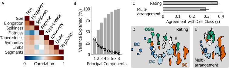
Humans excel at categorizing objects by shape. This facility involves identifying shape features that objects have in common with other members of their class and relies-at least in part-on semantic/cognitive constructs. For example, plants sprout branches, fish grow fins, shoes are moulded to our feet. Can humans parse shapes according to the processes that give shapes their key characteristics, even when such processes are hidden? To answer this, we investigated how humans perceive the shape of cells from the olfactory system of Xenopus laevis tadpoles. These objects are novel to most humans yet occur in nature and cluster into classes following their underlying biological function. We reconstructed three-dimensional (3D) cell models through 3D microscopy and photogrammetry, then conducted psychophysical experiments. Human participants performed two tasks: they arranged 3D-printed cell models by similarity and rated them along eight visual dimensions. Participants were highly consistent in their arrangements and ratings and spontaneously grouped stimuli to reflect the cell classes, unwittingly revealing the underlying processes shaping these forms. Our findings thus demonstrate that human perceptual organization mechanisms spontaneously parse the biological systematicities of never-before-seen, natural shapes. Integrating such human perceptual strategies into automated systems may enhance morphology-based analysis in biology and medicine.

Keywords: biological cell classification; generative models; perceptual organization; three-dimensional shape perception; visual similarity.


Conflict of interest statement

We declare we have no competing interests.

Figures

Using novel but natural stimuli to probe human 3D shape perception.
Figure 1.
Using novel but natural stimuli to probe human 3D shape perception. (A) Experimental stimuli were 3D reconstructions of cells from the olfactory epithelium of Xenopus laevis tadpoles. The cell types reconstructed included basal cells, dividing cells, olfactory sensory neurons, supporting cells and a set of indeterminate cells that could not be decisively assigned to any of the previous types. We reconstructed 30 individual stimuli, 6 stimuli per class. (B) For each of these cell types, 3D mesh model reconstructions were obtained through 3D microscopy. (C) These 3D mesh models were then 3D printed, and a spherical retroreflective marker was glued onto each object. (D) During the multi-arrangement task, participants were seated at a workbench imaged from multiple angles using passive marker tracking cameras. The experimenter placed the 3D-printed stimuli in random order in a circle on the workbench. Participants were asked to rearrange the stimuli by placing them anywhere on the workbench according to their shape similarity. The final object positioning was reconstructed and recorded using a position-tracking system. (E) In the rating task, participants viewed stimulus videos on a computer monitor. Videos were renderings of each object rotating in depth. Participants rated the stimuli along each of eight hand-selected feature dimensions, specified in (F).
Multi-arrangement results.
Figure 2.
Multi-arrangement results. (A) Multi-arrangement data for one example participant in the first (left) and second sessions (right) of the multi-arrangement task from Experiment 1. Data from the two sessions were aligned using Procrustes analysis. Different colours represent different cell classes: (BC) basal cells; (DC) dividing cells; (OSN) olfactory sensory neurons; (SC) supporting cells, (I) indeterminate cells. (B) Representational dissimilarity matrices (RDMs) for the first (left) and second sessions (right) from the same example participant. Dissimilarity was defined as the normalized Euclidian distance between stimulus pairs. Coloured bars along the x- and y-axis of the RDMs colour-code the cell classes. (C) Mean RDM averaged across participants and sessions (left) compared with the ground truth classification RDM derived from the cell classes. Note that we excluded indeterminate cells, as these may belong to different biological cell classes. (D) Within and between-participant agreement in the multi-arrangement task and agreement with the ground truth cell classes. Bars represent the mean across participants; error bars represent the 95% bootstrapped confidence intervals of the mean.
Between-participant agreement within and across experiments and tasks.
Figure 3.
Between-participant agreement within and across experiments and tasks. (A,B) Stimuli ranked 1st, 7th, 15th, 22nd and 30th from the average participant ratings of the ‘size’ and ‘spikiness’ dimensions. (C) Between-participant agreement for each of the rating dimensions in experiment 1. As reference, the darker grey bar displays the between-participant agreement for the same participants in the multi-arrangement task. (D) Agreement between rating and multi-arrangement data in experiment 1. (E) Agreement between participant ratings across experiments 1 and 2. (F) Agreement between rating and multi-arrangement data across experiments 1 and 2. Bars represent the mean across participants; error bars are the 95% confidence intervals of the mean. The grey-shaded region in D and F represents the noise ceiling (i.e. the upper and lower estimates of the between-participant agreement in the multi-arrangement task, corresponding to the upper bound of possible agreement across tasks).
The structure of the rating data.
Figure 4.
The structure of the rating data. (A) Multi-collinearity matrix showing the correlations between the eight feature dimensions probed by the rating task. (B) Scree plot displaying the variance explained (black curve) and cumulative variance explained (grey bars) the principal components of the rating data. (C) Agreement of the multi-arrangement and rating data with the ground truth cell classes. (D) The average rating data projected in the first two principal components separate the known cell classes. (E) The average multi-arrangement data are more clustered but do not separate cell class as clearly.
Classification analyses (A) Left: multi-arrangement data from naive participants from experiment 1, averaged across participants.
Figure 5.
Classification analyses (A) Left: multi-arrangement data from naive participants from experiment 1, averaged across participants. Right: cross-validated confusion matrix for a support vector machine trained to classify cell class from the multi-arrangement data. (B) Left: rating data from naive participants from experiment 1, projected in PCA space and averaged across participants. Right: cross-validated confusion matrix for a support vector machine trained to classify cell class from the PCA-projected rating data. (C,D) As A, B, except for expert biologists from experiment 3. (E) Cross-validated classification accuracy for all support vector machine classifiers trained on different data and participants. Bars are means, error bars are 95% confidence intervals. Dashed-line represents chance performance; grey lines show best-achievable classification accuracy.

Similar articles

References

    1. Phillips F, Fleming RW. 2020. The Veiled Virgin illustrates visual segmentation of shape by cause. Proc. Natl Acad. Sci. USA 117, 11735–11743. (10.1073/pnas.1917565117) - DOI - PMC - PubMed
    1. Spröte P, Fleming RW. 2016. Bent out of shape: the visual inference of non-rigid shape transformations applied to objects. Vis. Res. 126, 330–346. (10.1016/j.visres.2015.08.009) - DOI - PubMed
    1. Schmidt F, Spröte P, Fleming RW. 2016. Perception of shape and space across rigid transformations. Vis. Res. 126, 318–329. (10.1016/j.visres.2015.04.011) - DOI - PubMed
    1. Spröte P, Schmidt F, Fleming RW. 2016. Visual perception of shape altered by inferred causal history. Sci. Rep. 6, 36245. (10.1038/srep36245) - DOI - PMC - PubMed
    1. Schmidt F, Fleming RW. 2016. Visual perception of complex shape-transforming processes. Cogn. Psychol. 90, 48–70. (10.1016/j.cogpsych.2016.08.002) - DOI - PubMed

LinkOut - more resources