. 2025 May 20;16(1):4695.
doi: 10.1038/s41467-025-59463-1.

Human mitochondrial ferritin exhibits highly unusual iron-O2 chemistry distinct from that of cytosolic ferritins

Affiliations

Human mitochondrial ferritin exhibits highly unusual iron-O2 chemistry distinct from that of cytosolic ferritins

Justin M Bradley et al. Nat Commun. .

Abstract

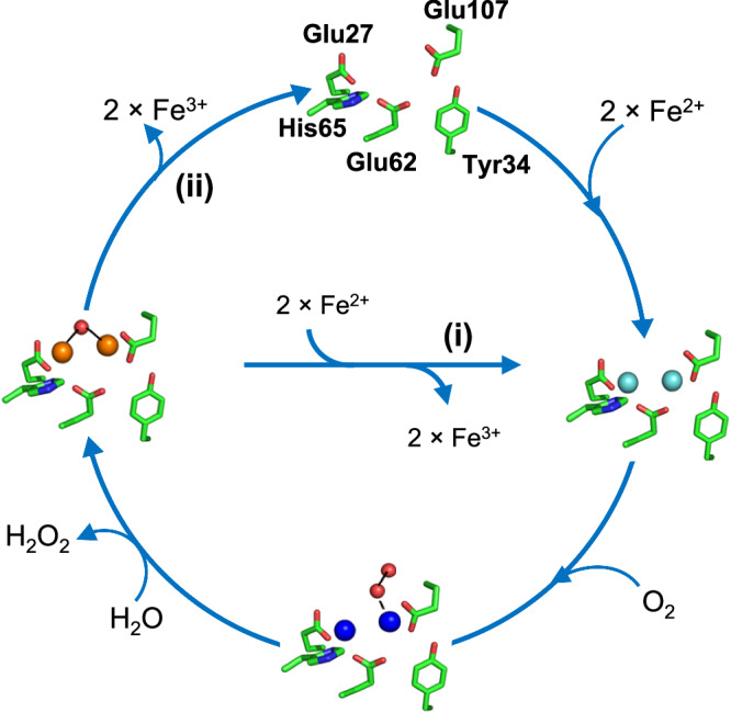
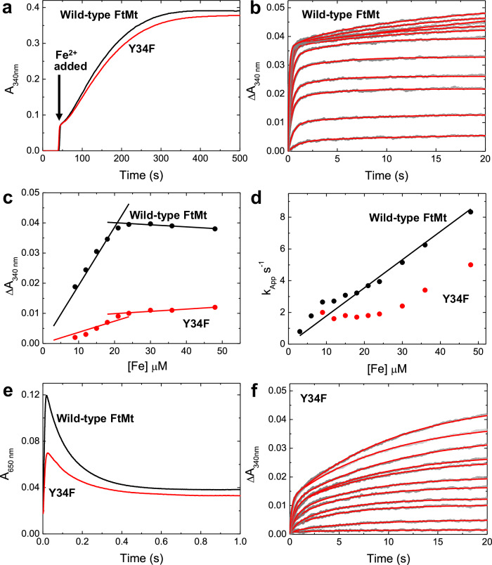
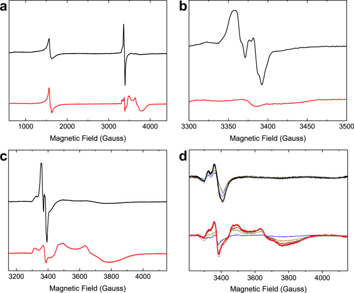
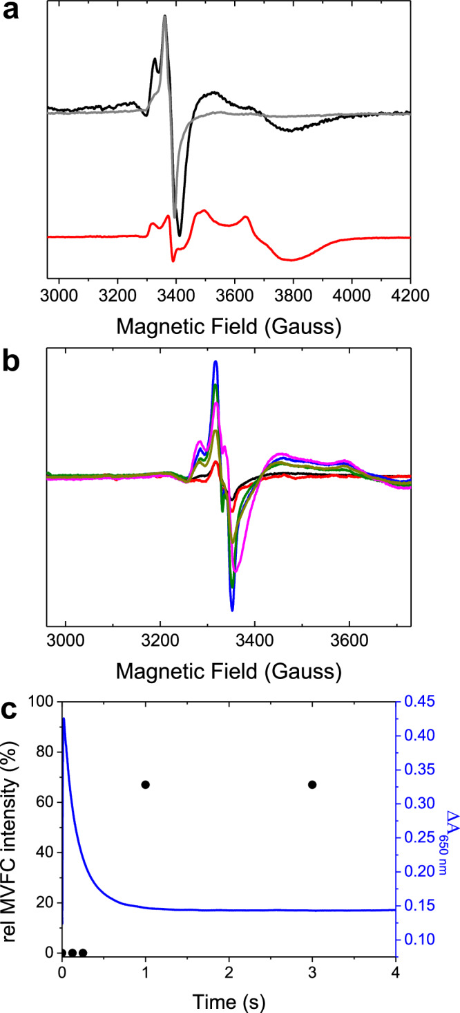
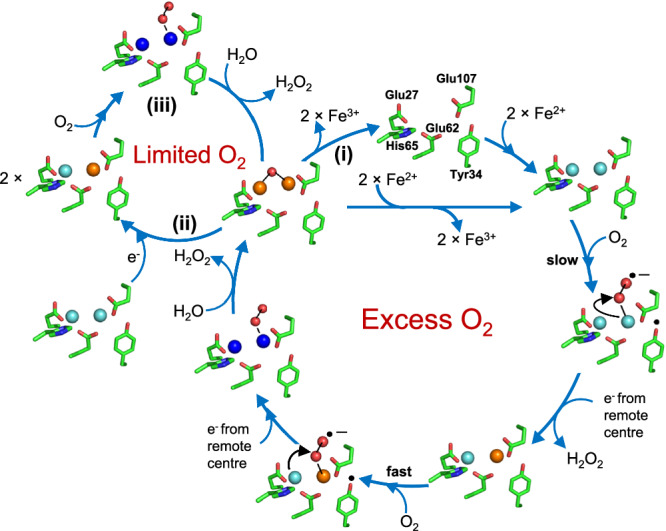
Ferritins are ubiquitous proteins that function in iron storage/detoxification by catalyzing the oxidation of Fe2+ ions and solubilizing the resulting Fe3+-oxo mineral. Mammalian tissues that are metabolically highly active contain, in addition to the widespread cytosolic ferritin, a ferritin that is localized to mitochondria. Mitochondrial ferritin (FtMt) protects against oxidative stress and is found at higher levels in diseases associated with abnormal iron accumulation, including Alzheimer's and Parkinson's. Here we demonstrate that, despite 80% sequence identity with cytosolic human H-chain ferritin, Fe2+ oxidation at the catalytic diiron ferroxidase center of FtMt proceeds via a distinct mechanism. This involves a mixed-valent ferroxidase center (MVFC) that is readily detected under the O2-limiting conditions typical of mitochondria, and formation of a radical on a strictly conserved Tyr residue (Tyr34) that is key for the activation of O2 and stability of the MVFC. The possible origin of the mechanistic differences exhibited by the highly-related human mitochondrial and cytosolic H-chain ferritins is explored.


Conflict of interest statement

Competing interests: The authors declare no competing interests.

Figures

Fig. 1
Fig. 1. Mechanism of Fe2+ oxidation at cytosolic ferritin ferroxidase centers (FCs).
Starting at the top, two equivalents of Fe2+ bind at the vacant (apo) FC to give di-Fe2+ FC (right). Transfer of a single electron from each onto O2 results in the formation of a di-Fe3+ peroxo intermediate (bottom) that is hydrolyzed to give a metastable di-Fe3+ oxo species and peroxide (left). In pathway (i) Fe3+ is then displaced by incoming Fe2+ substrate, regenerating the di-Fe2+ form of the FC and leading to the accumulation of a ferrihydrite-like mineral in the interior of the protein. In the absence of further Fe2+ substrate (pathway (ii)), the apo form of the FC is regenerated by the slower loss of Fe3+, into the interior of the protein, where it also contributes to the accumulation of a mineral core. Fe2+ ions are indicated in cyan, Fe3+ ions are in brown, and the DFP is in royal blue.
Fig. 2
Fig. 2. Iron oxidation and mineralization by FtMt.
a The increase in 340 nm absorbance as a function of time following the aerobic mixing of wild-type (black trace) and variant Y34F (red trace) FtMt with 400 equivalents of Fe2+. b The increase in 340 nm absorbance as a function of time following the aerobic mixing of wild-type FtMt with equal volumes of solutions containing 6–96 equivalents of Fe2+. Data are shown as gray circles; red traces represent fits to either mono- or bi-exponential functions. c The amplitude of the increase in 340 nm absorbance during the rapid phase of Fe2+ oxidation by wild-type (black) and variant Y34F (red) FtMt as a function of the concentration of added Fe2+. d The dependence of the apparent first-order rate constant for Fe2+ oxidation as a function of initial Fe2+ concentration for wild-type (black) and variant Y34F (red) FtMt. The black line represents the best fit through the origin to the data for the wild-type protein. e The change in 650 nm absorbance as a function of time following the aerobic mixing of wild-type (black trace) and variant Y34F (red trace) FtMt with 48 equivalents of Fe2+, reporting the formation and subsequent decay of the di-Fe3+ peroxo intermediate. f as (b) but for variant Y34F FtMt. Source data are provided as a Source Data file.
Fig. 3
Fig. 3. EPR spectra of the paramagnetic species formed during Fe2+ oxidation by FtMt.
a Spectra recorded at microwave power of 3.19 mW over the field range 600–4200 Gauss for 4.2 μm wild-type (black trace) and variant Y34F (red trace) FtMt frozen approximately 10 s following aerobic mixing with 72 equivalents of Fe2+. b as in (a) but spectra recorded over the field range 3300–3500 Gauss at microwave power 0.05 mW. c as (a) but spectra recorded over the field range 3200–4150 Gauss. d as (c) but with freezing times for the wild-type protein of 45 ms (blue), 125 ms (yellow), 245 ms (brown), 445 ms (heavy black trace with maximum radical intensity), or 10 s (gray) and freezing times for variant Y34F of 45 ms (blue), 245 ms (yellow), 445 ms (brown), 3 s (heavy red trace with maximum mixed-valent ferroxidase center intensity) or 10 s (gray). Source data are provided as a Source Data file.
Fig. 4
Fig. 4. O2-limited reaction of FtMt.
a The EPR spectrum of 4.17 μm wild-type FtMt anaerobically incubated with 200 μm Fe2+ then frozen 10 s after mixing with aerobic buffer, resulting in an O2 concentration of 50 μm post-mixing (black trace). The red trace shows the MVFC formed by variant Y34F when O2 is present in excess, and the gray trace shows the spectrum of wild-type protein when O2 is present in excess for comparison. b Accumulation of the MVFC intermediate with increasing freezing time following the mixing of 20.8 μm wild-type FtMt with 1 mm Fe2+ under aerobic conditions. Samples frozen 0.125 (black), 0.250 (red), 1 (blue), 3 (dark green), 6 (magenta) and 10 (dark yellow) s after mixing. c Comparison of the relative intensity of the MVFC of the samples from (b) (black circles, left axis) with the change in 650 nm absorbance as a function of time after mixing of an equivalent sample (blue trace, right axis). Source data are provided as a Source Data file.
Fig. 5
Fig. 5. Proposed mechanistic cycle of Fe2+ oxidation by FtMt.
Following binding of two Fe2+ ions to the apo FC (top of the main cycle), O2 binds and is activated by transfer of a single electron from Tyr34 to yield a tyrosyl radical and a superoxo adduct of the di-Fe2+ FC. The radical is quenched by electron transfer from a remote FC, and electron transfer from Fe2+ at the site with superoxide bound yields the peroxide product of O2 reduction and the MVFC intermediate. Binding of O2 and its activation by the MVFC is rapid in comparison to that occurring at the di-Fe2+ FC form, and hence, the MVFC intermediate does not accumulate under continuous turnover. This reaction results in the DFP intermediate, which is hydrolyzed to the metastable di-Fe3+ form. In the presence of excess O2, the metastable di-Fe3+ FC breaks down to release Fe3+ ions to the FtMt cavity, and the reaction cycle begins again (marked as (i)). Under limiting O2, where both di-Fe2+ and di-Fe3+ FCs are present, a disproportionation reaction occurs via long-range electron transfer, resulting in two MVFCs (marked as (ii) on the smaller left-hand cycle). When O2 is available again, these are oxidized to the di-Fe3+ form (marked as (iii)). The overall stoichiometry of the reaction is identical to that catalyzed by cytosolic ferritins, with each center coupling the oxidation of two equivalents of Fe2+ to Fe3+ with the reduction of O2 to peroxide. Fe2+ ions are indicated in cyan, Fe3+ are in brown, and the DFP is in royal blue.

Similar articles

References

    1. Beinert, H., Holm, R. H. & Munck, E. Iron-sulfur clusters: Nature’s modular, multipurpose structures. Science277, 653–659 (1997). - PubMed
    1. Gamba, I., Codola, Z., Lloret-Fillol, J. & Costas, M. Making and breaking of the O-O bond at iron complexes. Coord. Chem. Rev.334, 2–24 (2017).
    1. Poulos, T. L. Heme enzyme structure and function. Chem. Rev.114, 3919–3962 (2014). - PMC - PubMed
    1. Gayer, K. H. & Wootner, L. The hydrolysis of ferrous chloride at 25 degrees. J. Am. Chem. Soc.78, 3944–3946 (1956).
    1. Imlay, J. A. The molecular mechanisms and physiological consequences of oxidative stress: lessons from a model bacterium. Nat. Rev. Microbiol.11, 443–454 (2013). - PMC - PubMed

LinkOut - more resources