. 2025 May 20;3(1):35.
doi: 10.1038/s44276-025-00152-3.

Single-cell analysis reveals tumour size as a key driver of immune cell profile alterations in primary breast tumours and corresponding lymph nodes

Collaborators, Affiliations

Single-cell analysis reveals tumour size as a key driver of immune cell profile alterations in primary breast tumours and corresponding lymph nodes

Marit Otterlei Fjørtoft et al. BJC Rep. .

Abstract

Background: At diagnosis, 30-40% of women with breast cancer have metastases in sentinel (SN) or axillary lymph nodes (ALN). Nodal status is a strong prognostic factor and guides treatment decisions. Immune checkpoint inhibition has shown some efficacy, which can increase in the neoadjuvant setting. A better understanding of how tumour cells in primary tumours and metastatic lymph nodes shape the local immune microenvironment may provide clues for more individualized therapeutic interventions.

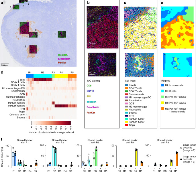
Methods: We conducted deep immunophenotypic analysis of 29 primary breast tumours and 36 lymph nodes from 38 patients with primary operable breast cancer.

Results: The immune profile of the primary tumour was not predictive of the lymph node immune profile or metastatic status. Primary tumours showed prominent CD8 T cell exhaustion and activated regulatory T cells, and the frequencies of these subsets were associated with tumour size. The immune cell profile in lymph nodes were different from the profile in primary tumours, except for the ALN+ nodes, which displayed a T-cell profile more similar to primary tumours. The frequencies of the T cell subsets in lymph nodeswere associated with metastatic size. Tumour cells from smaller metastases exhibited a distinct phenotype compared to those from larger tumour deposits, and the size of the tumour cell deposit impacted the local immune cell composition.

Conclusion: The tumour size of primary tumours and metastatic size in lymph nodes are the main drivers of changes in immune cell composition.


Conflict of interest statement

Competing interests: The authors declare no competing interests. Ethics approval and consent: The study was approved by the regional committee for research ethics (200606181-1, 538-07278a, 2009/4935, 2016/433). Informed consent was obtained from all participants in accordance with the Declaration of Helsinki.

Figures

Fig. 1
Fig. 1. Identification of cell types in primary tumours and lymph nodes.
Overview of the breast cancer cohort consisting of 29 primary tumours and 36 lymph nodes, of which 28 had paired samples from the same patient. The samples were analysed by single cell mass cytometry (CyTOF) and clustered into 40 cell types or metaclusters (MC) by FlowSOM analysis. a Cartoon of the 65 primary tumour and lymph node samples included in the analysis of immune cells. b The 40 metaclusters (MCs) identified by FlowSOM analysis were annotated and visualised by opt-SNE (top). The cells were also coloured by main cell type (middle) and tissue type (bottom). c Median event count of the 33 immune clusters grouped into main cell types presenting the cell composition in each tissue type, with bar encoding subtype and lymph node status.
Fig. 2
Fig. 2. Unique immune profiles in primary breast tumours and corresponding lymph nodes.
a Immune cell abundance (33 MCs) in the two tissues, coloured by main cell type. The line in the box plots denotes the median value, and error bars are included. b Forest plot showing the frequencies of different cell types occurring in lymph nodes compared to primary tumours. Values are log scaled with a 95% bootstrap confidence interval for each median value. c Hierarchical consensus clustering of paired primary tumour and lymph nodes (n = 28 pairs). Columns are normalised by total CD45+ cells per sample and scaled by subtracting half of the total range of each sample (centres the values around zero), then clustered by Pearson distance matrix and complete linkage. Rows (unnormalized) are clustered by Euclidean distance matrix and complete linkage. Fisher’s exact test was applied on categorical data and ANOVA was applied on numerical data for statistical testing of differences between clusters.
Fig. 3
Fig. 3. Immune cells clusters of primary tumour and lymph nodes.
Hierarchical consensus clustering of paired samples of (a) primary tumour (n = 28) and (b) lymph nodes (n = 28). Columns are normalised by total CD45+ cells per sample and scaled by subtracting half of the total range of each sample (centres the values around zero), then clustered by Pearson distance matrix and complete linkage. Rows (unnormalized) are clustered by Euclidean distance matrix and complete linkage. Fisher’s exact test was applied on categorical data and ANOVA was applied on numerical data for statistical testing of differences between clusters. Boxplot of clusters displaying association with (c) clinical tumour size in primary tumours, (d) TILs for primary tumours (TILs in PT = percentage of tumour bed infiltrated with lymphocytes, scored by pathologist), (e) primary tumour size in lymph nodes, and (f) metastasis size in lymph nodes. g Sankey plot illustrates the relationship between the primary tumour cluster and the lymph node cluster for each patient. There was no significant correlation between the primary tumour clusters and lymph node clusters (Fisher’s exact test, p = 0.19).
Fig. 4
Fig. 4. The T cell profile in ALN+ samples resemble the T cell profile in primary tumours.
a opt-SNE of T cells from all 65 samples using common T cell markers as indicated in the top row. In top row, all samples are merged and in bottom row, the samples of each sample type are merged. b The frequency of selected T cell clusters (all the T cell clusters are shown in Supplementary Fig. 5) in SN−, SN+, ALN+ and PT samples. (Kruskal Wallis with Dunn’s post hoc test: *p < 0.05, **p < 0.01, ***p < 0.001. **** p < 0.0001. Kruskal Wallis was Bonferroni corrected, 19 tests). PT = primary tumour sample. c Regression analysis showing the relationship between metastatic tumour size in lymph nodes and abundance of T cell clusters, assessed using Spearman’s correlation. d Regression analysis showing the relationship between primary tumour size and abundance of T cell clusters, assessed using Spearman’s correlation.
Fig. 5
Fig. 5. The phenotype of small tumour cell deposits differs from large tumour cell deposits.
a Tumour cells from primary tumours and lymph nodes were separated in an opt-SNE run on pan-keratin, EpCAM, PD-L1/L2, GATA3, CTLA-4, PD-1, ICOS, OX40, TIM-3, TIGIT, CCR5 and CCR7. b The median protein expression measured for tumour cells from primary tumours (PT) and metastatic lymph nodes. (Mann-Whitney, Bonferroni corrected for 8 tests: *p < 0.05, **p < 0.01). c Median protein expression in metastatic tumour cells from a separate cohort of lymph nodes from breast cancer patients (n = 22, 4 samples overlap with our cohort of paired samples). These samples were analysed by mass cytometry using an antibody panel that contained more tumour cell markers [23]. Small <5 mm, medium = 5–20 mm, large >20 mm. (Mann-Whitney U test, *p < 0.05). df Immunofluorescence with pan-keratin and E-cadherin was performed on 4 lymph node metastases, 2 large (BC05 and BC18) and 2 small (BC10 and BC34). The large metastases are shown in Supplementary Fig. 7C. Scale bar = 10 µm. d Median fluorescence intensity (MFI) for each sample. e To reveal spatial heterogeneity in pan-keratin and E-cadherin expression, fluorescence intensity was measured in small areas as indicated in (f, g).
Fig. 6
Fig. 6. Differences in the immune profile surrounding large and small metastatic tumour cell deposits.
a A metastatic lymph node (BC10) was stained with metal-tagged antibodies (Supplementary Table 4) and seven regions containing tumour cells were ablated by imaging mass cytometry. The images are overlaid with a corresponding AE1/AE3 IHC image of the same lymph node. b Image number 1 and 4 are shown pseudo-coloured by staining intensity of antibodies in the raw images as indicated and (c) as single-cell data with x and y position and colour-coded by cell population. d Using CytoMAP, a raster scan of 30 μM radium defined neighbourhoods, which were clustered based on the presence of cell types in each neighbourhood. This revealed 5 neighbourhood types called regions (R1-R5). e Image 1 and 4 are shown colour-coded by region type. f Interactions are calculated as the percentage of neighbourhoods sharing a border with a certain region type among all the neighbourhoods that share a border with another region type. These values are plotted separately for images with large tumour cell deposits (image 1–3) and images with small deposits (image 4–7).

References

    1. Bray F, Laversanne M, Sung H, Ferlay J, Siegel RL, Soerjomataram I, et al. Global cancer statistics 2022: GLOBOCAN estimates of incidence and mortality worldwide for 36 cancers in 185 countries. CA Cancer J Clin. 2024;74:229–63. - PubMed
    1. Chakraborty A, Bose CK, Basak J, Sen AN, Mishra R, Mukhopadhyay A. Determinants of lymph node status in women with breast cancer: a hospital based study from Eastern India. Indian J Med Res. 2016;143:45–51. - PMC - PubMed
    1. Giuliano AE, Edge SB, Hortobagyi GN. Eighth edition of the AJCC cancer staging manual: breast cancer. Ann Surg Oncol. 2018;25:1783–5. - PubMed
    1. Stanton SE, Adams S, Disis ML, Stranton S, Adams, SD LM. Variation in the incidence and magnitude of tumor-infiltrating lymphocytes in breast cancer subtypes a systematic review. JAMA Oncol. 2016;2:1354–60. - PubMed
    1. Denkert C, von Minckwitz G, Darb-Esfahani S, Lederer B, Heppner BI, Weber KE, et al. Tumour-infiltrating lymphocytes and prognosis in different subtypes of breast cancer: a pooled analysis of 3771 patients treated with neoadjuvant therapy. Lancet Oncol. 2018;19:40–50. - PubMed

LinkOut - more resources