

Findings on conventional ultrasonography and contrast-enhanced ultrasonography in different histopathological subtypes of ovarian thecoma-fibroma group
- PMID: 40394597
- PMCID: PMC12090646
- DOI: 10.1186/s12880-025-01693-2
Findings on conventional ultrasonography and contrast-enhanced ultrasonography in different histopathological subtypes of ovarian thecoma-fibroma group
Abstract
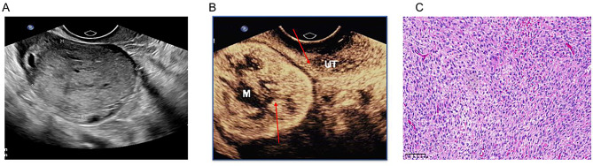
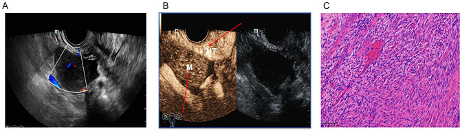
Background: Ovarian thecoma-fibroma group (OTFG) is an unusual type of ovarian cancer with three histopathologic subtypes, but their features on ultrasonography are still poorly understood. This study evaluated the features of different histopathological subtypes of OTFG on conventional ultrasonography and contrast-enhanced ultrasonography (CEUS).
Methods: This retrospective study enrolled sixty-nine women with pathologically confirmed OTFG who underwent preoperative CEUS. The characteristics of OTFG on conventional ultrasonography and CEUS, clinical manifestations, and laboratory findings were compared among subtypes.
Results: Fourteen patients were diagnosed with fibroma, fifty-one with thecofibroma, and four with thecoma. Although 69% of patients were post-menopausal, thecoma patients were significantly younger than those in other two groups. Laboratory examination revealed 21.7% (15/69) of patients had high carbohydrate antigen 125 (CA-125) level. On conventional ultrasonography, 72.5% (50/69) masses showed solid type, 24.6% (17/69) showed mixed cystic-solid type, and only 2.9% (2/69) showed cystic type. On CEUS, 50% (2/4) of thecoma lesions were rapid enhancement, 58.8% (30/51) of thecofibroma lesions and 78.6% (11/14) of fibroma lesions showed slow enhancement, 75% (3/4) of thecoma lesions showed isoenhancement during the descent process, and only 13.75% (7/51) of thecofibroma lesions and 7.1% (1/14) of fibroma lesions showed isoenhancement during the descent. they varied significantly among different histopathological subtypes.
Conclusions: The majority of OTFG is solid-like on conventional ultrasonography. Menopause is an important factor related to the subtype of OTFG. In postmenopausal patients with solid adnexal masses, slow hypoenhancement on CEUS is an important feature of fibroma. In premenopausal patients with solid or mixed cystic-solid adnexal masses, thecoma may be considered when rapid hyperenhancement, and isoenhancement or hypoenhancement during descent are observed on CEUS.
Clinical trial number: Not applicable.
Keywords: Contrast-enhanced ultrasonography; Fibroma; Ovarian thecoma-fibroma group; Thecoma.
© 2025. The Author(s).
Conflict of interest statement
Declarations. Ethics approval and consent to participate: This study was approved by the Ethics Committee of Ningbo University First Hospital ([2024]181RS, on September 3, 2024). The study was conducted according to the Declaration of Helsinki, and written informed consent was obtained from each patient. Consent for publication: Not applicable. Competing interests: The authors declare no competing interests.
Figures

Similar articles
-
Ovarian thecoma-fibroma groups: clinical and sonographic features with pathological comparison.J Ovarian Res. 2016 Nov 22;9(1):81. doi: 10.1186/s13048-016-0291-2. J Ovarian Res. 2016. PMID: 27876070 Free PMC article.
-
CT diagnosis in the thecoma-fibroma group of the ovarian stromal tumors.Cell Biochem Biophys. 2015 Mar;71(2):937-43. doi: 10.1007/s12013-014-0288-7. Cell Biochem Biophys. 2015. PMID: 25315640
-
Conventional ultrasound and high-frame-rate contrast-enhanced ultrasound characteristics of ovarian thecoma-fibroma groups.Quant Imaging Med Surg. 2025 May 1;15(5):3875-3890. doi: 10.21037/qims-24-2200. Epub 2025 Apr 17. Quant Imaging Med Surg. 2025. PMID: 40384658 Free PMC article.
-
The peripheral cysts sign of ovarian thecoma-fibroma groups: Incidence and diagnostic value.J Obstet Gynaecol Res. 2025 Jul;51(7):e16369. doi: 10.1111/jog.16369. J Obstet Gynaecol Res. 2025. PMID: 40610240
-
Comparison of conventional, doppler and contrast-enhanced ultrasonography in differential diagnosis of ovarian masses: a systematic review and meta-analysis.BMJ Open. 2021 Dec 24;11(12):e052830. doi: 10.1136/bmjopen-2021-052830. BMJ Open. 2021. PMID: 34952878 Free PMC article.
References
-
- Sex cord-stromal tumors of the ovary: a comprehensive review and update for radiologists - PubMed. https://pubmed.ncbi.nlm.nih.gov/26054417/ [Accessed August 29, 2024]. - PMC - PubMed
-
- Jung SE, Rha SE, Lee JM, Park SY, Oh SN, Cho KS, Lee EJ, Byun JY, Hahn ST. CT and MRI findings of sex Cord–Stromal tumor of the ovary. Am J Roentgenol. 2005;185:207–15. 10.2214/ajr.185.1.01850207. - PubMed
-
- Schoolmeester JK, Erickson LA. Ovarian fibrothecoma. Mayo Clin Proc. 2019;94:1652–3. 10.1016/j.mayocp.2019.04.019. - PubMed
-
- Cho YJ, Lee HS, Kim JM, Joo KY, Kim M-L. Clinical characteristics and surgical management options for ovarian fibroma/fibrothecoma: a study of 97 cases. Gynecol Obstet Invest. 2013;76:182–7. 10.1159/000354555. - PubMed
MeSH terms
Substances
LinkOut - more resources
Full Text Sources
Medical
Research Materials
Miscellaneous
