. 2025 May 20;20(1):488.
doi: 10.1186/s13018-025-05914-w.

Exploring the mechanism by which UCHL3 alleviates diabetic foot ulcers: FOXM1/NLRP3 inflammasome-mediated angiogenesis and endothelial cell pyroptosis

Affiliations

Exploring the mechanism by which UCHL3 alleviates diabetic foot ulcers: FOXM1/NLRP3 inflammasome-mediated angiogenesis and endothelial cell pyroptosis

Xincheng Liao et al. J Orthop Surg Res. .

Abstract

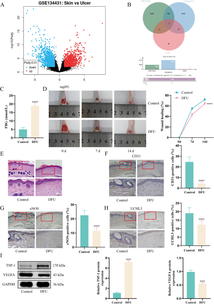
Background: This study investigated the role of ubiquitin C-terminal hydrolase L3 (UCHL3) in regulating endothelial cell (EC) pyroptosis and angiogenesis in diabetic foot ulcers (DFUs), with a focus on FOXM1 and NLRP3 inflammasomes.

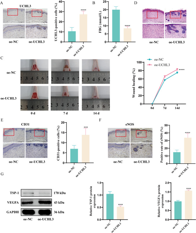
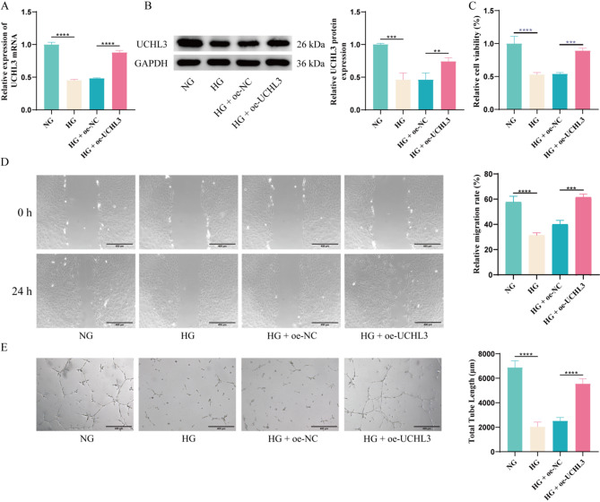
Methods: Differentially expressed genes in DFUs were identified using the GSE134431 dataset and cross-referenced with vascular formation-related factors from GeneCard and deubiquitinases from the UbiNet 2.0 database. A rat DFU model was used to evaluate wound healing, with or without UCHL3 overexpression and FOXM1 knockdown. Histological analysis and immunohistochemistry were employed to assess tissue morphology and the expression of CD31, eNOS, UCHL3, and FOXM1. In vitro, high glucose-induced human umbilical vein ECs (HUVECs) were transfected with UCHL3 overexpression and FOXM1 knockdown constructs. Cell viability, migration, and angiogenesis were assessed.

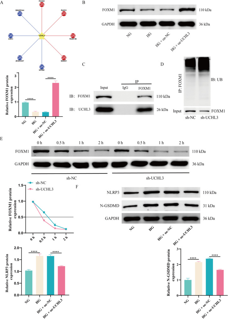
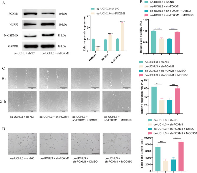
Results: UCHL3 expression was significantly reduced in DFU tissues. UCHL3 overexpression promoted wound healing in a rat model, while FOXM1 knockdown impaired wound healing and vascular formation. In HUVECs, UCHL3 overexpression enhanced cell viability, migration, and angiogenesis, accompanied by reduced NLRP3 and N-GSDMD levels. FOXM1 knockdown reversed these effects, but treatment with the NLRP3 inhibitor, MCC950, alleviated this damage.

Conclusion: UCHL3 enhances FOXM1 deubiquitination, inhibits NLRP3 inflammasome activation, and reduces EC pyroptosis, thereby contributing to DFU healing. UCHL3 and FOXM1 are potential therapeutic targets for DFU.

Keywords: Diabetic foot ulcers; Endothelial cells; FOXM1; NLRP3 inflammasome; UCHL3.


Conflict of interest statement

Declarations. Ethical approval: This experiment has been approved by the Animal Ethics Committee of Hunan Evidence-based Biotechnology Co., Ltd. (ABTZ24002). All procedures and reporting were performed according to the ARRIVE guidelines including the 3R concept. Competing interests: The authors declare no competing interests.

Figures

Fig. 1
Fig. 1
UCHL3 is lowly expressed in DFU. (A) Volcanic map of differentially expressed genes in GSE134431. (B) The intersection Venn diagram of DEGs with vascular formation related factors downloaded from GeneCard database and DUBs collected from UbiNet 2.0 database. (C) Detection of changes in blood glucose levels in rats before and after modeling. (D) Assessment of alterations in wound healing rate in rats before and after modeling. (E) HE staining was used to compare histological and morphological characteristics of the wound before and after modeling. (F) Immunohistochemical analysis of changes in CD31 expression in wound tissue before and after modeling. (G) Immunohistochemical analysis of changes in eNOS expression in wound tissue before and after modeling. (H) Immunohistochemical analysis of changes in UCHL3 expression in wound tissue before and after modeling. (I) WB detection of changes in angiogenesis-related proteins (VEGF and TSP-1) in rat wound tissues before and after modeling. n = 6. The magnification of images D, E, F and G is 100 times (scale = 400 μm) and 200 times (scale = 200 μm) respectively. **** P < 0.0001. The detection between the two groups was analyzed using t-test. Two factor analysis of variance (ANOVA) will be used for three or more sets of data, and Tukey’s will be used for post hoc testing
Fig. 2
Fig. 2
Overexpression of UCHL3 alleviates DFU. (A) Immunohistochemical detection of UCHL3 expression to verify overexpression efficiency. (B) Assessment of wound healing rate in rats after UCHL3 overexpression. (C) Detection of blood glucose levels in rats to evaluate metabolic effects of UCHL3 overexpression. (D) HE staining was used to analyze histological and morphological changes in the wound after UCHL3 overexpression. (E) Immunohistochemical detection of CD31 expression to assess angiogenesis following UCHL3 overexpression. (F) Immunohistochemical detection of eNOS expression to evaluate endothelial function after UCHL3 overexpression. (G) WB analysis of angiogenesis-related proteins (VEGF and TSP-1) in wound tissues after UCHL3 overexpression. n = 6. The magnification of images A, D, E and F is 100 times (scale = 400 μm) and 200 times (scale = 200 μm) respectively. *** P < 0.001, **** P < 0.0001. The detection between the two groups was analyzed using t-test. Two factor analysis of variance (ANOVA) will be used for three or more sets of data, and Tukey’s will be used for post hoc testing
Fig. 3
Fig. 3
UCHL3 inhibits endothelial cell damage and promotes angiogenesis. (A) RT-qPCR detection of UCHL3 mRNA expression levels in HUVECs. (B) WB detection of UCHL3 protein expression in HUVECs. (C) CCK-8 assay to measure HUVEC proliferation activity. (D) Scratch wound healing assay to evaluate HUVEC migration ability. (E) Tube formation assay to assess angiogenesis capacity of HUVECs. n = 3. The magnification of images D and E is 100 times (scale = 400 μm). ** P < 0.01, *** P < 0.001, **** P < 0.0001. Three or more sets of data were analyzed using one-way ANOVA and subjected to post hoc testing using Tukey’s
Fig. 4
Fig. 4
UCHL3 binds to FOXM1 and promotes FOXM1 deubiquitination while inhibiting NLRP3 inflammasome activation. (A) The targeting relationship of UCHL3 in Ubibrowser 2.0. (B) WB analysis of FOXM1 expression in HUVECs. (C) Immunoprecipitation detection of the binding relationship between UCHL3 and FOXM1. (D) Immunoprecipitation assay was used to detect the deubiquitination modification of FOXM1 by UCHL3. (E) CHX treatment was used to detect the effect of UCHL3 on the stability of FOXM1 protein. (F) WB detection of NLRP3 and N-GSDMD in HUVECs. n = 3. **** P < 0.0001. Three or more sets of data will be analyzed using one-way or two-way ANOVA, and Tukey’s will be used for post hoc testing
Fig. 5
Fig. 5
Knockdown of FOXM1 leads to pyroptosis of endothelial cells and inhibits angiogenesis. (A) WB detection of FOXM1, NLRP3, and N-GSDMD expression in HUVECs. (B) CCK-8 assay measuring proliferation activity of HUVECs. (C) Scratch wound healing assay detecting migration ability of HUVECs. (D) Tube formation assay evaluating angiogenesis capacity of HUVECs. n = 3. The magnification of images D and E is 100 times (scale = 400 μm). *** P < 0.001, **** P < 0.0001. Three or more sets of data will be analyzed using one-way or two-way ANOVA, and Tukey’s will be used for post hoc testing
Fig. 6
Fig. 6
UCHL3 alleviates DFU by promoting the expression of FOXM1. (A) Immunohistochemical detection of FOXM1 expression in rat wound tissue. (B) Wound healing rate assessment in rats. (C) Blood glucose level monitoring in rats. (D) HE staining analysis of wound histology and morphology. (E) Immunohistochemical detection of CD31 (angiogenesis marker) in wound tissue. (F) Immunohistochemical detection of eNOS (endothelial function marker) in wound tissue. (G) WB analysis of angiogenesis-related proteins (VEGF and TSP-1) in wound tissues. n = 6. The magnification of images A, D,E and F is 100 times (scale = 400 μm) and 200 times (scale = 200 μm) respectively. ** P < 0.01, *** P < 0.001. The detection between the two groups was analyzed using t-test. Two factor analysis of variance (ANOVA) will be used for three or more sets of data, and Tukey’s will be used for post hoc testing

Similar articles

References

    1. Li X, Xie X, Lian W, Shi R, Han S, Zhang H, et al. Exosomes from adipose-derived stem cells overexpressing Nrf2 accelerate cutaneous wound healing by promoting vascularization in a diabetic foot ulcer rat model. Exp Mol Med. 2018;50:1–14. - PMC - PubMed
    1. Ahluwalia R, Lazaro-Martinez JL, Reichert I, Maffulli N. Advances in pharmacotherapy for diabetic foot osteomyelitis. Expert Opin Pharmacother. 2021;22:2281–91. - PubMed
    1. Aicale R, Cipollaro L, Esposito S, Maffulli N. An evidence based narrative review on treatment of diabetic foot osteomyelitis. Surgeon. 2020;18:311–20. - PubMed
    1. Ramsey DJ, Kwan JT, Sharma A. Keeping an eye on the diabetic foot: the connection between diabetic eye disease and wound healing in the lower extremity. World J Diabetes. 2022;13:1035–48. - PMC - PubMed
    1. Yan C, Chen J, Wang C, Yuan M, Kang Y, Wu Z, et al. Milk exosomes-mediated miR-31-5p delivery accelerates diabetic wound healing through promoting angiogenesis. Drug Deliv. 2022;29:214–28. - PMC - PubMed

MeSH terms

LinkOut - more resources