. 2025 May 20;23(1):565.
doi: 10.1186/s12967-025-06558-4.

FACS-Proteomics strategy toward extracellular vesicles single-phenotype characterization in biological fluids: exploring the role of leukocyte-derived EVs in multiple sclerosis

Affiliations

FACS-Proteomics strategy toward extracellular vesicles single-phenotype characterization in biological fluids: exploring the role of leukocyte-derived EVs in multiple sclerosis

Maria Concetta Cufaro et al. J Transl Med. .

Abstract

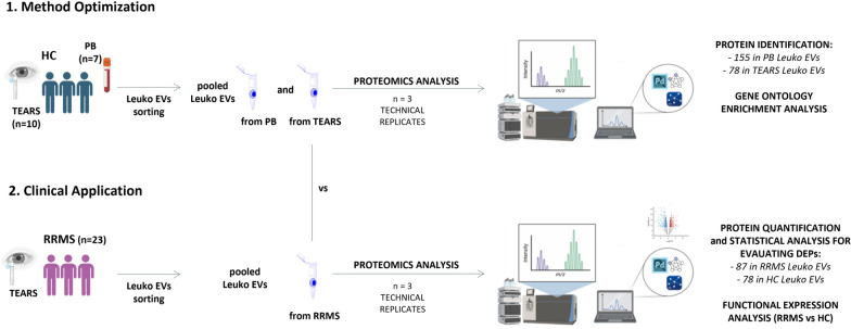
Background: The isolation and proteomics characterization of extracellular vesicles (EVs) from body fluids is challenging due to their vast heterogeneity. We have recently demonstrated that Fluorescence-activated Cell Sorting (FACS) efficiently isolates the whole EV circulating compartment directly from untouched body fluids enabling a comprehensive EV proteomics analysis.

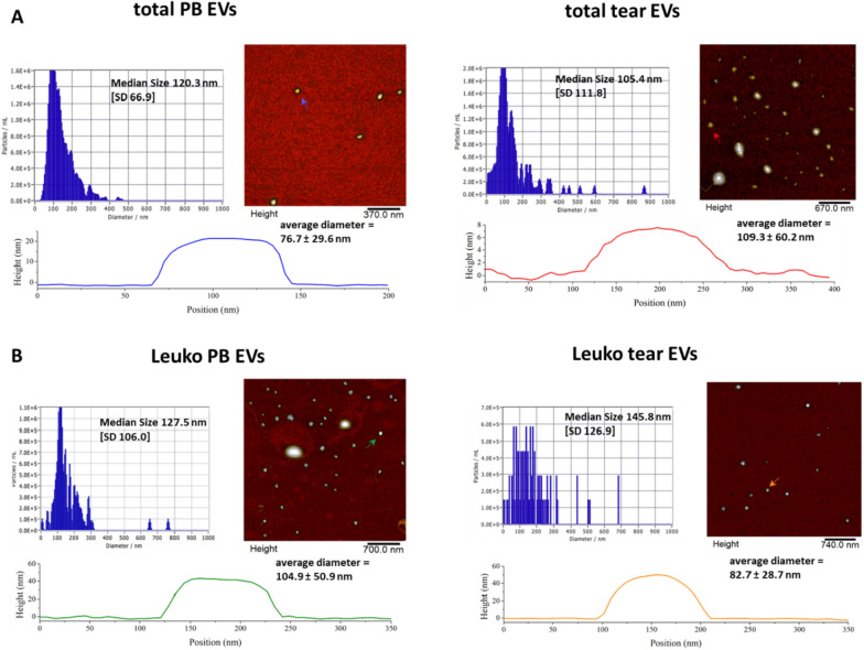
Results: Here, we characterized, for the first time, a single-phenotype EV subset by sorting leukocyte-derived EVs (Leuko EVs) from peripheral blood and tears of healthy volunteers. Using an optimized and patented staining protocol of the whole EV compartment we identified and excluded non-EV particles, debris and damaged EVs. We further isolated, using an anti-CD45 antibody, Leuko EVs (CD45+ EVs), reaching a high level of purity (> 90%). Purified Leuko EVs were characterized using atomic force microscopy, nanoparticle tracking, and shotgun proteomics analysis revealing a similar coded protein cargo in both biological fluids. Subsequently, the same workflow was applied to tears from Relapsing-Remitting Multiple Sclerosis (RRMS) patients, revealing a Leuko EVs protein cargo enrichment that reflects the neuroinflammatory condition characteristics of RRMS. This enrichment was evidenced by the activation of upstream regulators TGFB1 and NFE2L2, which are associated with inflammatory responses. Additionally, the analysis identified markers indicative of endothelial cell proliferation and the development of enhanced vascular networks, with AGNPT2 and VEGF emerging as activated upstream regulators. These findings indicate the complex interplay between inflammation and angiogenesis in RRMS.

Conclusions: In conclusion, our combined FACS-Proteomics strategy offers a promising approach for biomarker discovery, analysing cell-specific EV phenotypes directly from untouched body fluids, advancing the clinical value of tears EVs and improving the understanding of EV-mediated processes in vivo. Data are available via ProteomeXchange with the identifier PXD049036 and in EV-TRACK knowledgebase with ID: EV240150.

Keywords: EV fluorescence activated cell sorting isolation; Leukocyte-derived extracellular vesicles; Multiple sclerosis; Proteomics; Tears.


Conflict of interest statement

Declarations. Ethics approval and consent to participate: The protocol was approved on 29 December 2020 by the Ethic committee of “G. d’Annunzio”. Consent for publication: Not applicable. Informed consent: Informed consent was obtained from all subjects involved in the study. Competing interests: Not applicable.

Figures

Fig. 1
Fig. 1
Workflow, identification and isolation of tear and PB EVs. A EVs subtypes counts scheme in tears and PB samples of healthy subjects (HCs). The image was created by BioRender.com. B The scatter area containing EVs was firstly gated on FSC-H/SSC-H dot-plot; C Events positive to LCD (y-axis) and negativity to Phalloidin (x-axis) were identified as EVs; D EVs (LCD+/Phalloidin- events) were subtyped to identify leukocyte derived-EVs as events positive to CD45 (x-axis). EG The same gating strategy was used to identify and separate PB EVs. Data are representative of all analysed samples
Fig. 2
Fig. 2
Proteomics study workflow. PB: peripheral blood, Leuko EVs: leukocyte-derived EVs, HC: healthy controls, RRMS: relapsing–remitting multiple sclerosis, DEPs: differential expressed proteins. The image was created by BioRender.com
Fig. 3
Fig. 3
Protein expression markers detection. Typical EV markers (CD63 and CD81) were analysed on the whole EV compartment both in PB (panel A) and in tear (panel B) samples
Fig. 4
Fig. 4
Characterization of Leukocytes-derived EVs from healthy controls biofluids. A NTA tracking of size (diameter/nm) and concentration (particles/mL) of pure total EVs population sorted from PB (left) and tears (right). EVs derived from total PB or total tear showed a median diameter of 120.3 nm and 105.4 nm, respectively, falling within the size range detected by AFM reported in the figure. The height cross sectional profiles of representative vesicles of total PB (blue arrow) and total tear (red arrow) are also reported. B NTA tracking of size (diameter/nm) and concentration (particles/mL) of pure Leuko EVs sorted from PB (left) and tears (right). Leuko EVs derived from PB or tears showed a median diameter of 127.5 nm and 145.8 nm respectively, falling within the size range detected by AFM indicated in the figure. The height cross sectional profiles of representative Leuko EVs of PB samples (green arrow) and tears (orange arrow) are also reported
Fig. 5
Fig. 5
Proteomics assessment of Leuko EVs sorted from PB and tears of healthy subjects. A Bar diagrams depict the matching between Leuko EV proteins and Vesiclepedia, an online EV proteome repository. Only 8.92% and 5.2% of the Leuko EV proteins were unmatched in Vesiclepedia for the pooled PB and tear samples, respectively. Details of all proteins are shown in Tables S5 and S6. B The Venn diagram shows the common proteins between PB Leuko EVs or tears Leuko EVs. C, D Gene Ontology (GO) classification of proteins are reported as red and blue dots to underline “extracellular exosome” and “vesicle” as the most significant cellular component in PB and lacrimal Leuko EVs with an FDR of 6.55 × 10–89 and 2.07 × 10–63 for PB Leuko EVs (C), and 1.23 × 10–35 and 2.22 × 10–30 for tears Leuko EVs (D), respectively. White dots represent proteins not identified with the GO classifications (see Table S7, sheets 4 and 5, for protein details). E, F Mechanistic networks of “leukocyte migration” downstream in PB Leuko EVs (p-value = 6.31 × 10–25) and tears Leuko EVs (p-value = 5.95 × 10–8). Shapes and symbols of proteins are shown in Figure S4
Fig. 6
Fig. 6
Clinical application of single phenotype EV proteomics workflow to tear fluid of Multiple Sclerosis patients. A Venn diagram of quantified proteins in Leuko EVs sorted from tears of RRMS patients and healthy volunteers. B Proteomics PPI assessment of RRMS unique proteins. Yellow dots were reclassified as proteins involved in “Immune System HAS-168256 Reactome Pathway”. C Volcano Plot of proteins graphed by fold change (Difference) and -Log(p-value) by the comparison of RRMS vs HC subjects. Grey dots represent proteins that were not differentially expressed in the comparison carried out; red dots represent proteins that were significantly up-regulated and green dots indicate proteins that were significantly down-regulated in RRMS. D Regulator Effects network highlights the hypothesis of how “migration of endothelial cells” is activated by transforming growth factor beta 1 (TGFB1) as upstream regulator. E Networks of activated upstream regulators: Angiopoietin-2 (ANGPT2) and vascular endothelial growth factor group (VEGF). F Downstream effect analysis reveals the upregulation of “angiogenesis”. Orange and blue shapes represent predicted activation or inhibition, respectively. Instead, red and green shapes represent increased or decreased measurements of identified proteins, respectively, whose expression value is reported in the figure. Continuous lines represent direct relationships, dotted ones represent indirect relationships, whereas grey and yellow lines indicate the non-predicted and inconsistent relationships. Figure S4 reports an interpretation of IPA networks

Similar articles

References

    1. Krämer-Albers E-M, Werner HB. Mechanisms of axonal support by oligodendrocyte-derived extracellular vesicles. Nat Rev Neurosci. 2023;24:474–86. - PubMed
    1. Brocco D, Lanuti P, Pieragostino D, Cufaro MC, Simeone P, Bologna G, et al. Phenotypic and proteomic analysis identifies hallmarks of blood circulating extracellular vesicles in NSCLC responders to immune checkpoint inhibitors. Cancers. 2021;13:585. - PMC - PubMed
    1. Wang G, Li J, Bojmar L, Chen H, Li Z, Tobias GC, et al. Tumour extracellular vesicles and particles induce liver metabolic dysfunction. Nature. 2023;618:374–82. - PMC - PubMed
    1. Simeone P, Bologna G, Lanuti P, Pierdomenico L, Guagnano MT, Pieragostino D, et al. Extracellular vesicles as signaling mediators and disease biomarkers across biological barriers. Int J Mol Sci. 2020;21:2514. - PMC - PubMed
    1. van Niel G, D’Angelo G, Raposo G. Shedding light on the cell biology of extracellular vesicles. Nat Rev Mol Cell Biol. 2018;19:213–28. - PubMed