

Knee morphology and patella malalignment in neglected developmental dysplasia of the hip: a systematic review and meta-analysis
- PMID: 40394669
- PMCID: PMC12090680
- DOI: 10.1186/s13018-025-05877-y
Knee morphology and patella malalignment in neglected developmental dysplasia of the hip: a systematic review and meta-analysis
Abstract
Purpose: To quantitatively analyze the structural changes of the knee in patients with neglected developmental dysplasia of the hip (DDH).
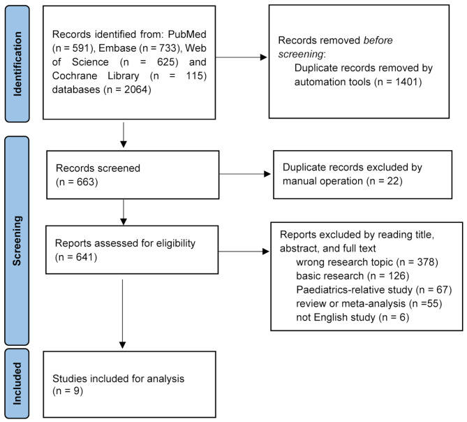
Methods: PubMed, Embase, Web of Science, and Cochrane Library databases were searched to identify studies comparing the morphological parameters of the knee between DDH patients and healthy individuals. Data on rotational and mechanical parameters of the lower limb, rate of occasional anterior knee pain (AKP), and knee morphological parameters, were extracted. Review Manager and R statistic software were used to perform the statistical analysis.
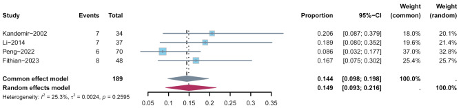
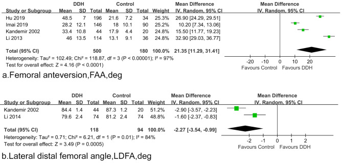
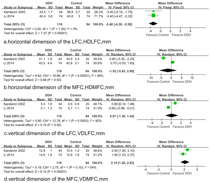
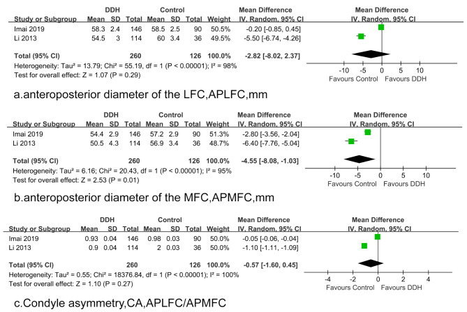
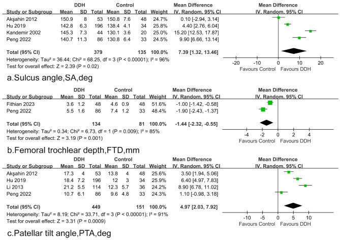
Results: Nine studies with a total of 790 legs in 521 neglected DDH patients and 431 legs in 303 health subjects were included. Patients were predominantly female (88.3%). The Crowe classification is most commonly used to assess the severity of DDH. The total incidence of occasional AKP ranged from 8.6 to 20.6%, with an overall pooled rate of 14.4% (95%CI = 9.8-19.8%). In patients with neglected DDH, significant increases (P < 0.0001) were observed in femoral anteversion (weighted mean: 39.1° vs. 17.7°), knee torsion (weighted mean: 9.0° vs. 1.6°), and the vertical dimension of the medial femoral condyle (weighted mean: 13.8 mm vs. 11.6 mm), along with a significant decrease in the lateral distal femoral angle (weighted mean: 82.1° vs. 84.8°), which can lead to torsion deformity of the lower limb and valgus inclination of the distal femoral articular surface. Compared with the intact subjects, DDH knees demonstrated an increased sulcus angle (weighted mean: 144.9° vs. 137.5°; P < 0.0001), decreased trochlear depth (weighted mean: 3.1 mm vs. 4.5 mm; P < 0.0001), increased lateral shift of the patella (5.1 mm vs. 3.8 mm, P = 0.06), and increased patellar tilt angle (weighted mean: 18.2° vs. 13.2°; P < 0.0001). These findings were associated with developmental dysplasia of femoral trochlear and patellar instability.
Conclusion: Developmental dysplasia of the hip leads to patellar malalignment and developmental changes in the bony anatomy of the knee joint, including the development of a valgus deformity of the lower extremity and trochlear dysplasia. These findings may be associated with patellar instability.
Level of evidence: III, systematic review.
Registration: This study was registered in the International Prospective Register of Systematic Reviews (PROSPERO) (CRD42025640292).
Keywords: Developmental dysplasia of the hip (DDH); Knee morphology; Patellar instability; Trochlear dysplasia; Valgus deformity.
© 2025. The Author(s).
Conflict of interest statement
Declarations. Competing interests: The authors declare no competing interests. Ethics approval and consent to participate: Not applicable. Consent for publication: Not applicable. PROSPERO registration number: CRD42025640292.
Figures
Similar articles
-
Patellar Instability.2023 Sep 4. In: StatPearls [Internet]. Treasure Island (FL): StatPearls Publishing; 2025 Jan–. 2023 Sep 4. In: StatPearls [Internet]. Treasure Island (FL): StatPearls Publishing; 2025 Jan–. PMID: 29494034 Free Books & Documents.
-
Femoral Trochlear Dysplasia Is Common in Lower Limbs With Hartofilakidis C2 Hip Dysplasia.Clin Orthop Relat Res. 2025 May 27. doi: 10.1097/CORR.0000000000003557. Online ahead of print. Clin Orthop Relat Res. 2025. PMID: 40434850
-
Changes in Alignment of Ipsilateral Knee on Computed Tomography after Total Hip Arthroplasty for Developmental Dysplasia of the Hip.Orthop Surg. 2019 Jun;11(3):397-404. doi: 10.1111/os.12462. Epub 2019 May 26. Orthop Surg. 2019. PMID: 31131564 Free PMC article.
-
Inconsistencies in Reporting Risk Factors for Medial Patellofemoral Ligament Reconstruction Failure: A Systematic Review.Am J Sports Med. 2022 Mar;50(3):867-877. doi: 10.1177/03635465211003342. Epub 2021 Apr 29. Am J Sports Med. 2022. PMID: 33914648
-
Osteoarthritic knees have a highly variable patellofemoral alignment: a systematic review.Knee Surg Sports Traumatol Arthrosc. 2021 Feb;29(2):483-490. doi: 10.1007/s00167-020-05928-3. Epub 2020 Mar 12. Knee Surg Sports Traumatol Arthrosc. 2021. PMID: 32162047
References
-
- Wedge JH, Wasylenko MJ. The natural history of congenital disease of the hip. J Bone Joint Surg Br. 1979;61–b:334–8. - PubMed
-
- Crawford AH, Mehlman CT, Slovek RW. The fate of untreated developmental dislocation of the hip: long-term follow-up of eleven patients. J Pediatr Orthop. 1999;19:641–4. - PubMed
-
- Kandemir U, Yazici M, Alpaslan AM, Surat A. Morphology of the knee in adult patients with neglected developmental dysplasia of the hip. J Bone Joint Surg Am. 2002;84:2249–57. - PubMed
-
- Wedge JH, Wasylenko MJ. The natural history of congenital dislocation of the hip: a critical review. Clin Orthop Relat Res. 1978;154:62. - PubMed
-
- Hao K, Li Z, Wang J, Huo Z, Niu Y, Wang F. Morphological improvement of the epiphyseal plate and trochlea after surgical correction in skeletally immature patients with patellar dislocation and trochlear dysplasia. Am J Sports Med. 2025;3635465241301775. - PubMed
Publication types
MeSH terms
Grants and funding
LinkOut - more resources
Full Text Sources
Research Materials
