. 2025 May 20;16(1):254.
doi: 10.1186/s13287-025-04358-7.

Combining sodium-glucose co-transporter-2 inhibitor with mesenchymal stem cells and brown adipose tissue (BAT) and white adipose tissue (WAT) transplantation to mitigate the progression of diabetic kidney disease: a pre-clinical approach

Affiliations

Combining sodium-glucose co-transporter-2 inhibitor with mesenchymal stem cells and brown adipose tissue (BAT) and white adipose tissue (WAT) transplantation to mitigate the progression of diabetic kidney disease: a pre-clinical approach

Stephany Beyerstedt et al. Stem Cell Res Ther. .

Abstract

Introduction: The increasing prevalence of Diabetes Mellitus (DM) correlates with a rising incidence of Diabetic Kidney Disease (DKD). DKD, a multifactorial condition, is characterized by activation of the renin-angiotensin-aldosterone system (RAAS), with angiotensin II playing a significant role in podocyte injury. While conventional treatments show potential in mitigating DKD progression, a combination of strategies is required to both impede its development and repair damaged structures.

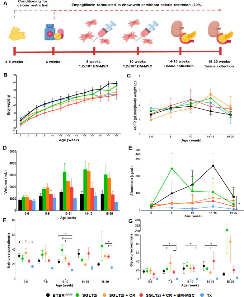
Methods: In this study, we explored the brown adipose tissue (BAT) and white adipose tissue (WAT) transplantation, and the use of bone marrow mesenchymal stem cell therapy (BM-MSC) combined with sodium-glucose co-transporter-2 (SGLT2) inhibitor treatment and calorie restriction in the BTBRob/ob model, recognized as a robust representation of DKD featuring hyperglycemia, obesity, time-dependent albuminuria, and histological changes.

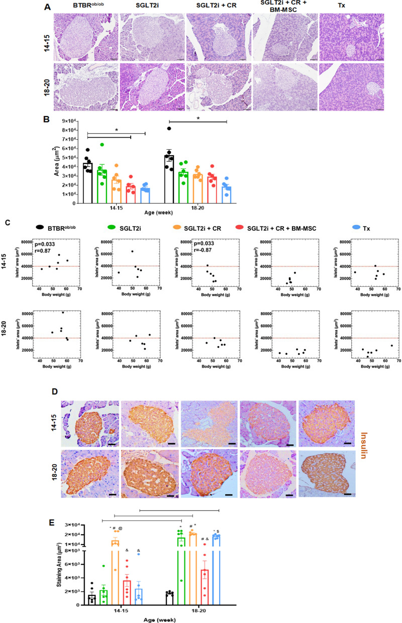
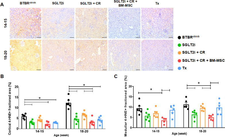
Results: Our primary findings revealed enhanced blood glucose control through combined cell therapy, diminished mesangial matrix expansion, alleviated tissue oxidative stress, preserved podocyte numbers, and an upregulation of podocyte structural markers and components of the RAAS renoprotective axis.

Conclusion: BM-MSC therapy demonstrates considerable promise as a combined treatment for mitigating DKD progression, with similar findings observed for BAT and WAT transplantation.

Keywords: Diabetic kidney disease; Mesenchymal stem cell; Podocyte; SGLT2i.


Conflict of interest statement

Declarations. Competing interests: The authors declare no competing interests. Consent for publication: Not applicable. Ethical approval and consent to participate: This research does not contain clinical experiments. All the experiments are approved under the projects “Treatment with empagliflozin, mesenchymal stem cells, and klotho as measures to slow down the progression of diabetic kidney disease and reduce the impact of acute kidney injury” and “Fat transplantation as a routine procedure to increase the number of BTBRob/ob mice in the animal facility” by the Institutional Animal Care and and Use Committee of Hospital Israelita Albert Einstein (HIAE). The approval numbers are SGPP 4900–21 (data of approval: August 27th, 2021) and SGPP 5171–22 (data of approval: April 29th, 2022), respectively. Our manuscript is reported following the ARRIVE guidelines.

Figures

Fig. 1
Fig. 1
Metabolic and renal functional parameters in the experimental groups. A Schematization of the treatment protocols. Conditioning of the groups receiving calorie restriction and initiation of empagliflozin in the chow for SGLT2i-tretaed gropus occurred at 4–5 weeks of age. In the groups receiving combined therapy with BM-MSC, injections were administered at the 8th and 10th weeks of age. Euthanasia was performed at 14–15 and 18–20 weeks of age. B Body weight variation. C Estimated glomerular filtration rate (eGFR) normalized by body weight. D Urinary volume in mL. E Albumin measured in μg/mL over time. F Natriuresis normalized by creatinuria. G Glycosuria normalized by creatinuria. (*p < 0.05). Error bars represent mean ± SEM; n = 6–12 animals
Fig. 2
Fig. 2
Pancreatic islet analysis. A Representative hematoxylin–eosin (HE) staining for morphological evaluation of pancreatic islets at 14–15 and 18–20 weeks of age. B Pancreatic islet area analysis across experimental groups. (* p < 0.05). C Spearman correlation between islets’ area (µm2) and body weight (g). D Representative images for insulin immunohistochemical analysis. E Quantification of insulin-positive cells per pancreatic islets across groups. (p < 0.05; * vs. BTBRob/ob; # vs. SGLT2i; & vs. SGLT2i + CR; $ vs. SGLT2i + CR + BM-MSCs; continuous line indicated difference between time points). Scale bars represent 100 μm in A and D. Error bars represent mean ± SEM; n = 6 animals
Fig. 3
Fig. 3
Immunohistochemical analysis of pancreatic islets. A Representative images of pancreatic sections from experimental groups stained for cleaved caspase-3 at 14–15 and 18–20 weeks of age. B Quantification of cleaved caspase-3-positive cells per pancreatic islets across groups. (p < 0.05; * vs. BTBRob/ob; continuous line indicates difference between time points). Scale bars represent 100 μm in A. Error bars represent mean ± SEM; n = 5 animals
Fig. 4
Fig. 4
Analysis of glomerular hypertrophy and mesangial expansion. A Representative periodic acid-Schiff (PAS) staining illustrating the morphological evaluation of glomeruli at 14–15 and 18–20 weeks of age. B Evaluation of glomerular tuft area. C Progression of glomerular tuft area at 18–20 weeks of age compared to 14–15 weeks. D Fractioned mesangial expansion. E Progression of fractioned mesangial expansion at 18–20 weeks of age compared to 14–15 weeks. (*p < 0.05). Scale bars represent 20 µm in A. Error bars represent mean ± SEM; n = 5–6 animals
Fig. 5
Fig. 5
Immunohistochemical analysis of podocyte count. A Representative images of kidney sections from the experimental groups stained for the podocyte nuclear marker WT-1. B Evaluation of positive WT-1 cells per glomerulus among the different groups at both time points. (*p < 0.05). Scale bars represent 10 µm in A. Error bars represent mean ± SEM; n = 5–6 animals
Fig. 6
Fig. 6
Immunohistochemical analysis of tissue oxidative stress via lipid peroxidation using 4-HNE. A Representative images of cortical sections from the experimental groups. B Evaluation of lipid peroxidation in the cortical region. C Evaluation of lipid peroxidation in the medullary region. (*p < 0.05). Scale bars represent 100 µm in A. Error bars represent mean ± SEM; n = 5–6 animals
Fig. 7
Fig. 7
Gene expression of podocyte markers and components of the RAAS and Ang II/Nox4/TRPC6 axis compared to BTBR wild type. A WT-1. B Nphs1. C Synpo2. D Actn4. E Nox4. F Trpc6. G Ace. H Ace2. I Ace2/Ace ratio. (*p < 0.05). Error bars represent mean ± SEM; n = 5–6 animals

References

    1. Sun H, Saeedi P, Karuranga S, Pinkepank M, Ogurtsova K, Duncan BB, Stein C, Basit A, Chan JCN, Mbanya JC, Pavkov ME, Ramachandaran A, Wild SH, James S, Herman WH, Zhang P, Bommer C, Kuo S, Boyko EJ, Magliano DJ. IDF Diabetes Atlas: Global, regional and country-level diabetes prevalence estimates for 2021 and projections for 2045. Diabetes Res Clin Pract. 2022;183: 109119. - PMC - PubMed
    1. Gnudi L. Renal disease in patients with type 2 diabetes: Magnitude of the problem, risk factors and preventive strategies. Presse Med. 2023;52: 104159. - PubMed
    1. Beyerstedt S, Casaro EB, Rangel EB. COVID-19: angiotensin-converting enzyme 2 (ACE2) expression and tissue susceptibility to SARS-CoV-2 infection. Eur J Clin Microbiol Infect Dis. 2021;40:905–19. - PMC - PubMed
    1. Verma S, Singh P, Khurana S, Ganguly NK, Kukreti R, Saso L, Rana DS, Taneja V, Bhargava V. Implications of oxidative stress in chronic kidney disease: a review on current concepts and therapies. Kidney Res Clin Pract. 2021;40:183–93. - PMC - PubMed
    1. Ilatovskaya DV, Blass G, Palygin O, Levchenko V, Pavlov TS, Grzybowski MN, Winsor K, Shuyskiy LS, Geurts AM, Cowley AW Jr, Birnbaumer L, Staruschenko A. A NOX4/TRPC6 pathway in podocyte calcium regulation and renal damage in diabetic kidney disease. J Am Soc Nephrol. 2018;29:1917–27. - PMC - PubMed

MeSH terms

Substances

LinkOut - more resources