. 2025 May;29(10):e70609.
doi: 10.1111/jcmm.70609.

Erk1/2 Orchestrates SSPH I-Induced Oxidative Stress, Mitochondrial Dysfunction and Ferroptosis in Hepatocellular Carcinoma

Affiliations

Erk1/2 Orchestrates SSPH I-Induced Oxidative Stress, Mitochondrial Dysfunction and Ferroptosis in Hepatocellular Carcinoma

Yuewen Sun et al. J Cell Mol Med. 2025 May.

Abstract

Although Erk1/2 has been linked to oxidative stress regulation in hepatocellular carcinoma (HCC), the interplay among Erk1/2, reactive oxygen species (ROS), and iron metabolism remains poorly characterised. The steroidal saponin SSPH I, a recognised ferroptosis inducer, exerts dual pharmacological effects via Erk1/2 and ROS-dependent pathways. This study aimed to investigate the regulatory mechanisms of Erk1/2 in ferroptosis and oxidative stress and analyse their feedback regulatory effects on Erk1/2 in HCC using SSPH I as a pharmacological probe, and further elucidate the anti-HCC effects and mechanisms of SSPH I in vitro and in vivo. Mechanistic studies utilised three inhibitors: U0126 (Erk1/2 phosphorylation inhibitor), Ferrostatin-1 (ferroptosis inhibitor), and N-acetyl cysteine (ROS scavenger), combined with SSPH I to delineate its effects on cell viability, mitochondrial dynamics, ferroptosis induction and oxidative stress. Mechanistically, SSPH I disrupted mitochondrial function and suppressed HCC cell survival through iron accumulation and ROS generation, while concurrently activating Erk1/2 signalling. Pharmacological inhibition of ROS or iron pathways partially attenuated SSPH I-induced ferroptosis and ROS generation, but failed to abrogate these effects. Erk1/2 inhibition completely abolished SSPH I-mediated regulation of the Nrf1/2-HO-1 axis and ferroptosis-related protein expression in cellular and animal models, identifying Erk1/2 as the upstream regulatory node. Notably, while both SSPH I and U0126 monotherapies inhibited xenograft growth, their combined use resulted in antagonistic effects. These findings establish Erk1/2 activation as the central molecular mechanism orchestrating SSPH I-driven oxidative stress amplification, mitochondrial dysfunction and ferroptosis execution in HCC.

Keywords: Erk1/2; Nrf1/2; ROS; SSPH I; ferroptosis.


Conflict of interest statement

The authors declare no conflicts of interest.

Figures
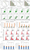
FIGURE 1
FIGURE 1
Fer, U0 and NAC interfered with the anti‐proliferation, apoptosis induction and cell cycle interruption effect of SSPH I in HCC cells. (A) SSPH I significantly inhibited the proliferation of HCC cells after 24 h treatment; both Fer, U0 and NAC showed antagonist effects towards SSPH I. (B) Apoptosis was analysed by Annexin V‐FITC/PI staining. (C) SSPH I induced apoptosis in HCC cells after 24 h treatment; Fer, U0 and NAC did not protect HCC cells from apoptosis. (D) Cell cycle analysis was performed by flow cytometry. (E) SSPH I induced cell cycle arrest in HCC cells after 24 h treatment; U0 and NAC rescued SMMC‐7721 cells from G2/M arrest but did not affect HepG2 cells. Data represent the mean ± SD, n = 3. *p < 0.05 compared with control; ^p < 0.05 compared with SSPH 4 μM.
FIGURE 2
FIGURE 2
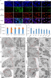
Fer, U0 and NAC alleviated the mitochondrial damage induced by SSPH I in HepG2 cells. (A) JC‐1 fluorescent probe detects MMP in HepG2 cells after 8 h treatment. (B) Ratio of fluorescence of JC‐1 monomer and polymer. (C) Length of mitochondria in HepG2 cells after 8 h treatment. (D) TEM observations of mitochondrial ultrastructure in HepG2 cells. Data represent the mean ± SD, B n = 3; C n = 20. *p < 0.05 compared with control; ^p < 0.05 compared with SSPH 4 μM.
FIGURE 3
FIGURE 3
Fer, U0 and NAC regulate protein expression induced by SSPH I. (A) The protein levels of HCC cells after 8 h treatment were measured. (B) SSPH I upregulated Nrf1/2‐HO‐1 and ferroptosis‐relative proteins; the combination of Fer, U0 and NAC further regulated the protein expression. (C) Immunofluorescence verified the expression of p‐Erk1/2, Nrf2 and HO‐1 in HepG2 cells. Cell shrinkage was observed after SSPH I treatment. The combination of Fer, U0 and NAC did not relieve the cell shrinkage. The increased fluorescence in the nucleus indicated that Fer and NAC facilitate the translocation of Nrf2. Data represent the mean ± SD, n = 2. *p < 0.05 compared with control; ^p < 0.05 compared with SSPH 4 μM.
FIGURE 4
FIGURE 4
The protective role of Fer, U0 and NAC in SSPH I induced oxidative stress and ferroptosis in HCC cells. Data represent the mean ± SD, n = 3. *p < 0.05 compared with control; ^p < 0.05 compared with SSPH 4 μM.
FIGURE 5
FIGURE 5
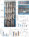
SSPH I and U0 inhibited the growth of HepG2 xenograft tumours. (A) Representative photographs of xenograft tumours from each dosage group in HepG2 nude mice models. (B) Gross morphology of excised HepG2 xenograft tumours. (C) Tumour growth curves of HepG2 xenografts. (D) Q value of volume and weight of tumours. (E) Final tumour weights of HepG2 xenografts. (F) Tumour growth inhibition rates across treatment groups. (G) Body weight changes of nude mice during treatment. Data represented the mean ± SD, n = 5. *p < 0.05 compared with control.
FIGURE 6
FIGURE 6
SSPH I and U0 modulate protein expression levels and mitochondrial morphology in HepG2 xenograft tumours. (A) Protein expression levels in tumour tissues were visualised by immunohistochemical staining. (B) Mitochondrial morphology in tumour tissues was examined by transmission electron microscopy. Red arrows: Autophagic vacuoles; blue arrows: Autophagic vacuoles containing lipid droplets.
FIGURE 7
FIGURE 7
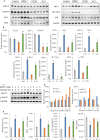
SSPH I and U0 modulate protein expression and the concentrations of ROS, MDA, GSH and Fe2+ in HepG2 xenograft tumours. (A) Protein expression in tumour tissues was analysed by Western blotting. (B) SSPH I activated Erk1/2 and upregulated Nrf1/2, HO‐1, SLC7A11 and TFR; U0 antagonised the effects of SSPH I by inhibiting Erk1/2 phosphorylation. (C and D) The duration and concentration of SSPH I influence Erk1/2 phosphorylation. (E) U0 attenuates SSPH I‐induced oxidative stress and ferroptosis in vivo. (B and E), data represent the mean ± SD, n = 3. *p < 0.05 compared with control; ^p < 0.05 compared with SSPH I group.

Similar articles

References

    1. Yang J. D., Hainaut P., Gores G. J., Amadou A., Plymoth A., and Roberts L. R., “A Global View of Hepatocellular Carcinoma: Trends, Risk, Prevention and Management,” Nature Reviews. Gastroenterology & Hepatology 16, no. 10 (2019): 589–604, 10.1038/s41575-019-0186-y. - DOI - PMC - PubMed
    1. Moon H. and Ro S. W., “MAPK/ERK Signaling Pathway in Hepatocellular Carcinoma,” Cancers 13, no. 12 (2021): 3026, 10.3390/cancers13123026. - DOI - PMC - PubMed
    1. Lei L., Zhao G.‐D., Shi Z., Qi L.‐L., Zhou L.‐Y., and Fu Z.‐X., “The Ras/Raf/MEK/ERK Signaling Pathway and Its Role in the Occurrence and Development of HCC,” Oncology Letters 12, no. 5 (2016): 3045–3050, 10.3892/ol.2016.5110. - DOI - PMC - PubMed
    1. Chen L., Shi Y., Jiang C. Y., Wei L. X., Wang Y. L., and Dai G. H., “Expression and Prognostic Role of Pan‐Ras, Raf‐1, pMEK1 and pERK1/2 in Patients With Hepatocellular Carcinoma,” European Journal of Surgical Oncology 37, no. 6 (2011): 513–520, 10.1016/j.ejso.2011.01.023. - DOI - PubMed
    1. Han L., Zhang C., Wang D., et al., “Retrograde Regulation of Mitochondrial Fission and Epithelial to Mesenchymal Transition in Hepatocellular Carcinoma by GCN5L1,” Oncogene 42, no. 13 (2023): 1024–1037, 10.1038/s41388-023-02621-w. - DOI - PubMed

MeSH terms