. 2025 May;21(5):e70239.
doi: 10.1002/alz.70239.

Choroid plexus free-water correlates with glymphatic function in Alzheimer's disease

Affiliations

Choroid plexus free-water correlates with glymphatic function in Alzheimer's disease

Xiaomeng Xu et al. Alzheimers Dement. 2025 May.

Abstract

Introduction: Free-water imaging of the choroid plexus (CP) may improve the evaluation of Alzheimer's disease (AD).

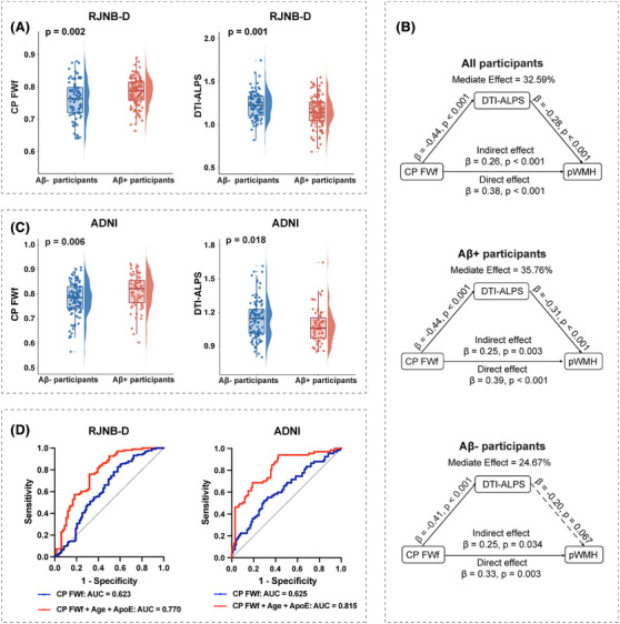
Methods: Our study investigated the role of free-water fraction (FWf) of CP in AD among 216 participants (133 Aβ+ participants and 83 Aβ- controls) enrolled in the NeuroBank-Dementia cohort at Ruijin Hospital (RJNB-D). The Alzheimer's Disease Neuroimaging Initiative dataset was used for external validation.

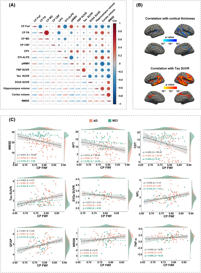
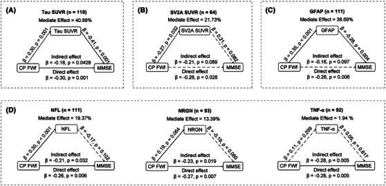
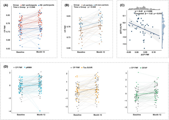
Results: At baseline, Aβ+ participants showed higher CP FWf, increased white matter hyperintensity (WMH) volume, and decreased diffusion tensor image analysis along the perivascular space (DTI-ALPS). In Aβ+ participants, DTI-ALPS mediated the association between CP FWf and periventricular WMH. CP FWf was associated with cortical tau accumulation, synaptic loss, hippocampal and cortical atrophy, and cognitive performance. During follow-up, CP FWf increased faster in Aβ+ participants than controls.

Discussion: Elevated CP FWf indicated impaired glymphatic function and AD neurodegeneration, and can be a sensitive biomarker for AD progression. The study was registered on ClinicalTrials.gov (NCT05623124).

Highlights: This cohort study found higher free-water fraction (FWf) of the choroid plexus (CP) in amyloid beta (Aβ)+ participants. CP FWf was related to glymphatic function, brain atrophy, tau burden, synaptic loss, and cognition. Aβ+ participants showed faster growth of CP FWf than Aβ- controls during follow-up. The growth rate of CP FWf exceeded that of white matter lesion and tau accumulation in Aβ+ participants. CP FWf can serve as a sensitive imaging marker of glymphatic function and Alzheimer's disease progression.

Keywords: Alzheimer's disease; choroid plexus; diffusion tensor image analysis along the perivascular space; free‐water mapping; white matter hyperintensity.


Conflict of interest statement

Fang Xie is an associate editor of Alzheimer's & Dementia. Other authors declare no conflicts of interest. Author disclosures are available in the supporting information.

Figures

FIGURE 1
FIGURE 1
Group difference and correlation between CP FWf and glymphatic markers. A, Increased CP FWf (p = 0.002) and decreased DTI‐ALPS (p = 0.001) were observed in Aβ+ participants in RJNB‐D cohort. B, Across all participants, mediation analysis revealed a partial mediation effect of DTI‐ALPS between CP FWf and pWMH. Within the Aβ+ group, a significant partial mediation effect of DTI‐ALPS between CP FWf and pWMH was observed. The Aβ– controls displayed the trend of mediation effect of DTI‐ALPS between CP FWf and pWMH. The log‐transformed pWMH volume was used. All the mediation analysis was adjusted for age. C, Increased CP FWf (p = 0.006) and decreased DTI‐ALPS (p = 0.018) were observed in Aβ+ participants in the ADNI dataset. D, Predictive values of CP FWf alone (blue) and with age and APOE genotype (red) for Aβ positivity calculated by ROC. The AUCs were similar for CP FWf alone (RJNB‐D AUC = 0.623; ADNI: AUC = 0.625), and with age and APOE genotype (RJNB‐D: AUC = 0.770; ADNI: AUC = 0.815). Aβ, amyloid beta; ADNI, Alzheimer's Disease Neuroimaging Initiative; AUC, area under the curve; APOE, apolipoprotein E; CP, choroid plexus; DTI‐ALPS, diffusion tensor image analysis along the perivascular space; FWf, free‐water fraction; pWMH, periventricular white matter hyperintensity; RJNB‐D, NeuroBank‐Dementia cohort at Ruijin Hospital; ROC, receiver operating characteristic.
FIGURE 2
FIGURE 2
CP FWf correlated with imaging metrics and blood biomarkers in AD. A, Spearman correlation analysis between the potential imaging biomarkers of CP, glymphatic function markers, AD imaging markers, and global cognitive performance within the Aβ+ group. The CP FWf showed significant association with CP FA, CPV, DTI‐ALPS, pWMH, cortical tau SUVR, cortical SV2A SUVR, hippocampus volume, cortex volume, and MMSE. The log‐transformed pWMH volume was used. B, The vertex‐wise GLM analysis revealed a negative correlation map between CP FWf and cortical thickness, and tau SUVR exhibited a positive correlation map with CP FWf. Age and sex were included as covariates in the GLM. Only the significant clusters with corrected p < 0.05 after permutations are colored. The color bar represents uncorrected vertex‐wise p values. C, CP FWf correlated with peripheral blood AD biomarkers and cognition. Within the Aβ+ group, there was a positive correlation between CP FWf and NfL (β = 3.117, p = 0.026), GFAP (β = 5.28, p < 0.001), NRGN (β = 5.70, p = 0.028), TNF‐α (β = 10.83, p = 0.009), and global tau (β = 9.27, p = 0.002). Subgroup analysis of AD (red) and MCI (green) stages were presented by dashed line. The log‐transformed levels of blood AD biomarkers were used. Moreover, CP FWf was negatively associated with cognitive performance, including MMSE, AFT, and CDT and SV2A. All the regressions were adjusted for age, sex, and APOE genotype. Aβ, amyloid beta; AD, Alzheimer's disease; AFT, animal fluency test; APOE, apolipoprotein E; CBF, cerebral blood flow; CDT, Clock Drawing Test; CP, choroid plexus; CPV, volume of choroid plexus; DTI‐ALPS, diffusion tensor image analysis along the perivascular space; FA, fractional anisotropy; FBP, 18F‐florbetapir; FWf, free‐water fraction; GFAP, glial fibrillary acidic protein; GLM, general linear model; MCI, mild cognitive impairment; MD, mean diffusivity; MMSE, Mini‐Mental State Examination; NfL, neurofilament light chain; NRGN, neurogranin; pWMH, periventricular white matter hyperintensity; SUVR, standardized uptake value ratios; SV2A, synaptic vesicle glycoprotein 2A; TNF‐α, tumor necrosis factor‐α.
FIGURE 3
FIGURE 3
Mediation effects of AD biomarkers on the relationship between CP FWf and MMSE. We observed a partial mediation effect of tau (A), and marginal effect of SV2A (B). Notably, GFAP exhibited a full mediation effect (C). On the other hand, NfL, NRGN, and TNF‐α did not significantly mediate the association between CP FWf and MMSE (D). All the mediation analysis was adjusted for age. AD, Alzheimer's disease; CP, choroid plexus; FWf, free‐water fraction; GFAP, glial fibrillary acidic protein; MMSE, Mini‐Mental State Examination; NfL, neurofilament light chain; NRGN, neurogranin; SUVR, standardized uptake value ratios; SV2A, synaptic vesicle glycoprotein 2A; TNF‐α, tumor necrosis factor‐α.
FIGURE 4
FIGURE 4
Longitudinal changes in CP FWf indicated neurodegeneration. The linear mixed model analysis indicated that Aβ+ participants exhibited a more rapid increase in CP FWf compared to Aβ– controls. Individual changes are represented by dashed lines, while the average changes in the group are depicted by solid lines (A). In the Aβ+ group, both APOE ε4 carriers and non‐carriers showed similar rates of CP FWf increase (B). The annual changes in CP FWf (ΔCP FWf) were significantly associated with alterations in DTI‐ALPS (ΔDTI‐ALPS) (C). Furthermore, linear mixed models with standardized preprocessing revealed that the growth rate of CP FWf surpassed that of pWMH, tau SUVR, and GFAP (D). Aβ, amyloid beta; APOE, apolipoprotein E; CP, choroid plexus; DTI‐ALPS, diffusion tensor image analysis along the perivascular space; FWf, free‐water fraction; GFAP, glial fibrillary acidic protein; pWMH, periventricular white matter hyperintensity; SUVR, standardized uptake value ratio.

Similar articles

Cited by

References

    1. Jack CR, Bennett DA, Blennow K, et al. A/T/N: an unbiased descriptive classification scheme for Alzheimer disease biomarkers. Neurology. 2016;87(5):539‐547. doi:10.1212/WNL.0000000000002923 - DOI - PMC - PubMed
    1. Garnier‐Crussard A, Cotton F, Krolak‐Salmon P, Chételat G. White matter hyperintensities in Alzheimer's disease: beyond vascular contribution. Alzheimers Dement. 2023;19(8):3738‐3748. doi:10.1002/alz.13057 - DOI - PubMed
    1. Choi JD, Moon Y, Kim HJ, Yim Y, Lee S, Moon WJ. Choroid Plexus Volume and Permeability at Brain MRI within the Alzheimer Disease Clinical Spectrum. Radiology. 2022;304(3):635‐645. doi:10.1148/radiol.212400 - DOI - PubMed
    1. Wang Y, Wang Q, Haldar JP, et al. Quantification of increased cellularity during inflammatory demyelination. Brain. 2011;134(Pt 12):3590‐3601. doi:10.1093/brain/awr307 - DOI - PMC - PubMed
    1. Chiu SY, Chen R, Wang WE, et al. Longitudinal Free‐Water Changes in Dementia with Lewy Bodies. Mov Disord. 2024;39(5):836‐846. doi:10.1002/mds.29763 - DOI - PMC - PubMed

Associated data