Review
. 2025 May 1.
doi: 10.1039/d5md00207a. Online ahead of print.

Research prospects and AI-driven strategies for metal-organic framework-hydrogel composite materials in cancer treatment

Affiliations
Review

Research prospects and AI-driven strategies for metal-organic framework-hydrogel composite materials in cancer treatment

Zijun Zhang et al. RSC Med Chem. .

Abstract

Metal-organic frameworks (MOFs) as emerging materials with highly tunable structures, large specific surface areas, and abundant pores show unique advantages in gas storage, catalysis, and drug delivery. Hydrogels are a class of materials consisting of polymers or small molecules that can trap a large amount of water and are thus widely used in biomedical applications owing to their high water content, good biocompatibility, biotissue-like softness and elasticity, and low immunogenicity. Consequently, composite materials formed by combining MOFs with hydrogels have been rapidly developed for cancer therapy. We have carried out considerable work on this type of composite material, exploring various combinations and investigating different treatment modalities, such as chemotherapy, immunotherapy, targeted therapy and combination therapy. Particularly, artificial intelligence (AI) technology was employed to characterize and enhance the therapeutic efficacy of the materials. This review first introduces the basic properties of MOFs and hydrogels and the role played by AI technology in their development. Subsequently, we describe the types, characteristics, applications, and challenges of MOF-hydrogel combinations, focusing on the research progress of various types of cancer treatments based on MOF hydrogels and the application of AI technology in this field. With the deepening of research, the development of smarter and more efficient MOF-hydrogel materials and the in-depth exploration of the application of AI technology in cancer therapy are expected to realize the precise treatment and effective control of cancer and bring new hope to cancer patients.


Conflict of interest statement

There are no competing interests.

Figures

Fig. 1
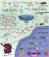
Fig. 1. Research progress in hydrogel MOFs applied in the cancer treatment.
Fig. 2
Fig. 2. Role of LLM in data-driven selection process. Springer, 2025.
Fig. 3
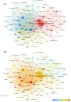
Fig. 3. Top co-occurring 100 keywords in AI research on MOFs, 2013–2024. (a) Keywords clustering map and (b) keywords clustering temporal evolution. JOM Technical Article, Elsevier, 2025.
Fig. 4
Fig. 4. Schematic of (a) ligand self-assembly formed MOGs, (b) MOGs produced via “MOF-first”, “polymer-first”, or “one-pot” strategies and (c) MOGs produced via MOFs and cross-linkers. RSC, 2023.
Fig. 5
Fig. 5. Novel injectable MOF@thermosensitive hydrogel (Dox/Cel/MOFs@Gel) for local dual drug delivery for treating oral cancer. Elsevier, 2020.
Fig. 6
Fig. 6. Cytotoxicity of IRMOF-3, Dox, Cel, Dox + Cel, Dox/MOFs, Dox/Cel/MOFs and Dox/Cel/MOFs@Gel in tumor cells A549 (a), HepG2 (b), KB (c) and SCC9 (d) (mean ± SD, *p < 0.05). Elsevier, 2020.
Fig. 7
Fig. 7. Design and preparation of the HA-BP-MOF@DOX hydrogel for injectable localized cancer treatment drug delivery. Elsevier, 2022.
Fig. 8
Fig. 8. Results of anticancer study of the HA-BP-MOF@DOX hydrogel in tumor-bearing mice. A) Individual kinetics of tumor growth in the PBS group and the tumour image after 15 days of PBS injection. B) Individual kinetics of tumor growth in the MOF@DOX group, and the tumor image after 21 days of MOF@DOX injection. C) Individual kinetics of tumor growth in the HA-BP-MOF group, and the tumor image after 15 days of HA-BP-MOF injection. D) Individual kinetics of tumor growth in the HABP-MOF@DOX group, and the tumor image after 35 days of HA-BP-MOF@DOX injection. E) Average growth of tumor in various treatment groups. F) Survival rates of mice in various treatment groups. G) Change in the body weight of mice in the various treatment groups (mean ± SD, N = 6–8). One-way ANOVA analysis was performed with a post hoc multiple comparison test. H) H&E staining images of the heart, liver, spleen, lung, and kidney after PCPD injection and of the lung and kidney after PBS, MOF@DOX, HA-BP-MOF, and HA-BP-MOF@DOX injection (scale bar = 1 mm). Elsevier, 2022.
Fig. 9
Fig. 9. Schematic representation of intratumoral delivery of the hydrogel system in a postoperative glioma mouse model that locally mimics the thermal tumour immune ecological feature and inhibits postoperative GBM recurrence. Springer, 2021.
Fig. 10
Fig. 10. Fabrication of niche-like hydrogel and its therapeutic pathway. a) Ti-MOF-Au, PEG-TK-DOX and PFD encapsulation by the HA-F127 hydrogel. b) Decrease in resistance and immune activation by the ultrasound-mediated niche-like hydrogel delivery system.
Fig. 11
Fig. 11. In vivo antitumor performances of HFTiDP + US. a) Schematic of PANC02 tumor-bearing mice for the treatment process. b) Average tumor growth curves for mice after different treatments. c) Average weight of excised tumors after various treatments. d) Photographs of the tumors collected after 14 days of treatment. Volume-based (e) and weight-based (f) tumor inhibition rates. g) Body weight profiles of the mice after various treatments. Area percentage of (h) apoptotic and (i) proliferative cells (n = 6). Area percentage of (j) collagen and (k) reticular fibers (n = 3). l) Staining of the collagen and reticular fibers of tumors. Scale bar = 100 μm (mean values ± SD). Two-tailed Student's test (t-test) (1.5 W cm−2, 1 MHz, 50% duty cycle, 5 min, n = 6). G1, control; G2, US; G3, HA-F127@Ti-MOF-Au; G4, HA-F127@Ti-MOF-Au + US; G5, HA-F127@Ti-MOF-Au + PFD + US; G6, HA-F127@Ti-MOF-Au + PEG-TK-DOX + US; G7, HA-F127@Ti-MOF-Au + PFD + PEG-TK-DOX + US. Wiley, 2024.
Fig. 12
Fig. 12. (a) Swelling behavior of P/MTX–MFM and P/MTX at pH of 1.2, 6.8 and 7.4. (b) Degradation behavior of P/MTX–MFM and P/MTX at pH 1.2, 6.8 and 7.4. (c) Drug release behavior of MTX–FM and MTX–MFM at pH 1.2 and 7.4 as a function of time. (d) P/MTX-MFM and P/MTX in simulated gastrointestinal tract passage (pH and time). MTT test results for (e) MTX, FM, MFM, MTX–MFM and MTX–FM and (f) pectin beads, P/MTX, and P/MTX–MFM against HT29 cells after 48 h incubation. Elsevier, 2024.
Fig. 13
Fig. 13. Preparation of CMC/CUR@bio-MOF and CUR release for the destruction of cancer cells. Elsevier, 2024.
Fig. 14
Fig. 14. (a) CUR release from CUR@bio-MOF and CMC/CUR@bio-MOF at pH 5.0 and 7.4. Effect of CUR, bio-MOF, and CUR@bio-MOF and CMC/CUR@bio-MOF at a concentration of 2.5–80 μM on the viability of various cell lines of (b) HEK 293 cells, (c) HeLa cells, and (d) SH-SY5Y cells. The cells were exposed to various amounts of each material and subjected to subsequent incubation for 24 h. Cell viability was assessed based on the activity of mitochondrial metabolic cells. The cell viability percentage of the samples was compared with the control (*significance between the sample and control in each cell line). (e) Confocal microscopy images of HEK 293 cells with curcumin uptake mediated by CUR@bio-MOF. Elsevier, 2024.
Fig. 15
Fig. 15. Preparation of pH-responsive injectable self-healing hydrogels containing Au/TF-MOF TNS and DOX (HAMD) and boosting acoustic-chemodynamic-starvation-chemotherapy for cancer. Elsevier, 2024.
Fig. 16
Fig. 16. (a) FT-IR spectra of SA and SADA. (b) FT-IR spectra of 2-FPBA, HPCS, SADA and SFH hydrogels. (c) SEM image of SFH. (d) Strain amplitude scanning test of G′ and G′′ of SFH. (e) Successive strain scanning steps of SFH in steps of 1% and 200% oscillatory strain lasting for three cycles. (f) SFH degradation behavior at various pH (n = 3, mean ± SD). Digital photographs of self-healing properties (g) and injectable properties (h) of SFH. Elsevier, 2024.
Fig. 17
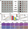
Fig. 17. (a) Digital photographs of tumours and major organs (heart, liver, spleen, lungs and kidneys) from various treatment groups on day 13. (b) Mean relative tumour volume during treatment (n = 5, mean ± SD, *p < 0.0001). (c) Mean tumour weight of different treatment groups on day 13 (n = 5, mean ± SD, *p < 0.001, *p < 0.0001). (d) Mean weight during drug administration (n = 5, mean ± SD). (e) H&E and TUNEL staining images of tumour tissues from nude mice in various treatment groups (scale bar: 200 μm). Elsevier, 2024.
Fig. 18
Fig. 18. MOF-based intelligent hydrogel nanobot for improving ferroptosis and activating immunotherapy. RSC, 2022.
Fig. 19
Fig. 19. (A) Description of FSMH-mediated chem-immunophotodynamic therapy for inhibiting tumor growth. Primary tumor growth (B) and distant tumor growth (C) in 4T1 tumor-bearing mice after different treatments (n = 6). (D) Matured DC proportions (CD11c+ CD86+) from the lymph nodes were analyzed using different therapies and flow cytometry. (E) T cell proportions (CD3+ CD8+) in primary experiments (*p < 0.05, **p < 0.01, ***p < 0.001, and t-test statistical analysis). RSC, 2022.

Similar articles

References

    1. Bray F. Laversanne M. Sung H. Ferlay J. Siegel R. L. Soerjomataram I. Jemal A. Ca-Cancer J. Clin. 2024;74(3):229–263. - PubMed
    1. He Q. T. Qian P. Yang X. Y. Kuang Q. Lin Y. T. Yi W. Tian T. Cai Y. P. Hong X. J. Chem. Eng. J. 2024;493:152760.
    2. Peng G. Tisch U. Adams O. Hakim M. Shehada N. Broza Y. Y. Billan S. Abdah-Bortnyak R. Kuten A. Haick H. Nat. Nanotechnol. 2009;4:669–673. - PubMed
    1. Pettinari C. Pettinari R. Di Nicola C. Tombesi A. Scuri S. Marchetti F. Coord. Chem. Rev. 2021;446:214121.
    2. Akhtar-Danesh G. G. Finley C. Akhtar-Danesh N. Pancreatology. 2016;16:259–265. - PubMed
    3. Yamakawa K. Ye J. Nakano-Narusawa Y. Matsuda Y. Cancers. 2021;13:686. - PMC - PubMed
    4. Halbrook C. J. Lyssiotis C. A. di Magliano M. P. Maitra A. Cell. 2023;186:1729–1754. - PMC - PubMed
    1. Coluccia M. Parisse V. Guglielmi P. Giannini G. Secci D. Eur. J. Med. Chem. 2022;244:114801. - PubMed
    2. Qin L. Li Y. Ma D. Y. Liang F. L. Chen L. D. Xie C. Y. Lin J. Y. Pan Y. N. Li G. Z. Chen F. R. Xu P. R. Hong C. L. Zhu J. X. J. Solid State Chem. 2022;312:123175.
    3. Horcajada P. Chalati T. Serre C. Gillet B. Sebrie C. Baati T. Eubank J. F. Heurtaux D. Clayette P. Kreuz C. Chang J. S. Nat. Mater. 2010;9:172–178. - PubMed
    4. Zhao Z. Wu Y. Liang X. Liu J. Luo Y. Zhang Y. Li T. Liu C. Luo X. Chen J. Wang Y. Wang S. Wu T. Zhang S. Yang D. Li W. Yan J. Ke Z. Luo F. Adv. Sci. 2023;10:2303872.
    1. Zhong X. F. Sun X. Acta Pharmacol. Sin. 2020;41:928–935. - PMC - PubMed
    2. Liang F. Qin L. Xu J. Li S. Luo C. Huan H. Ma D. Li Z. Xu J. J. Solid State Chem. 2020;289:121544.
    3. Wang J. Ma D. Liao W. Li S. Huang M. Liu H. Wang Y. Xie R. Xu J. CrystEngComm. 2017;19:5244.
    4. Zhang T. Sun Y. Cao J. Luo J. Wang J. Jiang Z. Huang P. J. Nanobiotechnol. 2021;19:1–14. - PMC - PubMed

LinkOut - more resources