. 2025 May 6:15:1430781.
doi: 10.3389/fendo.2024.1430781. eCollection 2024.

The Pregnane-X receptor regulates steroid synthesis in mouse Leydig cells

Affiliations

The Pregnane-X receptor regulates steroid synthesis in mouse Leydig cells

Emmanuelle Martinot et al. Front Endocrinol (Lausanne). .

Abstract

Introduction: Pregnane X Receptor (PXR, NR1I2) is a ligand-dependent transcription factor belonging to the nuclear receptor superfamily, that can be activated by a wide variety of endogenous and exogenous ligands. It is a major actor of the endo- and xeno-biotic detoxification process. It also regulates biological processes such as lipid metabolism in large number of tissues. Pxr was shown to be expressed in human, mouse, rat and pig testis, however its roles in the regulation of testicular functions have been little explored so far.

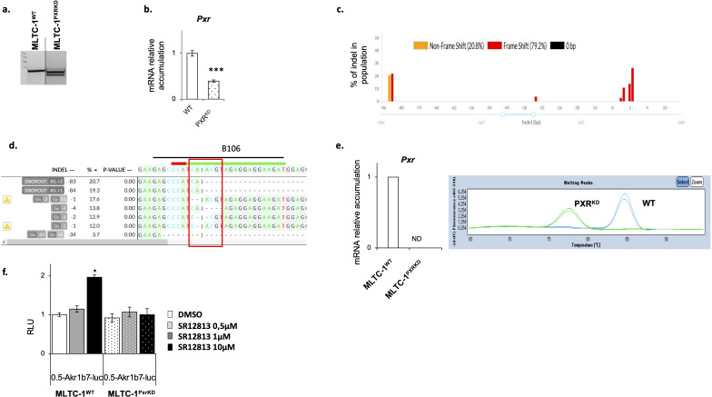
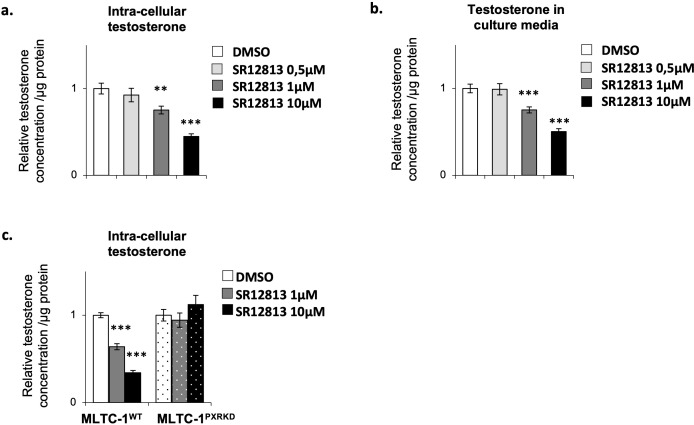
Methods: To determine the potential involvement of PXR in the regulation of steroidogenesis, experiments were performed on a wild type (MLTC-1WT) and a Pxr knock-down (MLTC-1PxrKD) mouse Leydig cell line (MLTC-1 cells), treated with a PXR agonist (SR-12813) in acute and chronic conditions.

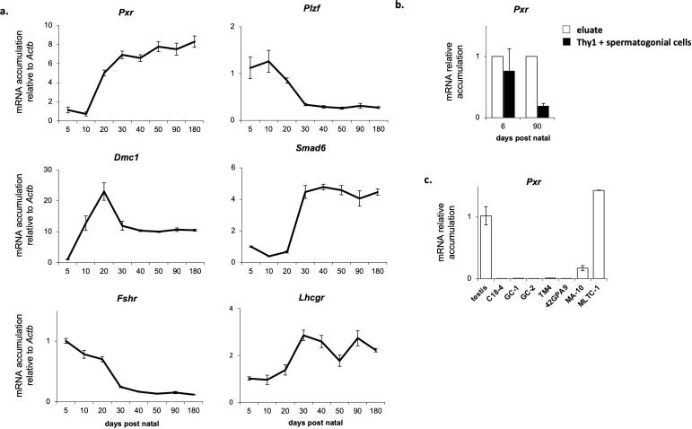
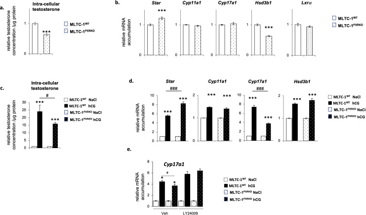
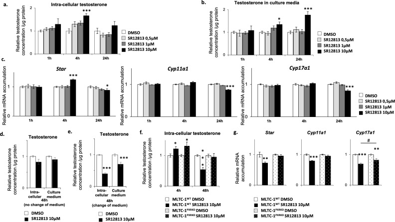
Results: Our analyses confirmed the presence of Pxr transcripts in the mouse testis, particularly in Leydig cells. In addition, A lower testosterone concentration was measured in MLTC-1PxrKD cells compared to wild type cells. Moreover, both acute and chronic stimulation of MLTC-1WT cells with SR-12813 led to a decrease in testosterone concentration, associated with a lower expression of some steroidogenic genes. This negative impact of SR-12813 on Leydig cell steroidogenesis was counteracted by Pxr knock down.

Discussion: Overall, these results support the involvement of PXR in the regulation of testosterone homeostasis in mouse Leydig cells and open new avenues of research into the involvement of this receptor in the deleterious effects of certain endocrine disruptors on the steroidogenic activity of Leydig cells.

Keywords: Leydig cells; PXR; mouse; testosterone; xenobiotics.


Conflict of interest statement

The authors declare that the research was conducted in the absence of any commercial or financial relationships that could be construed as a potential conflict of interest. The author(s) declared that they were an editorial board member of Frontiers, at the time of submission. This had no impact on the peer review process and the final decision.

Figures

Figure 1
Figure 1
Pxr is expressed in the Leydig cells of the mouse testis. (A) Ontogenic mRNA accumulation of Pxr, Plzf, Dmc1, Smad6, Fshr and Lhcgr in the mouse testis. (B) Pxr mRNA accumulation in Thy1+ spermatogonial cells and eluate from testis of neonate and adult mice. (C) Pxr mRNA accumulation in several immortalized testicular cell lines. (n = 5 per group). Data are means ± SEM.
Figure 2
Figure 2
Validation of Pxr gene editing in MLTC-1 cells. (A) Representative image of agarose gel showing genotyping PCR. (B) RT-qPCR analyses of Pxr mRNA levels in wild type (WT) and PXR knock-down (PXRKD) MLTC-1 cells using primers allowing amplification. (C) Quantification of the edits present in MLTC-1PxrKD cells sequencing data by DECODR software. (D) Sequence of the PXR genomic region at the site where CAS9 cuts (site of RNA guide, where CAS9 cuts). (E) RT-qPCR analyses of Pxr and representative image of melting peaks when primers (B106) at RNA guide induced sequence mutations [see panel (D)]. (F) Analysis of luciferase activity normalized to protein quantity in WT and PXRKD transfected with Akr1-B7-luciferase reporter plasmid and treated 24 hours with vehicle, SR1218 at 1μM or 10μM. n=15 per group. Vehicle groups were arbitrarily set at 1. Data are means ± SEM; statistical analysis: comparisons of two groups were made using Student’s T-test and comparisons of multiple groups were done using ANOVA2: *** p<0.001.
Figure 3
Figure 3
Pxr deficiency is associated with a deregulation of testosterone homeostasis in vitro. (A) Testosterone concentration in cells corrected to protein concentration in MLTC-1WT and MLTC-1PxrKD cells 24h after serum starvation. (B) mRNA accumulation of Star, Cyp11a1, Cyp17a1, Hsd3b1 and Lxrα normalized to b-actin in MLTC-1WT and MLTC-1PxrKD cells 24h after serum starvation. (C) Testosterone concentration in cells corrected to protein concentration in MLTC-1WT and MLTC-1PxrKD cells treated for 4h with vehicle or 0,125 IU hCG. (D) mRNA accumulation of Star, Cyp11a1, Cyp17a1 and Hsd3b1 in MLTC-1WT and MLTC-1PxrKD cells treated for 4h with vehicle or 0,125 IU hCG. (E) mRNA accumulation of Cyp17a1 in MLTC-1WT and MLTC-1PxrKD cells pre-treated for 1h with LY24009 at 10μM and then for 4h with vehicle or 0,125 IU hCG. n =3 triplicates per group; data means ± SEM; statistical analysis: comparisons of two groups were made using Student’s T-test and comparisons of multiple groups were done using ANOVA2: * p<0.05; *** p<0.001; # p<0.05; ### p<0.001.
Figure 4
Figure 4
SR-12813 leads to a decrease of testosterone concentration in Leydig cells in a PXR dependent manner. MLTC-1WT cells were treated for 1, 4 and 24h with vehicle or 0,5, 1 and 10 µM SR-12813 to determine testosterone concentration in (A) cells (corrected to protein concentration) and (B) culture media, and (C) expression of Star, Cyp11a1, Cyp17a1 and Hsd3b1 by RT-qPCR. MLTC-1WT cells were treated for 48h with vehicle or 10 µm SR-12813 to determine testosterone concentration in cells (corrected to protein concentration) and culture media (D) without and (E) with a medium change after 24h of treatment. (F) Intra-cellular testosterone concentration corrected to protein concentration from MLTC-1WT and MLTC-1PxrKD cells treated with vehicle or 10 µM SR-12813 for 4 or 48h (medium being changed after 24h). (G) mRNA accumulation of Star, Cyp11a1 and Cyp17a1 normalized to b-Actin from MLTC-1WT and MLTC-1PxrKD treated with vehicle or 10 µM SR-12813 for 24h. n =3 triplicates per group; data means ± SEM; statistical analysis: comparisons of two groups were made using Student’s T-test and comparisons of multiple groups were done using ANOVA2: *** p<0.001, # p<0.05.
Figure 5
Figure 5
Chronical activation of PXR leads to a decrease of testosterone concentration in Leydig cells. (A) Intra-cellular testosterone concentration corrected to protein concentration from MLTC-1WT cells treated every 24h for 3 days with vehicle or 0,5, 1 and 10 µM SR-12813. (B) Culture media testosterone concentration corrected to protein concentration from MLTC-1 cells treated every 24h for 3 days with vehicle or 0,5, 1 and 10 µM SR-12813. (C) Intra-cellular testosterone concentration corrected to protein concentration from MLTC-1WT and MLTC-1PxrKD cells treated every 24h for 3 days with vehicle or 0,5, 1 and 10 µM SR-12813. n =3 triplicates per group; data are means ± SEM; statistical analysis (Student’s T-test): ** p<0.01; *** p<0.001.

Similar articles

References

    1. Wilson JD, Griffin JE, George FW, Leshin M. The endocrine control of male phenotypic development. Aust J Biol Sci. (1983) 36:101−28. doi: 10.1071/BI9830101 - DOI - PubMed
    1. Singh J, O’Neill C, Handelsman DJ. Induction of spermatogenesis by androgens in gonadotropin-deficient (hpg) mice. Endocrinology. (1995) 136:5311−21. doi: 10.1210/endo.136.12.7588276 - DOI - PubMed
    1. Parker KL, Schimmer BP. Steroidogenic factor 1: a key determinant of endocrine development and function. Endocr Rev. (1997) 18:361−77. doi: 10.1210/edrv.18.3.0301 - DOI - PubMed
    1. Kim JW, Peng N, Rainey WE, Carr BR, Attia GR. Liver receptor homolog-1 regulates the expression of steroidogenic acute regulatory protein in human granulosa cells. J Clin Endocrinol Metab. (2004) 89:3042−7. doi: 10.1210/jc.2003-031599 - DOI - PubMed
    1. Zazopoulos E, Lalli E, Stocco DM, Sassone-Corsi P. DNA binding and transcriptional repression by DAX-1 blocks steroidogenesis. Nature. (1997) 390:311−5. doi: 10.1038/36899 - DOI - PubMed

LinkOut - more resources