

Giant Intracranial Chondroma
- PMID: 40395660
- PMCID: PMC12091629
- DOI: 10.1097/GOX.0000000000006726
Giant Intracranial Chondroma
Abstract
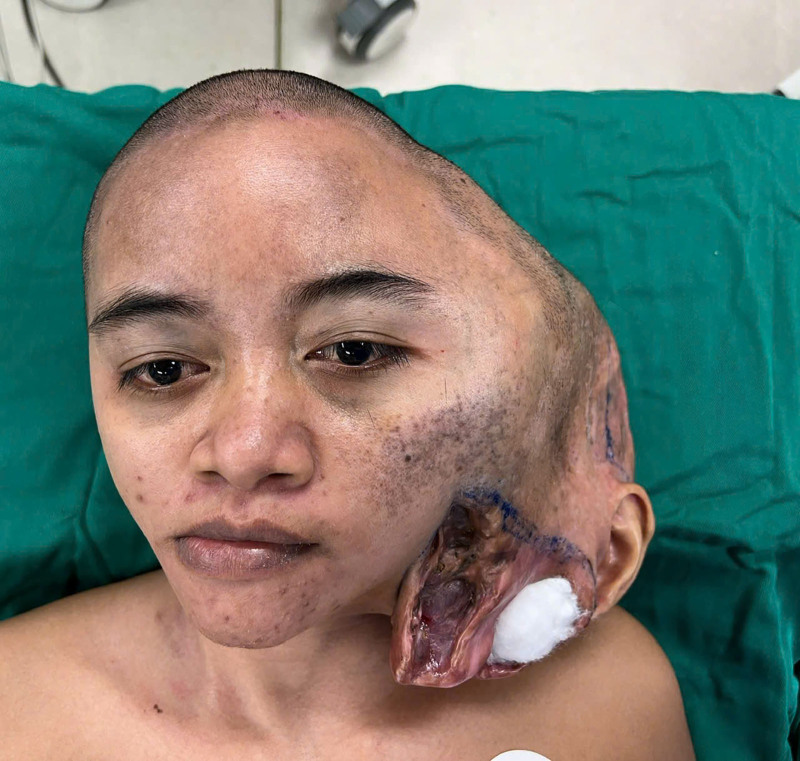
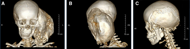
Intracranial chondroma is extremely rare. The treatment of choice is total tumor excision when resectable. A 30-year-old female patient presented with a giant intracranial chondroma that had enlarged for 18 years. In this case, we totally removed the tumor and reconstructed the meninges, cranium, and left ear using fascia lata, a combination of titan mesh and cement, and the posterior auricular artery axial flap, respectively. The aesthetic outcome was acceptable. No recurrence was identified after 7 months of follow-up. The patient was satisfied with the outcome. The lack of access to high-tech tools, such as 3-dimensional simulation, makes preoperative planning more difficult. Total tumor removal is currently the treatment of choice when the mass is resectable. It is a safe surgery, and with proper reconstruction procedures, an acceptable appearance can be achieved. Further multicenter studies with a greater sample size are needed to make a systematic treatment approach for intracranial chondroma.
Copyright © 2025 The Authors. Published by Wolters Kluwer Health, Inc. on behalf of The American Society of Plastic Surgeons.
Conflict of interest statement
The authors have no financial interest to declare in relation to the content of this article.
Figures
References
-
- Zülch KJ, Wechsler W. Pathology and classification of gliomas. In: Krayenbühl H, Maspes PE, Sweet WE, eds. Progress in Neurological Surgery. Vol 2. S.Karger AG; 1967:1–84.
-
- Berkmen YM, Blatt ES. Cranial and intracranial cartilaginous tumours. Clin Radiol. 1968;19:327–333. - PubMed
-
- Ozgen T, Pamir MN, Akalan N, et al. . Intracranial solitary chondroma. Case report. J Neurosurg. 1984;61:399–401. - PubMed
-
- El-Mofty SK. Chapter 9—bone lesions. In: Gnepp DR, ed. Diagnostic Surgical Pathology of the Head and Neck. 2nd ed. W.B. Saunders; 2009:729–784.
-
- Traflet RF, Babaria AR, Barolat G, et al. . Intracranial chondroma in a patient with Ollier’s disease. Case report. J Neurosurg. 1989;70:274–276. - PubMed
Publication types
LinkOut - more resources
Full Text Sources