. 2025 Feb 4;5(2):70004.
doi: 10.1002/EXP.70004. eCollection 2025 Apr.

Riding a Vascular Time Train to Spatiotemporally Attenuate Thrombosis and Restenosis by Double Presentation of Therapeutic Gas and Biomacromolecules

Affiliations

Riding a Vascular Time Train to Spatiotemporally Attenuate Thrombosis and Restenosis by Double Presentation of Therapeutic Gas and Biomacromolecules

Jingdong Rao et al. Exploration (Beijing). .

Abstract

Endothelial injury is a common occurrence following stent implantation, often leading to complications such as restenosis and thrombosis. To address this issue, we have developed a multi-functional stent coating that combines a dopamine-copper (DA-Cu) base with therapeutic biomolecule modification, including nitric oxide (NO) precursor L-arginine, endothelial glycocalyx heparin, and endothelial cell (EC) catcher vascular endothelial growth factor (VEGF). In our stent coating, the incorporated Cu acts as a sustainable catalyst for converting endogenous NO donors into NO, and the immobilized arginine serves as a precursor for NO generation under the effect of endothelial nitric oxide synthase (eNOS). The presence of heparin endows the stent coating with anticoagulant ability and enhances eNOS activity, whilst rapid capture of EC by VEGF accelerates re-endothelialization. After in vivo implantation, the antioxidant elements and produced NO alleviate the inflammatory response, establishing a favorable healing environment. The conjugated VEGF contributes to the formation of a new and intact endothelium on the stent surface to counteract inappropriate vascular cell behaviors. The long-lasting NO flux inhibits smooth muscle cell (SMC) migration and prevents its excessive proliferation, reducing the risk of endothelial hyperplasia. This innovative coating enables the dual delivery of VEGF and NO to target procedural vascular repair phases: promoting rapid re-endothelialization, effectively preventing thrombosis, and suppressing inflammation and restenosis. Ultimately, this innovative coating has the potential to improve therapeutic outcomes following stent implantation.

Keywords: anti‐restenosis; anti‐thrombosis; biomacromolecule; re‐endothelialization; therapeutic gas.


Conflict of interest statement

The authors declare no conflicts of interest.

Figures

FIGURE 1
FIGURE 1
Illustration of spatiotemporal therapeutic effect of the AHV coating. (A) The AHV coating is prepared by an immersion approach, where the DA‐Cu network adheres to the stent surface with functional molecular modifications. The grafting of VEGF facilitates the efficient capture of ECs. Simultaneously, Cu catalyzes the NO production when endogenous RSNOs were present, while in the scenarios where RSNOs are absent, the NO precursor arginine plays a vital role in NO synthesis. The conversion of arginine into NO is mediated by eNOS, whose level is influenced by the increasing number of ECs and can be further elevated by heparin. Such interplay ensures the production of sufficient NO under various physiological conditions. (B) After in vivo implantation of the AHV stent, the antioxidants DA, heparin and L‐arginine reduce ROS level, and then polarize macrophages into anti‐inflammatory M2 type with the assistance of generated NO, heparin and VEGF, alleviating local inflammation. Afterward, the grafted VEGF promotes rapid re‐endothelialization, enabling the restoration of endothelial functions and establishing a barrier against inappropriate cellular behaviors. The produced NO and anticoagulant heparin can synergistically inhibit platelet adhesion and activation, preventing thrombosis. The sustained NO production also suppresses SMC migration and proliferation at the later stage of stent implantation for a long time, reducing the risk of restenosis and promoting vessel repair. Altogether, the AHV coating provides a sequential treatment for the post‐implantation complications associated with stenting. EC: endothelial cell, VEGF: vascular endothelial growth factor, RSNO: S‐nitrosothiols, Cu: copper, DA: dopamine, eNOS: endothelial nitric oxide synthase, NO: nitric oxide, ROS: reactive oxygen species, M1: polarized M1 phenotype macrophages (pro‐inflammatory), M2: polarized M1 phenotype macrophages (anti‐inflammatory), SMC: smooth muscle cell.
FIGURE 2
FIGURE 2
Characterization of the AHV coating. (A) EPR and (B) MALDI‐TOF‐MS spectra of DA‐Cu. (C) XPS spectra of V and AHV. (D) GATR‐FTIR spectra of B, V and AHV. Immobilization of (E) arginine, (F) heparin and (G) VEGF onto the DA/HD‐Cu coating measured using QCM‐D. Approximately 346 ± 11 ng cm−2 arginine, 609 ± 7 ng cm−2 heparin and 232 ± 4 ng cm−2 VEGF had been conjugated onto the sample surface. (H) WCA of bare and coated samples. (I) Thickness of each coating. (J) Representative SEM micrographs showing the morphology of the AHV‐coating before and after stent dilation. Scale bars: 10, 50 and 500 µm. (K) Long‐term NO generation with NO donors. (L) NO production by arginine in cellular level without NO donors. (M) NO production (green) by arginine in HUVECs (with no NO donors). Scale bars: 50 µm. Data were displayed as mean ± standard deviation (SD) (n = 3). *p < 0.05 and **p < 0.01. B: bare 316L SS, DA: dopamine, Cu: copper, V: DA/hexamethylenediamine (HD)‐Cu‐VEGF, AV: DA/HD‐Cu‐arginine‐VEGF, HV: DA/HD‐Cu‐heparin‐VEGF, AH: DA/HD‐Cu‐arginine‐heparin, AHV: DA/HD‐Cu‐arginine‐heparin‐VEGF, PBS: phosphate buffered saline, WSC: water soluble carbodiimide, MES: 2‐(N‐morpholino) ethanesulfonic acid hydrate, NO: nitric oxide, DAPI: 4',6‐diamidino‐2‐phenylindole.
FIGURE 3
FIGURE 3
Inflammation alleviation. (A) Illustration of rapid inflammation alleviation. (B) Histogram and (C) fluorescence quantification of ROS scavenging. (D) Immunofluorescence staining of macrophage polarization (top: CD86, M1 marker; bottom: CD206, M2 marker) and (E) semi‐quantitative data. Scale bars: 100 µm. Data were displayed as mean ± standard deviation (SD) (n = 3). *p < 0.05, **p < 0.01, ***p< 0.001. NO: nitric oxide, ROS: reactive oxygen species, VASP: vasodilator stimulated phosphoprotein, M1: polarized M1 phenotype macrophages (pro‐inflammatory), M2: polarized M1 phenotype macrophages (anti‐inflammatory), B: bare 316L SS, V: DA/HD‐Cu‐VEGF, AV: DA/HD‐Cu‐arginine‐VEGF, HV: DA/HD‐Cu‐heparin‐VEGF, AH: DA/HD‐Cu‐arginine‐heparin, AHV: DA/HD‐Cu‐arginine‐heparin‐VEGF.
FIGURE 4
FIGURE 4
Enhanced re‐endothelialization and platelet repelling effect. (A) Illustration of EC growth and platelet inhibition. (B) The rapid adhesion and growth of HUVECs (green). Scale bars: 50 µm. (C) Number of adhered HUVECs on different coatings. (D) HUVEC proliferation using CCK‐8 assay. (E) eNOS expression and (F) NO production in HUVECs after incubation for 72 h. The increased EC adhesion, enhanced eNOS level and NO production contribute to the enhanced EC growth and rapid re‐endothelialization. (G) Fluorescence images of migrated HUVECs and (H) migration distance of HUVECs. Scale bars: 50 µm. (I) SEM images showing the adhered platelets (red arrows represent platelets) and (J) cGMP level of platelets with NO donors. Scale bars: 10 µm. (K) HUVEC and platelet (bottom) adhesion onto different coatings. AHV surface has the highest HUVEC adhesion with the lowest platelet adhesion. Scale bars: 25 µm. (L) The number of HUVECs and platelets cultivated on different coatings. Data were displayed as mean ± standard deviation (SD) (n = 3). *p < 0.05, **p < 0.01, ***p< 0.001. NO: nitric oxide, cGMP: cyclic guanylate monophosphate, eNOS: endothelial nitric oxide synthase, HUVEC: human umbilical vein endothelial cell, B: bare 316L SS, V: DA/HD‐Cu‐VEGF, AV: DA/HD‐Cu‐arginine‐VEGF, HV: DA/HD‐Cu‐heparin‐VEGF, AH: DA/HD‐Cu‐arginine‐heparin, AHV: DA/HD‐Cu‐arginine‐heparin‐VEGF, CCK‐8: cell counting kit‐8.
FIGURE 5
FIGURE 5
Long‐term SMC inhibition. (A) Illustration of SMC inhibition. (B) Adhesion and (C) proliferation of HUASMCs cultured in the presence of NO donors. Scale bars: 50 µm. (D) cGMP expression after incubation for 72 h with NO donors. (E) Images of competitive adhesion between HUVECs (green) and HUASMCs (red) for 72 h. Scale bars: 50 µm. (F) The ratio of HUVECs/HUASMCs on different coatings. *, **, ***(HUVECs vs HUASMCs) and ¶, ¶¶, ¶¶¶ (vs. 316 L SS) remarked p < 0.05, 0.01 and 0.001. The AHV coating has the highest HUVECs/HUASMCs ratio, indicating its potential to support HUVECs and suppress HUASMCs. (G) Fluorescence images of migrated HUASMCs and (H) migration distance of HUASMCs. Scale bars: 50 µm. Data were displayed as mean ± standard deviation (SD) (n = 3). *p < 0.05, **p < 0.01, ***p < 0.001. NO: nitric oxide, cGMP: cyclic guanylate monophosphate, HUASMC: human umbilical artery smooth muscle cells, B: bare 316L SS, V: DA/HD‐Cu‐VEGF, AV: DA/HD‐Cu‐arginine‐VEGF, HV: DA/HD‐Cu‐heparin‐VEGF, AH: DA/HD‐Cu‐arginine‐heparin, AHV: DA/HD‐Cu‐arginine‐heparin‐VEGF, HUVEC: human umbilical vein endothelial cell, CCK‐8: cell counting kit‐8.
FIGURE 6
FIGURE 6
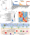
Underlying mechanism investigation by gene sequencing. (A) Volcano graphs. (B) GO classification of DEGs. (C) KEGG enrichment scatter plot. (D) Heatmap evaluation of DEGs (AHV vs. bare). (E) The mechanisms of AHV coating to reestablish a healthy endothelial environment. Data were displayed as mean ± standard deviation (SD) (n = 3). AHV: DA/HD‐Cu‐arginine‐heparin‐VEGF, B: bare 316L SS, NO: nitric oxide, O2: oxygen gas, NADPH: nicotinamide adenine dinucleotide phosphate hydrogen, NADP: nicotinamide adenine dinucleotide phosphate, eNOS: endothelial nitric oxide synthase, GTP: guanosine triphosphate, sGC: soluble guanylyl cyclase, cGMP: cyclic guanylate monophosphate, Rap1: ras‐proximate‐1, EC: endothelial cell.
FIGURE 7
FIGURE 7
Ex vivo anti‐thrombosis investigation of the AHV coatings. (A) Ex vivo arteriovenous circulation device. (B) Cross‐sections of foil tubes coated with different coatings show the thrombosis formation. (C) Representative SEM images of adherent platelets. Scale bars: 20 µm. (D) Percent occlusion of circuits and (E) weight of thrombus. (F) Blood flow rate. Data were displayed as mean ± standard deviation (SD) (n = 4). *p < 0.05, **p < 0.01, ***p< 0.001. B: bare 316L SS, V: DA/HD‐Cu‐VEGF, AV: DA/HD‐Cu‐arginine‐VEGF, HV: DA/HD‐Cu‐heparin‐VEGF, AH: DA/HD‐Cu‐arginine‐heparin, AHV: DA/HD‐Cu‐arginine‐heparin‐VEGF.
FIGURE 8
FIGURE 8
Long‐term in vivo anti‐restenosis effect of AHV coating. (A) Representative image showing stent implantation in a rabbit model. (B) Representative SEM images of implanted stents after implantation for 1 week. Scale bars: 20 µm. (C) CD31 immuno‐fluorescence (green), phalloidin (red) and 4′,6‐diamidino‐2‐phenylindole (DAPI, blue) staining showing re‐endothelialization of 1‐week implanted stents. Scale bars: 50 µm. (D) Representative SEM images and Van Gieson's staining on restenosis of different CVSs. Scale bars: 20 µm, 0.5 mm and 200 µm. (E) Neointimal area and (F) stenosis analysis. Data were displayed as mean ± standard deviation (SD) (n = 7). *p < 0.05 and **p < 0.01. AHV: DA/HD‐Cu‐arginine‐heparin‐VEGF, CD31: cluster of differentiation 31.

Similar articles

References

    1. (a) Palmerini T., Benedetto U., Biondi‐Zoccai G., et al., “Long‐Term Safety of Drug‐Eluting and Bare‐Metal Stents,” Journal of the American College of Cardiology 65 (2015): 2496–2507. - PubMed
    2. (b) Frieden T. R. and Jaffe M. G., “Saving 100 Million Lives by Improving Global Treatment of Hypertension and Reducing Cardiovascular Disease Risk Factors,” The Journal of Clinical Hypertension 20 (2018): 208–211. - PMC - PubMed
    1. (a) Yang Y., Gao P., Wang J., et al., “Endothelium‐Mimicking Multifunctional Coating Modified Cardiovascular Stents via a Stepwise Metal‐Catechol‐(Amine) Surface Engineering Strategy,” Research 2020 (2020): 9203906. - PMC - PubMed
    2. (b) Yang Z., Zhao X., Hao R., et al., “Bioclickable and Mussel Adhesive Peptide Mimics for Engineering Vascular Stent Surfaces,” PNAS 117 (2020): 16127–16137. - PMC - PubMed
    1. (a) Habib A. and Finn A. V., “Endothelialization of Drug Eluting Stents and Its Impact on Dual Anti‐platelet Therapy Duration,” Pharmacological Research 93 (2015): 22–27. - PMC - PubMed
    2. (b) Lee H., Dellatore S. M., Miller W. M., and Messersmith P. B., “Mussel‐Inspired Surface Chemistry for Multifunctional Coatings,” Science 318 (2007): 426–430. - PMC - PubMed
    1. (a) Straub A. C., Lohman A. W., Billaud M., et al., “Endothelial Cell Expression of Haemoglobin α Regulates Nitric Oxide Signalling,” Nature 491 (2012): 473–477. - PMC - PubMed
    2. (b) Li P., Cai W., Wang K., et al., “Selenium‐Functionalized Polycarbonate‐Polyurethane for Sustained In Situ Generation of Therapeutic Gas for Blood‐Contacting Materials,” Smart Materials in Medicine 3 (2022): 361–373.
    1. Lee W. J., Tateya S., Cheng A. M., et al., “M2 Macrophage Polarization Mediates Anti‐Inflammatory Effects of Endothelial Nitric Oxide Signaling,” Diabetes 64 (2015): 2836–2846. - PMC - PubMed

LinkOut - more resources