

PCR-Based Genotyping of Zebrafish Genetic Mutants
- PMID: 40395840
- PMCID: PMC12086339
- DOI: 10.21769/BioProtoc.5248
PCR-Based Genotyping of Zebrafish Genetic Mutants
Abstract
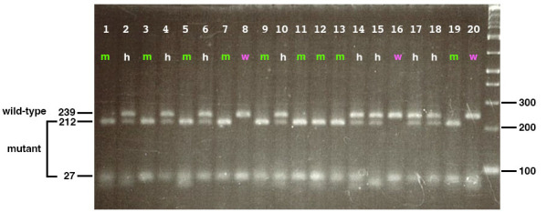
Zebrafish genetic mutants have emerged as a valuable model system for studying various aspects of disease and developmental biology. Mutant zebrafish embryos are generally identified based on phenotypic defects at later developmental stages, making it difficult to investigate underlying molecular mechanisms at earlier stages. This protocol presents a PCR-based genotyping method that enables the identification of wild-type, heterozygous, and homozygous zebrafish genetic mutants at any developmental stage, even when they are phenotypically indistinguishable. The approach involves the amplification of specific genomic regions using carefully designed primers, followed by gel electrophoresis. This genotyping method facilitates the investigation of the molecular mechanisms driving phenotypic defects that are observed at later timepoints. This protocol allows researchers to perform analyses such as immunofluorescence, RT-PCR, RNA sequencing, and other molecular experiments on early developmental stages of mutants. The availability of this protocol expands the utility of zebrafish genetic mutants for elucidating the molecular underpinnings of various biological processes throughout development. Key features • Enables genotyping of zebrafish genetic mutants at any developmental stage, even before the onset of phenotypic defects. • Utilizes PCR amplification and restriction enzyme digestion to distinguish wild-type, mutant, and heterozygous genotypes.
Keywords: Developmental biology; Embryos; Genotyping; Model organism; Mutant analysis; PCR; Phenotypic defects; Restriction enzyme digestion; Zebrafish.
©Copyright : © 2025 The Authors; This is an open access article under the CC BY license.
Conflict of interest statement
Competing interestsThe authors declare that they have no competing interests.
Figures


Similar articles
-
Rapid genotyping of mutant mice using dried blood spots for polymerase chain reaction (PCR) analysis.Brain Res Brain Res Protoc. 1997 May;1(2):117-23. doi: 10.1016/s1385-299x(96)00019-0. Brain Res Brain Res Protoc. 1997. PMID: 9385073
-
Automated multi-sample DNA extraction for genotyping live Xenopus embryos.Dev Dyn. 2023 Mar;252(3):429-438. doi: 10.1002/dvdy.544. Epub 2022 Oct 19. Dev Dyn. 2023. PMID: 36217575 Free PMC article.
-
Combining genotypic and phenotypic analyses on single mutant zebrafish larvae.MethodsX. 2018 Mar 14;5:244-256. doi: 10.1016/j.mex.2018.03.002. eCollection 2018. MethodsX. 2018. PMID: 30090702 Free PMC article.
-
Using zebrafish models to explore genetic and epigenetic impacts on evolutionary developmental origins of aging.Transl Res. 2014 Feb;163(2):123-35. doi: 10.1016/j.trsl.2013.10.004. Epub 2013 Oct 25. Transl Res. 2014. PMID: 24239812 Free PMC article. Review.
-
Zebrafish as a Model for Cardiovascular and Metabolic Disease: The Future of Precision Medicine.Biomedicines. 2024 Mar 20;12(3):693. doi: 10.3390/biomedicines12030693. Biomedicines. 2024. PMID: 38540306 Free PMC article. Review.
References
-
- Babu S., Takeuchi Y. and Masai I.(2022). Banp regulates DNA damage response and chromosome segregation during the cell cycle in zebrafish retina. eLife. 11: e74611. https://doi.org/10.7554/elife.74611 - DOI - PMC - PubMed
-
- Babu S.(2022). Banp regulates DNA damage response and chromosome segregation to promote cell-cycle progression and cell survival in zebrafish retina. Dissertation. OIST Japan.
LinkOut - more resources
Full Text Sources
