. 2025 Mar 20;15(6):e5249.
doi: 10.21769/BioProtoc.5249.

Generation of FECD Phenotypes in the Mouse Cornea by UVA Exposure and Surgical Removal of its Corneal Endothelial Layer

Affiliations

Generation of FECD Phenotypes in the Mouse Cornea by UVA Exposure and Surgical Removal of its Corneal Endothelial Layer

Susmita Sharma et al. Bio Protoc. .

Abstract

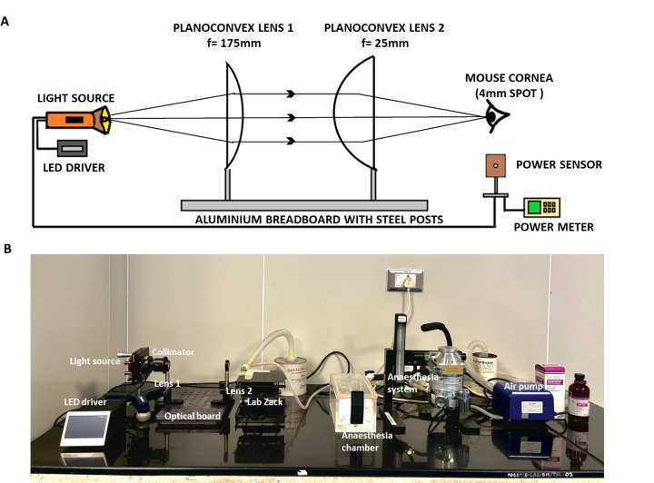
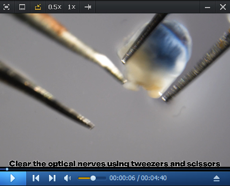
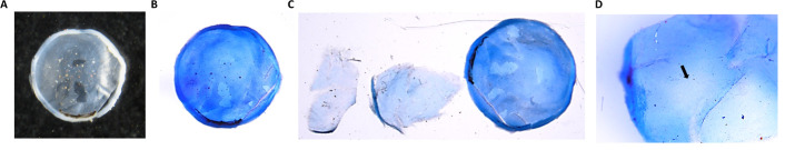
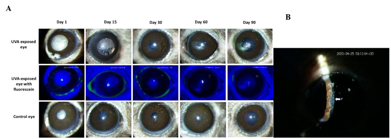
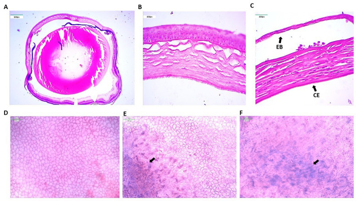
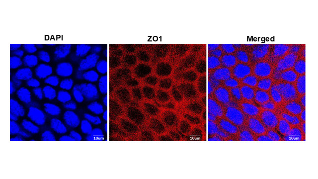
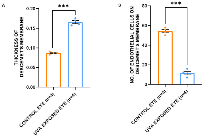
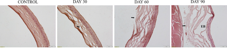
Fuchs endothelial corneal dystrophy (FECD) is a rare and multifactorial disorder leading to cell death in the innermost layer of the cornea, i.e., the endothelium; UV radiation is reported as the major environmental risk for the disease. Establishing an animal model for this disease has remained challenging in FECD research. We have developed a detailed protocol for the establishment of a UVA-induced FECD mouse model and removal of corneal endothelium from the eye for further molecular and histological studies by taking references from previous studies. UVA light of 500 J/cm2 was focused on the C57BL/6J female mouse cornea and kept for an observation period of 90 days. The animal developed corneal scarring by the end of three months. Slit-lamp microscopy and alizarin red-trypan blue staining confirmed endothelial cell death and formation of corneal guttae in the endothelium. Surgical removal of the endothelial layer was successfully done in the diseased mouse, and the result was confirmed by immunofluorescence. This study is relevant for in-depth research using a FECD mouse model, which will surpass the limitation of human tissue scarcity and can be used for in vivo drug targeting to develop therapeutics to cure FECD. Key features • UVA radiation induces FECD only in the exposed eye of female mice. • Females are more affected and develop the FECD phenotype. • This protocol will help dissect the endothelium layer with Descemet's membrane (DM) from the mouse cornea, which is equivalent to human surgical tissue.

Keywords: Cornea; Corneal endothelium; Dissection; Fuchs dystrophy; Mouse model; Slit-lamp microscopy; UVA irradiation.


Conflict of interest statement

Competing interestsThe authors declare no competing interests.

Figures

Figure 1.
Figure 1.. UVA exposure setup.
A. Design for UVA exposure setup. B. Different parts of the UVA exposure setup with the isoflurane anesthesia system.
Figure 2.
Figure 2.. Irradiation of mice’s eyes with UVA.
A. C57BL/6J mice were used in the experiment. B. The mouse was positioned on the lab jack with a continuous supply of isoflurane during UVA exposure to the left eye. The experiment was done in dark room conditions. C. Slit-lamp microscopic examination of the mouse eye by an animal handling expert.
Figure 3.
Figure 3.. Surgical instruments used for the dissection of the cornea from the eyeball.
A. A zero-size brush, curved-end micro-dissecting forceps, pointed-end forceps, and micro-suture cutting scissors were used to remove the cornea. B. A 10 mL syringe needle with the end slightly bent was used to remove the corneal endothelium from the cornea.
Video 1.
Video 1.. Endothelial layer removal from mice cornea
Figure 4.
Figure 4.. Dissection of the mouse eyeball.
A. Dissected transparent corneal cup from the mouse eyeball. B. The dissected mouse cornea is stained with trypan blue. C. Dissected corneal endothelium layer and the corneal cup without endothelium layer. The central portion of the corneal cup appears without the endothelium layer (marked with an arrow). D. Corneal endothelium layer at 40× magnification.
Figure 5.
Figure 5.. Effect of UVA exposure on female mice.
A. After UVA exposure, both eyes of the mice were examined by slit-lamp microscope at regular periodic intervals for three months. Images show that the female mice developed corneal scarring in the UVA-exposed left eye, whereas the control right eye was in a healthy condition. Fluorescein staining of the exposed eye confirmed the unaffected corneal epithelium. B. The slit image taken on day 30 confirmed that the scar is on the corneal endothelium.
Figure 6.
Figure 6.. Fuchs endothelial corneal dystrophy (FECD) phenotypes in the mouse cornea.
A. Histological image of a whole control mouse eyeball. B. The control eye shows a healthy cornea. The corneal endothelium layer is intact with no cell death. C. UVA-exposed eye shows severe FECD phenotypes such as loss of endothelial cells (CE) due to cell death and epithelial bullae (EB) formation due to detachment of corneal epithelium from the stroma. D. Alizarin red and trypan blue staining of the control corneal cup shows a hexagonal sheet of corneal endothelial cells with no abnormalities. E. Trypan blue staining of dead corneal endothelium cells in the corneal cup of the exposed eye. F. Higher magnification shows that the hexagonal corneal endothelial cells lost their shape and integrity.
Figure 7.
Figure 7.. Mouse corneal endothelium with ZO-1 staining.
ZO-1 is present exclusively in the tight junctions of epithelial and endothelial cells. The immunofluorescence image reveals the presence of ZO-1 at the cellular junctions of the corneal endothelial cells, which shows the morphology of the cell. The endothelium appears as a single sheet of nearly hexagonal cells. This experiment is only done with the corneal endothelium of the control eye of the mouse.
Figure 8.
Figure 8.. Quantification of histopathology data, namely the thickness of the Descemet’s membrane (DM) and the number of endothelial cells, to access phenotype development.
A. The thickness of the DM in the UVA-exposed eye (0.16 ± 0.02 μm) is significantly higher (p = 9.84 × 10⁻⁵) than that of the control eye (0.08 ± 0.01 μm). B. The number of endothelial cells present in the DM is significantly reduced in the cornea of the exposed eye (p = 2.92 × 10⁻⁶). A t-test was used to measure the statistical significance level. ***p < 0.005.
Figure 9.
Figure 9.. Histopathological analysis of the progression of abnormalities in the cornea of UVA-exposed mice for three months.
Results show normal cornea in the control eyeball. After the first month of UVA exposure, the disruption of collagen fibers begins. Epithelial bullae formation starts in the cornea after the second month, disrupting the stroma in the central cornea. Prominent epithelial bullae formation with severe stromal fiber disruption occurs at the end of the third month. The stromal disruption area is marked with a black arrow mark. EB, epithelial bullae.

Similar articles

References

    1. Bonanno J.(2003). Identity and regulation of ion transport mechanisms in the corneal endothelium. Prog Retin Eye Res. 22(1): 69 94 94. 10.1016/s1350-9462 (02)00059-9 - DOI - PubMed
    1. Elhalis H., Azizi B. and Jurkunas U. V.(2010). Fuchs endothelial corneal dystrophy. Ocul Surf. 8:173 84 84 . https://www.ncbi.nlm.nih.gov/books/NBK545248/ - PMC - PubMed
    1. Ong Tone S., Kocaba V., Böhm M., Wylegala A., White T. L. and Jurkunas U. V.(2021). Fuchs endothelial corneal dystrophy: The vicious cycle of Fuchs pathogenesis. Prog Retin Eye Res. 80: 100863 10.1016/j.preteyeres .2020.100863 - DOI - PMC - PubMed
    1. Nanda G. G. and Alone D. P.(2019). REVIEW: Current understanding of the pathogenesis of Fuchs’ endothelial corneal dystrophy. Mol Vis. 25:295 310 310. 10.1016/j.exer .2014.10.023 - DOI - PMC - PubMed
    1. Adamis A. P., Filatov V., Tripathi B. J. and Tripathi R. C.(1993). Fuchs' endothelial dystrophy of the cornea. Surv Ophthalmol. 38(2): 149 168 168. 10.1016/0039-6257 (93)90099-s - DOI - PubMed

LinkOut - more resources