Decreased expression of cytokeratin 15 and tropoelastin in men with androgenetic alopecia and its relationship with increased expression of p15/p16
- PMID: 40395887
- PMCID: PMC12087341
- DOI: 10.5114/aoms.2020.94914
Decreased expression of cytokeratin 15 and tropoelastin in men with androgenetic alopecia and its relationship with increased expression of p15/p16
Abstract
Introduction: Androgenetic alopecia (AGA) or male pattern baldness is a polygenic disease with a high prevalence in the Caucasian population, with psychosocial implications. However, the molecular mechanisms involved are unknown. The aim of this study was to assess the possible changes in protein expression of cytokeratin 15 (CK15), tropoelastin (TE) and cellular senescence markers (p15/p16) in AGA patients.
Material and methods: An observational, analytical and prospective cohort study was performed in 57 men with AGA (42.31 ±3.01 years). Two tissue biopsies were taken: the control area (C) and the alopecia area (A). Cell viability was assessed using scalp explants from the patients, maintaining explants long term (90 days). Protein expression analysis was performed by immunohistochemistry by detecting antigen-antibody reactions (avidin-biotin complex).
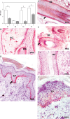
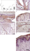
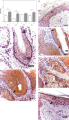
Results: The results showed a significantly higher percentage of dead cells in area A (17.00 ±1.09% C vs. 28.50 ±1.41% A, *p < 0.05). The area affected by alopecia showed significantly lower CK15 and TE protein expression in hair follicles, sebaceous glands and epidermis (*p < 0.05). Expression of the senescence marker p15/p16 was significantly higher in hair follicles, hair bulbs and the epidermis (*p < 0.05).
Conclusions: The results suggest that patients with AGA suffer tissue damage that affects different components of hair follicle stem cells as well as the extracellular matrix itself.
Keywords: androgenetic alopecia; cellular senescence; cytokeratin 15; extracellular matrix; tropoelastin.
Copyright: © 2020 Termedia & Banach.
Conflict of interest statement
The authors declare no conflict of interest.
Figures


Similar articles
-
Innovative strategies for the discovery of new drugs against androgenetic alopecia.Expert Opin Drug Discov. 2025 Apr;20(4):517-536. doi: 10.1080/17460441.2025.2473905. Epub 2025 Mar 11. Expert Opin Drug Discov. 2025. PMID: 40029254 Review.
-
Stem cells from human hair follicles: first mechanical isolation for immediate autologous clinical use in androgenetic alopecia and hair loss.Stem Cell Investig. 2017 Jun 27;4:58. doi: 10.21037/sci.2017.06.04. eCollection 2017. Stem Cell Investig. 2017. PMID: 28725654 Free PMC article.
-
High-frequency ultrasonography of the scalp: A comparison between androgenetic alopecia and healthy volunteers.Skin Res Technol. 2024 Aug;30(8):e13863. doi: 10.1111/srt.13863. Skin Res Technol. 2024. PMID: 39081105 Free PMC article.
-
Quantitative analysis of the anatomical changes in the scalp and hair follicles in androgenetic alopecia using magnetic resonance imaging.Skin Res Technol. 2021 Jan;27(1):56-61. doi: 10.1111/srt.12908. Epub 2020 Jun 28. Skin Res Technol. 2021. PMID: 32596954
-
Cellular Senescence: Ageing and Androgenetic Alopecia.Dermatology. 2023;239(4):533-541. doi: 10.1159/000530681. Epub 2023 Apr 22. Dermatology. 2023. PMID: 37088073 Review.
References
-
- Paus R, Olsen EA, Messenger AG. Hair growth disorders. In: Fitzpatrick’s Dermatology in General Medicine. 7th ed. Wolff K, Goldsmith LA, Katz SI, Gilchrest BA, Paller As, Leffell DJ (eds.). USA: The McGraw-Hill Companies Inc. 2008; 753-77.
-
- Lolli F, Pallotti F, Rossi A, et al. . Androgenetic alopecia: a review. Endocrine 2017; 57: 9-17. - PubMed
-
- Severi G, Sinclair R, Hopper JL, et al. . Androgenetic alopecia in men aged 40–69 years: prevalence and risk factors. Br J Dermatol 2003; 149: 1207-13. - PubMed
-
- Khumalo NP, Jessop S, Gumedze F, Ehrlich R. Hairdressing and the prevalence of scalp disease in African adults. Br J Dermatol 2007; 157: 981-8. - PubMed
LinkOut - more resources
Full Text Sources
Research Materials
Miscellaneous
