. 2025 May 6:13:1593226.
doi: 10.3389/fcell.2025.1593226. eCollection 2025.

Generation and functional characterization of tuft cells in non-human primate pancreatic ducts through organoid culture systems

Affiliations

Generation and functional characterization of tuft cells in non-human primate pancreatic ducts through organoid culture systems

Kosuke Sakaguchi et al. Front Cell Dev Biol. .

Abstract

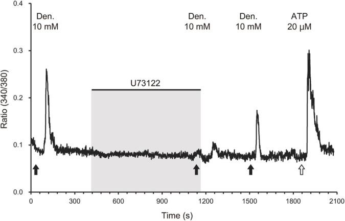
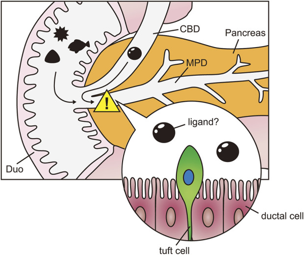
The pancreatic duct plays a key role in collecting pancreatic juice, which is rich in digestive enzymes. The fluid flows unidirectionally into the duodenum, where it mixes with partially digested food to further facilitate digestion. In this study, we report the generation of pancreatic ductal organoids from non-human primates for the first time, aimed at investigating the role of tuft cells that reside in the pancreatic duct since no studies have addressed the role of tuft cells in the pancreas. The organoids were maintained in a medium supplemented with Wnt3a, Noggin, R-spondin, and other factors that support pancreatic duct proliferation. These pancreatic organoids expressed the stem cell marker LGR5 mRNA and the ductal marker protein CK19, although tuft cell markers were not detectable at this stage. Upon stimulation with IL-4/13, tuft cell differentiation was confirmed by immunohistochemistry and transcriptomic analysis. We observed induction of DCLK1, as well as taste signaling molecules such as TRPM5 and PLCβ2, which are markers of type II taste cells. Additionally, upregulation of LYZ and DEFB1 mRNA indicated the expression of antimicrobial peptide markers, alongside molecules associated with inflammation. Furthermore, the differentiated organoids specifically responded to a bitter compound, suggesting that pancreatic tuft cells may play a role in detecting potentially harmful chemicals. Finally, immunohistochemical analysis identified tuft cells in the non-human primate pancreas, supporting their involvement in sensing harmful compounds and regulating protective responses within the pancreas.

Keywords: organoid; pancreas; primate; tuft cells; type 2 immunity.


Conflict of interest statement

The authors declare that the research was conducted in the absence of any commercial or financial relationships that could be construed as a potential conflict of interest.

Figures

FIGURE 1
FIGURE 1
Tuft cells in the pancreatic ducts and small intestine of mice. Wild-type (WT) mice were treated with 100 mM succinate or water (H2O) for 7 days. Immunofluorescence staining was performed to detect Dclk1 (green), a tuft cell marker, in the pancreatic duct (A, B) and ileum (E, F) of wild-type mice. Dclk1 expression was examined in both the pancreatic duct and ileum of Pou2f3-KO mice treated with 100 mM succinate for 7 days (C, G). Small intestinal epithelial cells were stained with Ep-CAM (red). Nuclei were stained with DAPI (blue). Scale bars: 50 µm. The number of Dclk1-expressing tuft cells in each condition was quantified (D, H). Data are presented as means ± SEM (n = 3).
FIGURE 2
FIGURE 2
Generation of macaque pancreatic ductal organoids. Schematic illustration depicting the process of generating pancreatic ductal organoids (A). Representative images of primary organoid cultures at days 2, 4, and 5 (B–D) and clusters of growing organoids at days 2, 5, and 8 after passage (E–G). Day 10 pancreatic ductal organoids cultured with proliferation medium were stained with anti-CK19 antibodies (green, (H). Nuclei were stained with Hoechst 33342 (blue). The pancreatic ducts are outlined with dashed lines. Scale bars: B-D, 200 μm; E-G, 500 μm; H, 50 µm.
FIGURE 3
FIGURE 3
Transcriptome analysis of macaque ductal tuft cells induced by IL-4 and IL-13 stimulation. DCLK1 immunoreactive tuft cells were not detected before induction with IL-4 or IL-13 (A). After stimulated with IL-4 or IL-13 for 5 days, DCLK1-expressing tuft cells (green) were observed (B, C). Nuclei were stained with Hoechst 33342 (blue). Scale bars: A-C, 100 µm. Quantitative analysis of the DCLK1-expressing tuft cells under three culture conditions (D). Principle component analysis (PCA) showed that individual culture condition fell into the same cluster (E). The heatmap illustrates the tuft cell markers that were significantly upregulated in organoids induced by IL-4 and IL-13 (FC ≥ 2 and FDR p-value <0.05) (F). Color bars represent Log10-transformed TPM values (F). Gene expression of representative tuft cell markers (POU2F3, GFI1B, DCLK1, TRPM5, CHAT, AVIL, PLCB2, GNAT3, ALOX5, and ALOX5AP), genes related to tuft cell function (GNG13, IL4R, and SUCNR1), and taste receptors (TAS1R1, TAS1R3, TAS2R1, TAS2R3, TAS2R4, TAS2R5, and TAS2R46) is shown (G). All data are presented as means ± SEM (n = 3). The p-value was determined by a one-way ANOVA followed by Tukey’s HSD test (*P < 0.05, **P < 0.01, ***P < 0.001).
FIGURE 4
FIGURE 4
Phospholipase C-mediated calcium response of IL-4-treated organoids to a bitter compound. A representative waveform of a cell derived from IL-4-treated organoids exposed to 10 mM denatonium benzoate (black arrows) is shown. The response to denatonium benzoate was attenuated by the phospholipase C inhibitor U73122 (40 µM). The response recovered after the inhibitor was removed via perfusion for 6 min. Black bars indicate the timing of application of the phospholipase C inhibitor U73122 (40 µM) and following washing out. ATP (20 μM, a white arrow) was applied as a positive control to validate the cell viability.
FIGURE 5
FIGURE 5
Tuft cells reside in the main pancreatic duct and papilla of Vater in monkey The schematic illustration shows location stained with anti-DCLK1 antibody (A). Fluorescent immunostaining images of DCLK1-positive tuft cells through PV to Tail region (B, E–G). Fluorescent immunohistochemical analysis show that DCLK1 (green) and CHAT (red, (C) or DCLK1 (green) and GNAT3 (red, (D) are partially co-localized around PV. Nuclei were stained with DAPI (blue). Scale bars: 50 µm. PV, papilla of Vater; CBD, common bile duct; MPD, main pancreatic duct; Gb, gallbladder; Duo, Duodenum; H, head of pancreas; B, body of pancreas; T, tail of pancreas.
FIGURE 6
FIGURE 6
Speculation of tuft cell function in pancreas. Schematic diagram of the hypothesis that tuft cells in the pancreatic ducts detect harmful agents from the duodenum and bile ducts. Duo, Duodenum; CBD, common bile duct; MPD, main pancreatic duct.

Similar articles

References

    1. Behrens M., Meyerhof W. (2006). Bitter taste receptors and human bitter taste perception. Cell Mol. Life Sci. 63, 1501–1509. 10.1007/s00018-006-6113-8 - DOI - PMC - PubMed
    1. Bernstein Z. J., Shenoy A., Chen A., Heller N. M., Spangler J. B. (2023). Engineering the IL-4/IL-13 axis for targeted immune modulation. Immunol. Rev. 320, 29–57. 10.1111/imr.13230 - DOI - PubMed
    1. Billipp T. E., Nadjsombati M. S., von Moltke J. (2021). Tuning tuft cells: new ligands and effector functions reveal tissue-specific function. Curr. Opin. Immunol. 68, 98–106. 10.1016/j.coi.2020.09.006 - DOI - PMC - PubMed
    1. Broutier L., Andersson-Rolf A., Hindley C. J., Boj S. F., Clevers H., Koo B. K., et al. (2016). Culture and establishment of self-renewing human and mouse adult liver and pancreas 3D organoids and their genetic manipulation. Nat. Protoc. 11, 1724–1743. 10.1038/nprot.2016.097 - DOI - PubMed
    1. Delgiorno K. E., Chung C. Y., Vavinskaya V., Maurer H. C., Novak S. W., Lytle N. K., et al. (2020a). Tuft cells inhibit pancreatic tumorigenesis in mice by producing prostaglandin D (2). Gastroenterology 159, 1866–1881.e8. 10.1053/j.gastro.2020.07.037 - DOI - PMC - PubMed

LinkOut - more resources