. 2025 May 13:2025:1212255.
doi: 10.1155/sci/1212255. eCollection 2025.

Effects of Bariatric Surgery-Related Weight Loss on the Characteristics, Metabolism, and Immunomodulation of Adipose Stromal/Stem Cells in a Follow-Up Study

Affiliations

Effects of Bariatric Surgery-Related Weight Loss on the Characteristics, Metabolism, and Immunomodulation of Adipose Stromal/Stem Cells in a Follow-Up Study

Amna Adnan et al. Stem Cells Int. .

Abstract

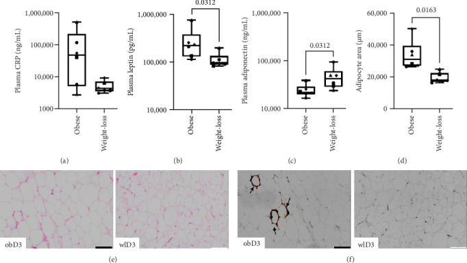
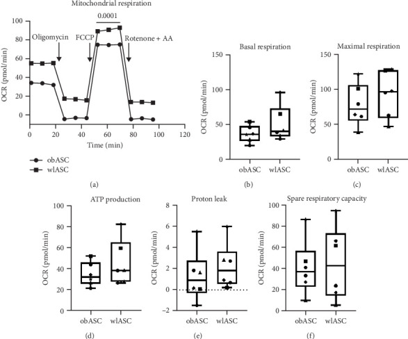
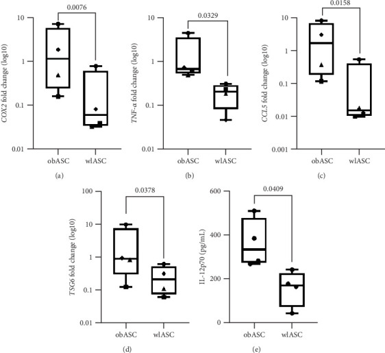
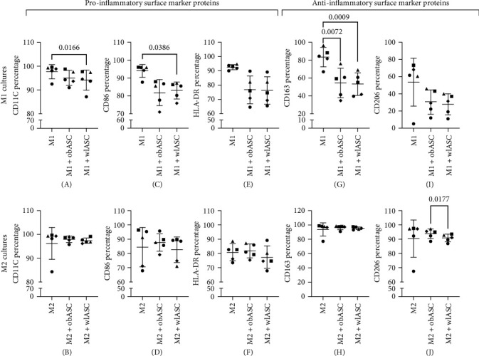
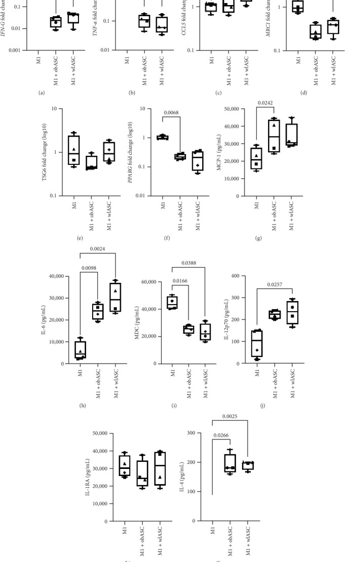
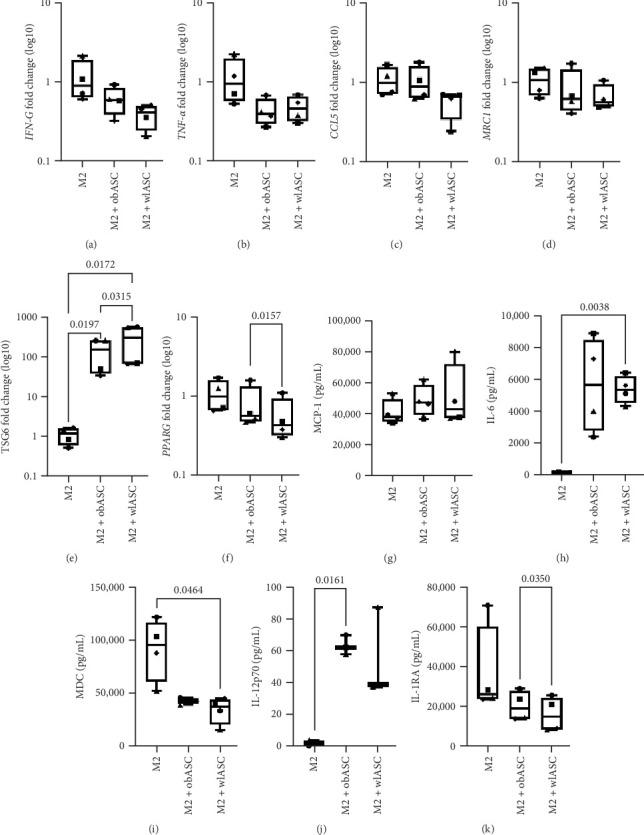
Background: The success of adipose stromal/stem cell (ASC)-based therapies may depend on donor characteristics such as body mass index (BMI). A high BMI may negatively impact the therapeutic potential of ASCs, but the effects of weight loss on ASC-mediated immunoregulation have not been extensively studied. Methods: ASCs were obtained from donors with obesity (obASCs) undergoing bariatric surgery and from the same donors after weight loss (wlASCs). Plasma samples, adipose tissue histology, and ASC characteristics, such as mitochondrial respiration and inflammatory factors, were studied before and after weight loss. The immunomodulatory capacity of ob/wlASCs was evaluated in cocultures with prepolarized and preactivated proinflammatory (M1) and anti-inflammatory (M2) macrophages by determining macrophage surface markers, gene expression, and cytokine secretion. Results: Weight loss significantly decreased plasma leptin levels and increased adiponectin levels. After weight loss, crown-like structures (CLSs) were undetectable, and the adipocyte size decreased. Weight loss significantly improved mitochondrial respiration in ASCs and resulted in a notable increase in their proliferative capacity. The proinflammatory marker genes tumor necrosis factor alpha (TNF-α), chemokine ligand 5 (CCL5), and cyclooxygenase-2 (COX2), as well as the proinflammatory cytokine interleukin 12p70 (IL-12p70), were significantly downregulated, while the anti-inflammatory gene tumor necrosis factor-inducible gene 6 (TSG6) was also significantly downregulated in ASC monocultures after weight loss. Following weight loss, ASCs exhibited increased proinflammatory properties when cocultured with macrophages, characterized by the downregulation of anti-inflammatory factors, along with the upregulation of several proinflammatory factors, compared with the effects of macrophage monocultures. Conversely, wlASCs demonstrated improved immunosuppressive functions in coculture with macrophages, as indicated by the upregulation of TSG6 gene expression and interleukin 4 (IL-4) secretion. Conclusions: Weight loss improved donors' metabolic health and partially recovered ASCs' anti-inflammatory gene expression and cytokine secretion profiles in monocultures. However, it was inadequate to fully restore the immunosuppressive functions of ASCs in cocultures with macrophages. Therefore, not only donor BMI but also weight loss history, among other donor characteristics, might be considered for optimal ASC-based therapy.

Keywords: adipose stromal/stem cells; bariatric surgery; inflammation; macrophages; obesity; weight loss.


Conflict of interest statement

The authors declare no conflicts of interest.

Figures

Figure 1
Figure 1
Effects of weight loss on plasma inflammatory (CRP, leptin) and anti-inflammatory (adiponectin) marker levels, adipocyte areas, and crown-like structures (CLSs). Decreased plasma CRP and leptin levels and increased adiponectin levels were detected after weight loss; n = 6. The Wilcoxon test for paired samples was used. p Values < 0.05 were considered significant. (A) CRP, (B) leptin, and (C) adiponectin levels. Hematoxylin and eosin (HE)-stained histological samples of AT after weight loss presented a decreased adipocyte area and a lack of CLS following CD68 chromogenic staining. (D) Adipocyte area in µm before and after weight loss. (E) Representative HE histological samples before and after weight loss from one donor. (F) Representative CD68-positive chromogenic histological samples before and after weight loss from one donor. Arrows indicate the accumulation of CLS around adipocytes. CRP, C-reactive protein; obD, donor before weight loss; wlD, donor after weight loss. See also Figure S2.
Figure 2
Figure 2
Improved oxygen consumption rates of ASCs after weight loss; n = 6. (A) Mitochondrial respiration, (B) basal respiration, (C) maximal respiration, (D) ATP production, (E) proton leakage, and (F) spare respiratory capacity. Paired t-test. p Values < 0.05 were considered significant. Data are presented as the medians for mitochondrial respiration and as the minimum to maximum values for other measured individual factors. obASCs, ASCs obtained before weight loss; OCR, oxygen consumption rate; wlASCs, ASCs obtained after weight loss.
Figure 3
Figure 3
Comparison of gene expression and cytokine secretion between obASCs and wlASCs; n = 4. (A)–(D) Gene expression. Box plots of the fold change values and statistical analysis of the delta CT values. (E) Cytokine secretion. (A) TNF-α, (B) CCL5, (C) COX2, (D) TSG6, and (E) IL-12p70. The Wilcoxon test was used for paired samples for gene expression, and the paired t-test was used for cytokine secretion. p Values < 0.05 were considered significant. The data are presented as the minimum to maximum values. obASCs, ASCs derived before weight loss; wlASCs, ASCs derived after weight loss.
Figure 4
Figure 4
Percentages of CD markers in M1 and M2 cells in monoculture and coculture with obASCs and wlASCs; n = 5. (A) CD11C, (C) CD86, (E) HLA-DR, (G) CD163, and (I) CD206 in M1 mono-/cocultures. (B) CD11C, (D) CD86, (F) HLA-DR, (H) CD163, and (J) CD206 expression in M2 mono-/cocultures. For RM ANOVA, p values < 0.05 were considered significant. The data are presented as the means and SDs. M1, proinflammatory macrophages; M2, anti-inflammatory macrophages; obASCs, ASCs derived before weight loss; wlASCs, ASCs derived after weight loss. See also Figure S13.
Figure 5
Figure 5
Comparison of gene expression and cytokine secretion between mono- and cocultures of M1 macrophages with obASCs and wlASCs; n = 4. (A)–(F) Gene expression in M1 mono-/cocultures. (G)–(L) Cytokine secretion in M1 mono-/cocultures. (A) IFNɣ, (B) TNF-α, (C) CCL5, (D) MRC1, (E) TSG6, (F) PPAR-ɣ, (G) MCP-1, (H) IL-6, (I) MDC, (J) IL-12p70, (K) IL-1RA, and (L) IL-4. For RM ANOVA, p values < 0.05 were considered significant. The data are presented as the minimum to maximum values. M1, proinflammatory macrophages; obASCs, ASCs derived before weight loss; wlASCs, ASCs derived after weight loss. See also Figure S14.
Figure 6
Figure 6
Comparison of gene expression and cytokine secretion between mono- and cocultures of M2 macrophages with obASCs and wlASCs; n = 4. (A)–(F) Gene expression in M2 mono-/cocultures. (G)–(K) Cytokine secretion in M2 mono-/cocultures. (A) IFNɣ, (B) TNF-α, (C) CCL5, (D) MRC1, (E) TSG6, (F) PPAR-ɣ, (G) MCP-1, (H) IL-6, (I) MDC, (J) IL-12p70, and (K) IL-1RA. For RM ANOVA, p values < 0.05 were considered significant. The data are presented as the minimum to maximum values. M2, anti-inflammatory macrophages; obASCs, ASCs derived before weight loss; wlASCs, ASCs derived after weight loss. See also Figure S15.

Similar articles

References

    1. Al-Ghadban S., Bunnell B. A. Adipose Tissue-Derived Stem Cells: Immunomodulatory Effects and Therapeutic Potential. Physiology . 2020;35(2):125–133. doi: 10.1152/physiol.00021.2019. - DOI - PubMed
    1. De Francesco F., Ricci G., D’Andrea F., Nicoletti G. F., Ferraro G. A. Human Adipose Stem Cells: From Bench to Bedside. Tissue Engineering Part B: Reviews . 2015;21(6):572–584. doi: 10.1089/ten.teb.2014.0608. - DOI - PubMed
    1. Kurki A., Paakinaho K., Hannula M., et al. Promoting Cell Proliferation and Collagen Production With Ascorbic Acid 2-Phosphate-Releasing Poly(l-Lactide-Co-ε-Caprolactone) Membranes for Treating Pelvic Organ Prolapse. Regenerative Biomaterials . 2024;11 doi: 10.1093/rb/rbae060.rbae060 - DOI - PMC - PubMed
    1. Czerwiec K., Zawrzykraj M., Deptuła M., et al. Adipose-Derived Mesenchymal Stromal Cells in Basic Research and Clinical Applications. International Journal of Molecular Sciences . 2023;24(4) doi: 10.3390/ijms24043888.3888 - DOI - PMC - PubMed
    1. Hutchings G., Janowicz K., Moncrieff L., et al. The Proliferation and Differentiation of Adipose-Derived Stem Cells in Neovascularization and Angiogenesis. International Journal of Molecular Sciences . 2020;21(11) doi: 10.3390/ijms21113790.3790 - DOI - PMC - PubMed

LinkOut - more resources