. 2025 May 6:16:1522495.
doi: 10.3389/fimmu.2025.1522495. eCollection 2025.

Differential activation of monocytes and PMNs in orofacial granulomatosis patients induced by bacterial and non-bacterial stimuli

Affiliations

Differential activation of monocytes and PMNs in orofacial granulomatosis patients induced by bacterial and non-bacterial stimuli

Francesco Palestra et al. Front Immunol. .

Abstract

Introduction: Orofacial Granulomatosis (OFG) is a rare chronic inflammatory disorder characterized by persistent or recurrent swelling of the lips and oral mucosa, often accompanied by granulomatous inflammation in the orofacial region with limited effective treatment options available. Emerging evidence suggests an immune dysregulation in the development and progression of OFG. Immune cells, including monocytes and neutrophils (PMNs), are involved in autoimmune and inflammatory diseases by releasing pro-inflammatory and immunomodulatory molecules.

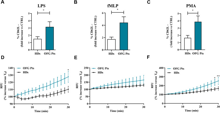
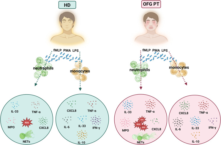
Methods: Considering that microbial agents have been suggested as potential triggers for OFG, in this study we evaluated the effect of LPS, fMLP and PMA on the activation of monocytes and PMNs purified by 11 OFG patients and 11 sex-and age-matched healthy donors (HDs).

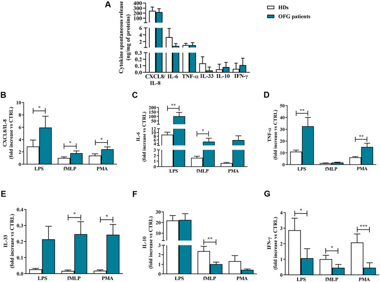
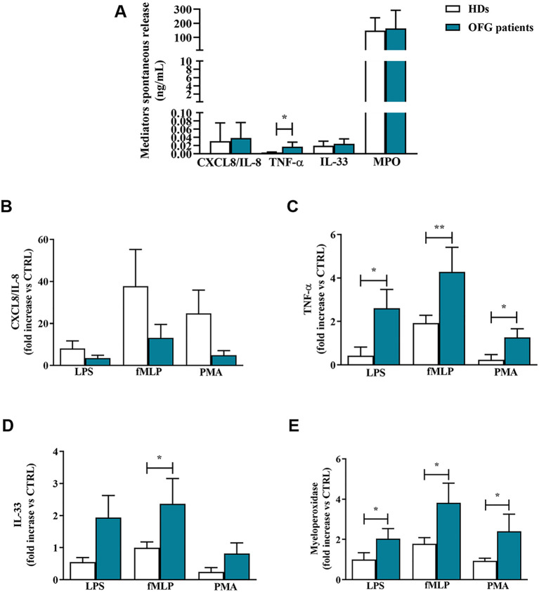
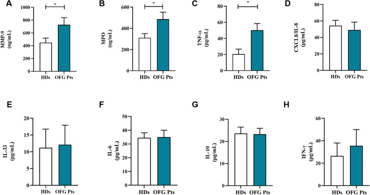
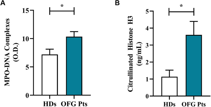
Results: Upon stimulation, OFG-derived monocytes displayed a higher release of pro-inflammatory cytokines (CXCL8/IL-8, IL-6, TNF-α, IL-33) compared to HDs. Conversely, OFG-derived monocytes showed a lower release of IL-10, IFN-γ compared to HDs. Upon stimulation, peripheral PMNs from OFG patients released large amounts of TNF-α and MPO compared to HDs. In addition, OFG-derived PMNs showed high percentages of activated PMNs (CD62L-) and increased ROS production compared to HDs. Compared to HDs, OFG patients presented higher serum levels of MMP-9, MPO and TNF-α, together with MPO-DNA complexes and citrullinated histone H3 (CitH3) (two biomarkers for neutrophil extracellular traps).

Discussion: These preliminary data suggest that in presence of various stimuli, monocytes and PMNs of OFG patients displayed an activated phenotype compared to HDs. Unraveling the interplay between bacterial triggers and immune cell function in OFG will be necessary to elucidate mechanisms driving this complex disease and identify novel therapeutic targets for improved management of OFG patients.

Keywords: NETs; ROS; bacterial stimuli; cytokines/chemokines; immunopathogenesis; inflammation.


Conflict of interest statement

The authors declare that the research was conducted in the absence of any commercial or financial relationships that could be construed as a potential conflict of interest. The author(s) declared that they were an editorial board member of Frontiers, at the time of submission. This had no impact on the peer review process and the final decision.

Figures

Figure 1
Figure 1
(A) Monocytes (1 x 10⁶ cells/well) were incubated for 18 hrs at +37°C with 5% CO2 in complete medium. At the end of incubation, the concentrations of CXCL8/IL-8, IL-6, TNF-α, IL-33, IL-10, and IFN-γ proteins in supernatants were evaluated by ELISA and expressed as ng of mediators for mg of total proteins. (B-G) Monocytes (1 x 10⁶ cells/well) were incubated for 18 hrs at +37°C with 5% CO2 with complete medium, LPS (100 ng/mL), fMLP (1 µM) or PMA (10 ng/mL). CXCL8/IL-8 (B), IL-6 (C), TNF-α (D), IL-33 (E), IL-10 (F) and IFN-γ (G) proteins in supernatants were evaluated by ELISA and normalized for the total protein content in each well. Effect of LPS, fMLP, and PMA on the mediators release were expressed as fold increase vs control. Data are expressed as mean ± SD. All the experiments were run in duplicate. *p < 0.05; **p < 0.01; ***p < 0.005 vs. the same condition of respective HDs.
Figure 2
Figure 2
(A) PMNs (1.5 x 10⁵ cells/well) were incubated for 1 h at +37°C with 5% CO2, in complete medium. At the end of incubation the supernatants were harvested and the extracellular levels of CXCL8/IL-8, TNF-α, IL-33 and MPO were measured by ELISA. (B–E) PMNs (1,5 x 10⁵ cells/well) were incubated with complete medium, LPS (100 ng/mL), fMLP (1 µM) or PMA (10 ng/mL) for 1 h at +37°C with 5% CO2. At the end of incubation the supernatants were harvested and the extracellular levels of CXCL8/IL-8 (B), TNF-α (C), IL-33 (D) and MPO (E) were evaluated by ELISA. Effect of LPS, fMLP, and PMA on the mediator release was expressed as fold increase vs control. Results were expressed as mean ± SD. All the experiments were run in duplicate. *p < 0.05; **p < 0.01 vs. the same condition of respective HDs.
Figure 3
Figure 3
Serum concentrations of MMP-9 (A), MPO (B), TNF-α (C), CXCL8/IL-8 (D), IL-33 (E), IL-6 (F), IL-10 (G) and IFN-γ (H) in OFG patients (green bars) and HDs (white bars) were measured by ELISA. Results were expressed as mean ± SD. All the experiments were run in duplicate. *p < 0.05. Student’s t test or Mann-Whitney U test according to the parametric or nonparametric distribution of the variables.
Figure 4
Figure 4
Serum levels of NET biomarkers, MPO-DNA Complex (A) and CitH3 (B) in OFG patients (green bars) and HDs (white bars) were measured by two different ELISA assays. Results were expressed as mean ± SD. All the experiments were run in duplicate. *p < 0.05. Student’s t test or Mann-Whitney U test according to the parametric or nonparametric distribution of the variables.
Figure 5
Figure 5
(A-C) PMNs (1.5 x 10⁵ cells) from peripheral blood of OFG patients (green bars) and HDs (white bars) were stimulated with complete medium, LPS (100 ng/mL) (A), fMLP (1 µM) (B) or PMA (10 ng/mL) (C) for 1h at +37°C with 5% CO2, then stained for the neutrophil activation marker CD62L (A-C) and subjected to cytofluorimetric analysis. Results were expressed as mean ± SD. *p < 0.05. (D-F) PMNs (2 × 10⁶ cells) from peripheral blood of OFG patients (green line) and HDs (black line) were incubated (30 min, +37°C) with 2’,7’-dichlorodihydrofluorescein diacetate (H2DCFDA, 10 µM), washed with PBS and then stimulated with complete medium, LPS (100 ng/mL) (D), fMLP (1 µM) (E) or PMA (10 ng/mL) (F). Immediately after stimulation, PMNs were seeded in 96-well microplate (2 × 10⁵ cells/well) and were analyzed with a multimode microplate reader (EnSpire Multimode Plate reader, PerkinElmer), DCF fluorescence was measured for 30 min at 2 min intervals. The results were expressed as Relative Fluorescence Unit (RFU) and percentage increase versus time 0 (mean ± SD); All the experiments were run in duplicate. *p < 0.05; **p < 0.01.
Figure 6
Figure 6
Graphical summary showing the differential activation of PMNs and monocytes of OFG patients and HDs under bacterial stimulation.

Similar articles

References

    1. Galdiero MR, Maio F, Arcoleo F, Boni E, Bonzano L, Brussino L, et al. . Orofacial granulomatosis: Clinical and therapeutic features in an Italian cohort and review of the literature. Allergy. (2021) 76:2189–200. doi: 10.1111/all.14799 - DOI - PubMed
    1. Phillips F, Verstockt B, Sladek M, de Boer N, Katsanos K, Karmiris K, et al. . Orofacial granulomatosis associated with crohn’s disease: a multicentre case series. J Crohns Colitis. (2022) 16:430–5. doi: 10.1093/ecco-jcc/jjab158 - DOI - PubMed
    1. Tilakaratne WM, Freysdottir J, Fortune F. Orofacial granulomatosis: review on aetiology and pathogenesis. J Oral Pathol Med. (2008) 37:191–5. doi: 10.1111/j.1600-0714.2007.00591.x - DOI - PubMed
    1. Gale G, Ostman S, Saalman R, Telemo E, Jontell M, Hasseus B. Immunophenotype in orofacial granulomatosis with and without Crohn’s disease. Med Oral Patol Oral Cir Bucal. (2014) 19:e584–91. doi: 10.4317/medoral.20187 - DOI - PMC - PubMed
    1. Patel P, Barone F, Nunes C, Boursier L, Odell E, Escudier M, et al. . Subepithelial dendritic B cells in orofacial granulomatosis. Inflammation Bowel Dis. (2010) 16:1051–60. doi: 10.1002/ibd.21169 - DOI - PubMed

LinkOut - more resources