. 2025 Oct 22;46(40):4105-4119.
doi: 10.1093/eurheartj/ehaf324.

Empagliflozin enhances metabolic efficiency and improves left ventricular hypertrophy in a hypertrophic cardiomyopathy mouse model

Affiliations

Empagliflozin enhances metabolic efficiency and improves left ventricular hypertrophy in a hypertrophic cardiomyopathy mouse model

Tomas Baka et al. Eur Heart J. .

Abstract

Background and aims: Hypertrophic cardiomyopathy (HCM) is a genetic cardiac disorder characterized by left ventricular hypertrophy (LVH), diastolic dysfunction, and impaired metabolic efficiency. This study investigates the therapeutic potential of the sodium-glucose cotransporter 2 inhibitor (SGLT2i) empagliflozin (EMPA) in ameliorating these pathological features in a mouse model carrying the myosin R403Q mutation.

Methods: Male mice harbouring the R403Q mutation were treated with EMPA for 16 weeks. Multi-nuclear magnetic resonance spectroscopy (31P, 13C, and 23Na MRS), echocardiography, transcriptomic, proteomic, and phosphoproteomic profiling were utilized to assess metabolic, structural, and functional changes.

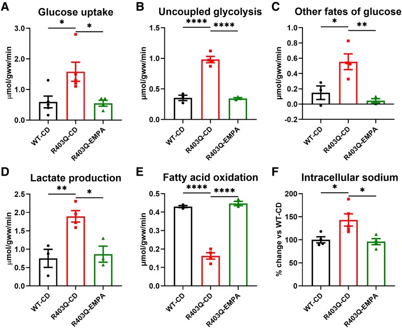
Results: Empagliflozin facilitated the coupling of glycolysis with glucose oxidation and normalized elevated intracellular sodium levels. Treatment resulted in a significant reduction in LVH and myocardial fibrosis as evidenced by echocardiography and histopathology. These structural improvements correlated with enhancements in mitochondrial adenosine triphosphate (ATP) synthesis, fatty acid oxidation, and branched-chain amino acid catabolism. Furthermore, EMPA improved left ventricular diastolic function and contractile reserve, underscored by improved ATP production and reduced energy cost of contraction. Notably, these benefits were linked to down-regulation of the mammalian target of rapamycin signalling pathway and normalization of myocardial substrate metabolic fluxes.

Conclusions: Empagliflozin significantly mitigates structural and metabolic dysfunctions in a mouse model of HCM, underscoring its potential as a therapeutic agent for managing this condition. These findings suggest broader applicability of SGLT2i in cardiovascular diseases, including those due to myocardial-specific mutations, warranting further clinical investigation.

Keywords: Branched-chain amino acids; Cardiac energetics; Cardiac metabolism; Hypertrophic cardiomyopathy; Metabolic reprogramming; SGLT2 inhibition; Uncoupled glycolysis; mTOR.


Figures

Structured Graphical Abstract
Figure 1
Figure 1
Study protocol. Mice harbouring R403Q mutation in cardiac myosin heavy chain or their wild-type littermates were randomized by body weight into three groups: (i) wild-type-control diet (WT-CD); (ii) R403Q-CD; and (iii) R403Q-EMPA. Nine-to-10-weeks-old mice were started on a control diet or the same diet enriched with empagliflozin. After 2 weeks of treatment echocardiography was performed and subset of mice was sacrificed for myocardial RNA sequencing analysis. At the end of the 16-week treatment period, mice underwent echocardiography and heart isolation followed by 31P, 13C, and 23Na NMR spectroscopy. Hearts from a separate set of animals were used for histology, western blot analysis, cardiac branched-chain amino acid levels measurement, and both proteomics and phosphoproteomics assessments. Created with BioRender.com
Figure 2
Figure 2
Cardiac proteomic and phosphoproteomic analysis performed in 25-week-old mice after 16 weeks of treatment. Cardiac proteomics showed (A) down-regulation of pathways associated with fatty acid oxidation and metabolism, pyruvate metabolism, and tricarboxylic acid and respiratory electron transport in the R403Q mice; up-regulation of pathways associated with collagen formation and the regulation of insulin-like growth factor activity in the R403Q mice. Treatment with empagliflozin countered these changes. Cardiac phosphoproteomics showed (B) down-regulation of pathways associated with oxidative phosphorylation, citric acid cycle and respiratory electron transport, adenosine triphosphate metabolic process, and regulation of the force of heart contraction. Treatment with empagliflozin countered these changes. The n = 3 (WT-CD), 4 (R403Q-CD), and 3 (R403Q-EMPA). Proteome and phosphor-proteome gene set enrichment analyses depict top differential pathways and select heatmaps with hierarchically clustered (Euclidean distance) profiles of corresponding annotated protein components. Pathways with FDR <0.05 in the R403Q-CD comparison was considered significant. Differential expression of pathways was assessed via a normalized enrichment score, in which a positive normalized enrichment score represents up-regulation and negative normalized enrichment score down-regulation in the first comparison group (e.g. positive normalized enrichment score in the plot comparing R403Q-WT signifies up-regulated in R403Q). Volcano plots are shown in Supplementary data online, Figure S2
Figure 3
Figure 3
Empagliflozin decreases glucose uptake, improves glycolysis/glucose oxidation coupling and increases fatty acid oxidation and normalizes intracellular sodium levels in R403Q hearts. R403Q heart presented increased glucose uptake (A) associated with glycolysis/glucose oxidation uncoupling (B) with increase in ‘other fates of glucose’ (C) and increased lactate production (D). Furthermore, R403Q heart showed decreased fatty acid oxidation (E) and increased levels of intracellular sodium ([Na+]i) (F). A 16-week empagliflozin treatment switched cardiac metabolism back to fatty acid oxidation (E), decreased glucose uptake (A) and improved glycolysis/glucose oxidation coupling (B) as also evidenced by reducing ‘other fates of glucose’ (C), decreasing lactate production (D), and normalizing [Na+]i (F). Uncoupled glycolysis = glycolysis-glucose oxidation. Data shown are mean ± SEM. n = 3–4. P-values were obtained by one-way analysis of variance with Bonferroni multiple comparisons tests (adjusted P-value threshold <.05). *P < .05, **P < .01, and ****P < .0001
Figure 4
Figure 4
Empagliflozin improves cardiac contractile function, energetics, and efficiency in R403Q hearts. At low workload, R403Q hearts exhibited elevated rate pressure product (A) and developed pressure (B) and similar end-diastolic pressure (C) compared with control hearts. At high workload, while control hearts increased rate pressure product (A) and developed pressure (B) without significant changes in end-diastolic pressure (C), R403Q hearts failed to increase rate pressure product (A), displayed decreased developed pressure (B), and markedly increased end-diastolic pressure (C). After 16-weeks of empagliflozin treatment, R403Q contractile reserve improved as evidenced by increased rate pressure product (A) and developed pressure (B) at high workload, while maintaining stable end-diastolic pressure (C). R403Q hearts showed impaired cardiac energetics: R403Q hearts failed to adequately increase adenosine triphosphate synthesis (E) had elevated ADP levels (D) and showed a significant reduction in the free energy of adenosine triphosphate hydrolysis (|ΔG∼ATP|) (F), which was already lower than control at low workload. Empagliflozin treatment effectively increased adenosine triphosphate synthesis (E) decreased ADP levels (D), and stabilized |ΔG∼ATP| (F) under high workload conditions. The graph (G) demonstrates the interplay between cardiac energetics and contractile function, indicating that empagliflozin treatment enabled significant increases in rate pressure product at high workload with minimal impact on |ΔG∼ATP|. Additionally, empagliflozin treatment reduced the energy cost of contraction (H) at both low workload and high workload, indicating enhanced energetic efficiency. Data shown are mean ± SEM. n = 5–9. P-values were obtained by two-way repeated measures analysis of variance with Bonferroni multiple comparisons tests (adjusted P-value threshold <.05) to analyse the contractile function and 31P NMR data from the isolated beating heart, and one-way analysis of variance with Bonferroni multiple comparisons tests (adjusted P-value threshold <.05) to analyse energy cost of contraction. *P < .05, **P < .01, ***P < .001, and ****P < .0001 to denote significant differences between study groups; ##P < .01, ###P < .001, and ####P < .0001 to denote significant differences between high workload and low workload within a study group
Figure 5
Figure 5
Empagliflozin prevents cardiac hypertrophy and improves diastolic function in R403Q hearts. R403Q hearts developed left ventricular hypertrophy as evidenced by increased total wall thickness (A) on echocardiogram and increased cardiomyocyte cross-section area (E) and fibrosis (F) in histopathology. Hypercontractility in R403Q hearts was evidenced by increased left ventricular fractional shortening (B). Furthermore, deteriorated left ventricular diastolic function was evidenced by reduced E/A ratio (C) and Em (D). A 16-week empagliflozin treatment reduced cardiac hypertrophy and fibrosis (A, E, and F). Additionally, empagliflozin enhanced left ventricular diastolic function (C and D) without affecting hypercontractility (B). Data shown are mean ± SEM. n = 6 (for echocardiography) and 8 (for histopathology). P-values were obtained by one-way analysis of variance with Bonferroni multiple comparisons tests (adjusted P-value threshold <.05). *P < .05, **P < .01, ***P < .001, and ****P < .0001. Representative photomicrographs, hematoxylin–eosin for myocyte cross-section area analysis (E) or picrosirius red for cardiac fibrosis analysis (F), transmitted light, scale bar indicates 20 µm for myocyte cross-section area analysis (E) and 50 µm for cardiac fibrosis analysis (F). Additional histopathological images are shown in Supplementary data online, Figure S9
Figure 6
Figure 6
Empagliflozin attenuates mammalian target of rapamycin pathway activation and decreases cardiac branched-chain amino acids levels in R403Q hearts. Correspondingly to hypertrophy development, p-mTOR/t-mTOR ratio (A), a key regulator of growth, and p-S6/t-S6 ribosomal protein ratio (B), its major downstream effector, were increased in R403Q hearts as shown by western blot analysis. A 16-week empagliflozin treatment attenuated mammalian target of rapamycin pathway activation (A and B) and decreased cardiac branched-chain amino acids levels (C), a well-known mammalian target of rapamycin pathway activator. Data shown are mean ± SEM. The n = 5 (for western blot) and 4–6 (for cardiac branched-chain amino acid levels measurement). Uncropped western blot images are depicted in Supplementary data online, Figure S13. P-values were obtained by one-way analysis of variance with Bonferroni multiple comparisons tests (adjusted P-value threshold <.05). ns, not significant. *P < .05 and **P < .01, and ****P < .0001

References

    1. Maron BJ, Desai MY, Nishimura RA, Spirito P, Rakowski H, Towbin JA, et al. Diagnosis and evaluation of hypertrophic cardiomyopathy: JACC state-of-the-art review. J Am Coll Cardiol 2022;79:372–89. 10.1016/J.JACC.2021.12.002 - DOI - PubMed
    1. Marian AJ, Braunwald E. Hypertrophic cardiomyopathy: genetics, pathogenesis, clinical manifestations, diagnosis, and therapy. Circ Res 2017;121:749–70. 10.1161/CIRCRESAHA.117.311059 - DOI - PMC - PubMed
    1. Toepfer CN, Garfinkel AC, Venturini G, Wakimoto H, Repetti G, Alamo L, et al. Myosin sequestration regulates sarcomere function, cardiomyocyte energetics, and metabolism, informing the pathogenesis of hypertrophic cardiomyopathy. Circulation 2020;141:828–42. 10.1161/CIRCULATIONAHA.119.042339 - DOI - PMC - PubMed
    1. Sequeira V, Bertero E, Maack C. Energetic drain driving hypertrophic cardiomyopathy. FEBS Lett 2019;593:1616–26. 10.1002/1873-3468.13496 - DOI - PubMed
    1. Yotti R, Seidman CE, Seidman JG. Advances in the genetic basis and pathogenesis of sarcomere cardiomyopathies. Annu Rev Genomics Hum Genet 2019;20:129–53. 10.1146/ANNUREV-GENOM-083118-015306 - DOI - PubMed

MeSH terms