. 2025 Dec;14(1):2509742.
doi: 10.1080/22221751.2025.2509742. Epub 2025 Jun 16.

Pathogenicity and transmissibility of bovine-derived HPAI H5N1 B3.13 virus in pigs

Affiliations

Pathogenicity and transmissibility of bovine-derived HPAI H5N1 B3.13 virus in pigs

Taeyong Kwon et al. Emerg Microbes Infect. 2025 Dec.

Abstract

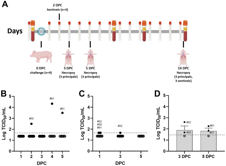
Since the first emergence of highly pathogenic avian influenza (HPAI) H5N1 viruses in dairy cattle, the virus has continued to spread, reaching 17 states and at least 1000 dairy herds in the United States. Subsequently, spillovers of the virus from dairy cattle to humans have been reported. Pigs are an important host in influenza ecology because they serve as a mixing vessel in which novel reassortant viruses with pandemic potential can be generated. Here, we show that oro-respiratory infection of pigs resulted in productive replication of a bovine-derived HPAI H5N1 B3.13 virus. Infectious virus and viral antigen were mainly identified in the lower respiratory tract of principal infected pigs, and sero-conversion was observed in most of the principal pigs at later time points, suggesting replication of the bovine-derived HPAI H5N1 B3.13 virus in pigs. In one animal, we detected the emergence of a mutation in the hemagglutinin (HA) previously associated with increased affinity for "mammalian-type" α2,6-linked sialic acid receptors, but this mutation did not reach majority consensus levels. Sentinel contact pigs remained sero-negative throughout the study, indicating lack of transmission. These results support that pigs are susceptible to a bovine-derived HPAI H5N1 B3.13 virus, but this virus did not replicate as robustly in pigs as swine-adapted influenza viruses.

Keywords: Cattle; H5N1 genotype B3.13; highly pathogenic avian influenza; mammalian-like mutation; pathogenicity; pig; transmissibility.


Conflict of interest statement

The J.A.R. laboratory received support from Tonix Pharmaceuticals, Xing Technologies, Esperovax, and Zoetis, outside of the reported work. J.A.R. is inventor on patents and patent applications on the use of antivirals and vaccines for the treatment and prevention of virus infections, owned by Kansas State University.

Figures

Figure 1.
Figure 1.
Study design and virus replication in pigs infected with a bovine-derived HPAI H5N1 B3.13 virus. (A) Nine principals were challenged with 5 × 106 TCID50 per pig via intra-tracheal (2 mL), intranasal (1 mL), and oral (1 mL) routes. Three sentinels were commingled with principals in a single pen at 2 DPC. Six pigs were randomly selected and humanely euthanized at 3 (n = 3, pig ID: #50, #52, and #54) and 5 DPC (n = 3, #51, #53, and #61), and the remaining three principals (#57, #59, and #60) and three sentinels (#55, #56, and #58) were humanely euthanized at 14 DPC. Nasal swabs, oropharyngeal swabs, and serum were collected throughout the study. Nasal swab (B), and oropharyngeal swab (C), and bronchoalveolar lavage fluid or BALF (D). Virus titration was performed on MDCK cells and virus-infected cells were visualized by immunofluorescence assay. The positive samples were shown as closed circles with pig ID; open circles represent below the limit of detection (LOD). The dashed line is the LOD of assay (3.29 × 101 TCID50/mL for BALF and 4.64 × 101 TCID50/mL for swabs).
Figure 2.
Figure 2.
Macroscopic (A) and microscopic (B) lung scores in pigs infected with a bovine-derived HPAI H5N1 B3.13 virus. Macroscopic lung scores were calculated based on the percentage of the surface area showing lobular atelectasis and hyperemia. Microscopic scores were calculated by the average scores of six different criteria.
Figure 3.
Figure 3.
Histological features in lungs of pigs infected with a bovine-derived HPAI H5N1 B3.13 virus. Histologically, pulmonary lesions were characterized by multifocal to coalescing interstitial pneumonia with limited necrotizing bronchiolitis. At 3 DPC (A and lower right inset), multifocal bronchopulmonary segments were characterized by infrequent bronchiolar epithelial cell degeneration and necrosis (lower right inset) and associated areas of pulmonary parenchyma with collapsed alveolar spaces (atelectasis) and expanded alveolar septa by lymphocytes and histiocytes (A) and alveolar epithelial necrosis (A-lower right inset). Influenza A virus nucleoprotein intracytoplasmic antigen was detected within pneumocytes and alveolar macrophages (B) and segmentally or individually within bronchiolar epithelial cells (C). Infrequent yet similar epithelial necrosis was noted at 5 DPC (E, upper and lower insets), which is accompanied by multifocal to coalescing interstitial pneumonia characterized by alveolar septal thickening and cellular debris filling alveolar spaces (E and F). Influenza A virus nucleoprotein antigen was much less abundant and mostly in cells lining and within alveoli (G). At 14 DPC, there are multifocal to coalescing areas of mild to moderate interstitial pneumonia. Alveolar septa are thickened by infiltrates of lymphocytes and macrophages and there is multifocal thickening of lobular septa with non-suppurative inflammation (H and I). Sporadic epithelial cells in bronchioles and alveoli as well as septal macrophages contained viral antigen (J). Magnifications of images: 20× for A, E and H, 100× for B, C, F, G, I and J. Magnifications of inserts: 400× for A, E and H and 100× for lower right (A).
Figure 4.
Figure 4.
Antibody responses in pigs infected with a bovine-derived HPAI H5N1 B3.13 virus. Virus neutralization (VN) test (A) and commercial NP-based competition ELISA (B). (A) The virus neutralization test was performed with the H5N1 challenge strain on MDCK cells to determine the 50% inhibition of virus growth. (B) Commercial NP-based competition ELISA (BioStone, USA) was used to determine serological responses. The positive samples were shown as closed circles with pig ID; open circles represent negative samples. The dashed line is the initial dilution for the VN test (1:10) and the cut-off value of the ELISA, < 40%: negative, 40-50%: doubtful, > 50% positive.
Figure 5.
Figure 5.
Majority (variants at ≥50% frequency) and minority (variants <50% frequency) variants across the influenza virus genome in pigs infected with a bovine-derived HPAI H5N1 B3.13 virus. Nonsynonymous and synonymous variants are shown in relation to our analytical pipeline’s standard reference sequence, A/Bovine/Texas/24-029328-01/2024. Amino acid location and identity are labeled. We used the isolate A/Cattle/Texas/063224-24-1/2024 (EPI_ISL_19155861) in this study. The original cattle isolate sequence as deposited in GISAID (GISAID sequence) and the virus stock used to inoculate pigs (inoculum) are also shown in the figure. The inoculum was propagated on bovine uterine epithelial cells for 3 passages at Cornell University and then propagated on MDCK cells for 1 passage at Kansas State University prior to inoculating animals in this study. The following sites did not meet the iSNV frequency threshold and are therefore not shown in the heatmap – PB2 R175R in pig 50 (2 DPC NS) and pig 51 (5 DPC BALF), and NA Q5Q in pig 52 (1 DPC OP). NS = nasal swab, BALF = bronchoalveolar lavage fluid, and OP = oropharyngeal swab.

Update of

References

    1. Stieneke-Grober A, Vey M, Angliker H, et al. Influenza virus hemagglutinin with multibasic cleavage site is activated by furin, a subtilisin-like endoprotease. EMBO J. 1992;11(7):2407–2414. doi: 10.1002/j.1460-2075.1992.tb05305.x - DOI - PMC - PubMed
    1. World Health Organization . Cumulative number of confirmed human cases for avian influenza A(H5N1) reported to WHO, 2003-2023, 5 January 2023. 2023.
    1. WHO/OIE/FAO H5N1 Evolution Working Group. Toward a unified nomenclature system for highly pathogenic avian influenza virus (H5N1) . Emerg Infect Dis. 2008;14(7):e1. doi: 10.3201/eid1407.071681 - DOI - PMC - PubMed
    1. World Health Organization . Evolution of the influenza A(H5) haemagglutinin: WHO/OIE/FAO H5 Working Group reports a new clade designated 2.3.4.4 2015. Available from: https://www.who.int/publications/m/item/evolution-of-the-influenza-a(h5)....
    1. Adlhoch C, Fusaro A, Gonzales JL, et al. Avian influenza overview April - June 2023. EFSA J. 2023 Jul;21(7):e08191. - PMC - PubMed

MeSH terms

Substances