. 2025 Jun 17;99(6):e0035425.
doi: 10.1128/jvi.00354-25. Epub 2025 May 21.

Cullin 3-mediated ubiquitination restricts enterovirus D68 replication and is counteracted by viral protease 3C

Affiliations

Cullin 3-mediated ubiquitination restricts enterovirus D68 replication and is counteracted by viral protease 3C

Yan Li et al. J Virol. .

Abstract

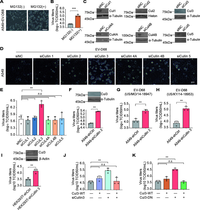
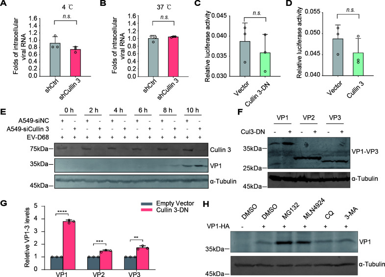
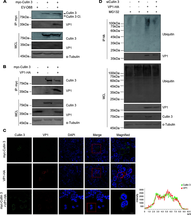
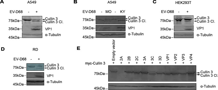
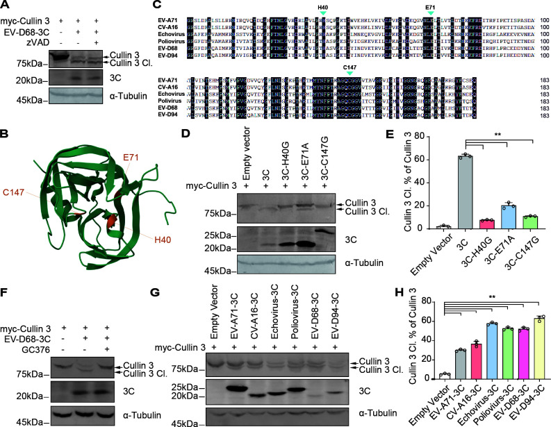
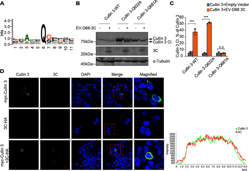
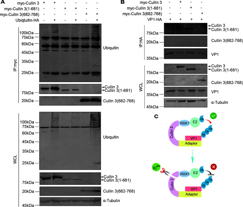
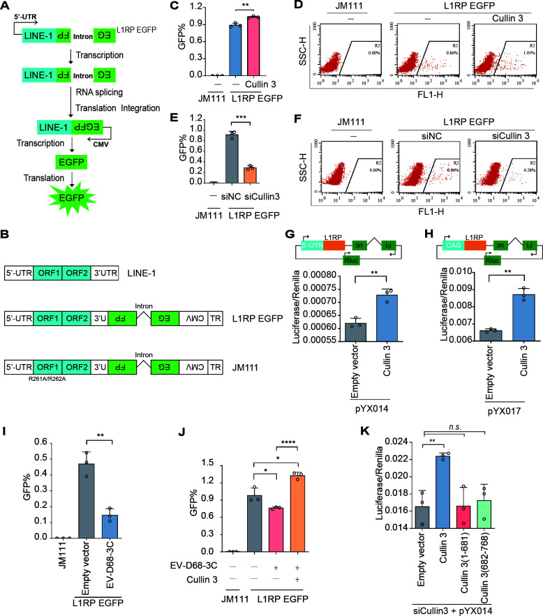
Enterovirus D68 (EV-D68) has emerged as a significant threat to public health because of its association with respiratory illnesses and neurological complications, including acute flaccid myelitis. However, the molecular mechanisms underlying EV-D68 replication and pathogenesis remain unclear. Here, we revealed a novel interaction between EV-D68 and the host Cullin-RING E3 ligase system, specifically Cullin 3, which was reported to restrict viral replication. We initially demonstrated that proteasome inhibition enhanced EV-D68 replication, suggesting an important role for the ubiquitin-proteasome system in viral restriction. Cullin 3 was further identified as a key factor that inhibits EV-D68 replication, and the downregulation of its expression increased viral titers. Mechanistically, Cullin 3 was observed to target the viral capsid protein VP1 for ubiquitination and degradation. However, EV-D68 was determined to utilize its protease 3C to cleave Cullin 3 at the Q681 residue, thereby inhibiting E3 ligase activity and facilitating resistance to Cullin 3-mediated VP1 degradation. This study uncovered a host-virus arms race, wherein the ubiquitin-proteasome system of the host actively targets viral proteins for degradation, and viral proteases counteract this defense mechanism. Accordingly, these findings could lead to more effective antiviral treatments.

Importance: The ubiquitin-proteasome system (UPS) is a critical cellular pathway involved in the regulation of protein stability and has been implicated in the regulation of viral infections. However, its role in EV-D68 infection has not been extensively explored. Our study proves that the host UPS, through the scaffold protein Cullin 3, can restrict EV-D68 replication, representing a previously unrecognized antiviral mechanism. Furthermore, we describe a viral strategy used to evade this host defense mechanism comprising Cullin 3 cleavage, which has broad implications for understanding virus-host interactions and could inform the development of novel therapeutic strategies against EV-D68 and other enteroviruses.

Keywords: Cullin 3; enterovirus D68; protein cleavage; protein degradation; ubiquitination.


Conflict of interest statement

The authors declare no conflict of interest.

Figures

Fig 1
Fig 1
Cullin 3, a scaffold protein for Cullin-RING E3 ligases, promotes EV-D68 replication. (A) CPEs in EV-D68-infected A549 cells. Cells were treated with MG132 (10 μΜ) or DMSO 36 h after infection with the EV-D68 virus (Fermon, MOI = 0.01). CPEs were observed 48 h post-infection. (B) Titers of progeny virions. Supernatants were gathered 48 h post-infection, and viral titers were measured via a standard plaque assay. Data are expressed as the mean ± standard deviation (SD). ***P < 0.001. (C) Validation of Cullin family member knockdown efficiency using immunoblotting. (D) CPEs after the Cullin family member knockdown in A549 cells. A549 cells pre-transfected with siRNAs were infected with EV-D68 (Fermon, MOI = 0.01). CPEs were observed 48 h post-infection. (E) Viral titers of progeny virions in Cullin family member-knockdown A549 cells. Viral titers were determined via a standard plaque assay. Data are expressed as the mean ± SD. **P < 0.01. n.s., not significant. (F through H) Viral titers in stable Cullin 3-silenced A549 cells. A549 cells were infected with the EV-D68 prototype Fermon (2014) and isolated US/MO/14-18947 (MO) and US/KY/14-18953 (KY) at an MOI of 0.01. Viral titers were determined using a standard plaque assay 48 h post-infection. Data are expressed as the mean ± SD. **P < 0.01. (I) Viral titers in stable Cullin 3-knockdown HEK293T cells. Stable Cullin 3-knockdown or control HEK293T cells were infected with EV-D68 (Fermon, MOI = 0.01). CPEs were observed 48 h post-infection. Data are expressed as the mean ± SD. **P < 0.01. (J, K) Viral titers in Cullin 3-inhibited HEK293T cells. HEK293T cells were transfected with siRNAs or dominant-negative Cullin 3. Wild-type Cullin 3 or an empty vector was co-transfected. Cells of all groups were infected with EV-D68 (Fermon, MOI = 0.01) 24 h post-transfection. Viral titers were determined 48 h post-infection. Data are expressed as the mean ± SD. **P < 0.01. n.s., not significant.
Fig 2
Fig 2
Cullin 3 negatively regulates EV-D68 VP1 protein level. (A and B) Virus attachment and entry assays. Stable Cullin 3-knockdown A549 cells were incubated with EV-D68 (Fermon, MOI = 1) at 4 ℃ or 37 ℃ for 2 h, and cells were washed with DMEM to remove unbound viruses. Viral RNA was quantified using qRT-PCR. Data are expressed as the mean ± SD. n.s., not significant. (C and D) EV-D68 5′ UTR activity. Plasmids with the EV-D68 5′ UTR were transfected into HEK293T cells, along with dominant-negative Cullin 3 (C) or wild-type Cullin 3 (D) plasmids. Control groups were transfected with a control vector. Renilla luciferase activity served as an internal control. The activities of Firefly and Renilla luciferases were measured 48 h post-transfection. The bar graph represents the ratio of Firefly and Renilla luciferase. Data are expressed as the mean ± SD. n.s., not significant. (E) EV-D68 VP1 levels in Cullin 3-knockdown A549 cells. A549 cells pre-transfected with the indicated siRNAs were infected with EV-D68 (Fermon, MOI = 0.05). Cells were harvested 2, 4, 6, 8, and 10 h after infection. VP1 expression levels were detected using an immunoblotting assay. (F) VP1–VP3 protein levels in Cullin 3-inhibited HEK293T cells. HEK293T cells were transfected with dominant-negative Cullin 3 or control vectors and VP1–VP3 plasmids. VP1–VP3 levels were measured 48 h after transfection. (G) The abundance of VP1–VP3 was quantified using ImageJ software. Data are expressed as the mean ± SD. ****P < 0.0001. ***P < 0.001. **P < 0.01. (H) Immunoblot analysis of VP1 level. HEK293T cells pre-transfected with the VP1 plasmids were treated with MG132 (10 μΜ), MLN4924 (2 μΜ), CQ (50 μΜ), or 3-MA (5 mΜ). The VP1 levels were measured 12 h after treatment.
Fig 3
Fig 3
Cullin 3 promotes EV-D68 VP1 ubiquitination. (A) Analysis of VP1 in Cullin 3-pull-down precipitates. HEK293T cells pre-transfected with Cullin 3 or empty control constructs were infected with EV-D68 (Fermon, MOI = 0.01). Cells were harvested 48 h post-infection and analyzed via co-IP and immunoblotting. (B) Analysis of VP1 in Cullin 3-pull-down precipitates. HEK293T cells pre-transfected with Cullin 3 or empty control constructs, along with VP1-expressing vectors, were harvested 48 h post-transfection and analyzed using co-IP and immunoblotting. (C) Subcellular localization of Cullin 3 and VP1. HEK293T cells were transfected with Cullin 3 and VP1 plasmids for 48 h. The cells were then analyzed by performing immunofluorescence and confocal microscopy. Scale bar, 10 µm. (D) VP1 ubiquitination analysis. HEK293T cells pre-transfected with siRNAs were transfected with EV-D68 VP1 or empty control vectors. Cells were treated with MG132 (10 μΜ) for 12 h before harvesting and lysis.
Fig 4
Fig 4
EV-D68 infection triggers the cleavage of Cullin 3 via the enzymatic activity of 3C. (A, B) Analysis of the host Cullin 3 level. A549 cells were infected with the EV-D68 prototype Fermon (2014) (A) or the isolated US/MO/14-18947 (MO) and US/KY/14-18953 (KY) (B) at an MOI of 0.01. Cullin 3 was detected via immunoblotting 48 h post-infection. (C and D) Immunoblotting analysis of Cullin 3. HEK293T and RD cells were infected with EV-D68 (Fermon, MOI = 0.01). Cullin 3 was detected 48 h post-infection. (E) Screening of Cullin 3 cleavage by EV-D68-encoded proteins. HEK293T cells were transfected with myc-tagged Cullin 3-expressing vectors and indicated EV-D68-encoded proteins. Cell lysates were analyzed 48 h post-transfection.
Fig 5
Fig 5
Enterovirus 3C with conserved enzyme activity sites induces Cullin 3 cleavage. (A) Cullin 3 cleavage treatment with zVAD. HEK293T cells transfected with Cullin 3 and EV-D68 3C plasmids were cultured in a medium containing Z-VAD-FMK (20 µmol/L) for 48 h. (B) Structure of EV-D68 3C protease. (C) Sequence alignment of the enteroviral 3C proteases. H40, E71, and C147 indicate the conserved enzyme activity sites. (D) Cullin 3 cleavage by 3C protease-defective mutants. (E) Relative intensities of cleaved Cullin 3 were quantified using ImageJ software. Data are expressed as the mean ± SD. **P < 0.01. (F) Cullin 3 cleavage treatment with GC376. HEK293T cells pre-transfected with Cullin 3 and EV-D68 3C vectors were cultured in a medium containing GC376 (1 µmol/L) for 24 h. (G) Cullin 3 cleavage by the indicated enterovirus 3C proteases. (H) Relative abundances of cleaved Cullin 3, quantified using ImageJ software. Data are expressed as the mean ± SD. **P < 0.01.
Fig 6
Fig 6
EV-D68 cleaves Cullin 3 at the Q681 residue. (A) Sequence logo analysis of the predicted EV-D68 3C protease cleavage site. (B) Cullin 3 mutants cleaved by EV-D68 3C. HEK293T cells were transfected with EV-D68 3C and the indicated myc-tagged Cullin 3 plasmids. Cells were harvested 48 h post-transfection. Cullin 3 was detected via immunoblotting. (C) Relative densities of cleaved Cullin 3 quantified using ImageJ software. Data are expressed as the mean ± SD. ***P < 0.001. (D) Subcellular localization of Cullin 3 and EV-D68 3C. HEK293T cells pre-transfected with Cullin 3 and 3C plasmids were cultured for 48 h. The subcellular localization of these proteins was examined by performing immunofluorescence and confocal microscopy. Scale bar, 10 µm.
Fig 7
Fig 7
Cullin 3 cleavage disrupts VP1 ubiquitination. (A) Immunoblotting analysis of Cullin 3-mediated ubiquitination. HEK293T cells were transfected with the indicated vectors. Whole-cell lysates and myc pull-down products were obtained from HEK293T cell lysates. (B) Analysis of Cullin 3 and its cleaved products in VP1-pull-down precipitates. HEK293T cells were transfected with the indicated vectors. Whole-cell lysates and HA pull-down products were analyzed. (C) Working model illustrating how VP1 interacts with Cullin 3.
Fig 8
Fig 8
EV-D68 3C suppresses L1 mobilization via Cullin 3 cleavage. (A) Schematic representation of EGFP reporter-based retrotransposition assay. A positive signal can only be detected after L1 has been transcribed, spliced, translated, reverse-transcribed, and integrated into the host genome. (B) Schematic of LINE-1 plasmids. (C, D) L1 retrotransposition mediated by Cullin 3 overexpression, as determined via flow cytometry. HEK293T cells were transfected with L1RP EGFP plasmids, along with Cullin 3 or an empty vector. Transfected cells were selected with puromycin (3 µg/mL for 2 days) 48 h post-transfection. The percentage of GFP (+) cells was measured using flow cytometry. The data are expressed as the mean ± SD. **P < 0.01. (E, F) L1 mobility following Cullin 3 knockdown based on flow cytometry. Data are expressed as the mean ± SD. ***P < 0.001. (G, H) L1 mobility was determined using a dual luciferase assay. HEK293T cells were transfected with pYX014 or pYX017 plasmids, and L1 mobility was measured using a dual-luciferase assay after 4 days. Data are expressed as the mean ± SD. **P < 0.01. (I) L1 mobilization mediated by the EV-D68 3C protease. (J) L1 transposition in EV-D68 3C-overexpressing HEK293T cells. HEK293T cells were transfected with L1RP EGFP plasmids, along with EV-D68 3C or an empty vector. Cullin 3 or an empty vector was co-transfected. L1 mobility was measured using flow cytometry after 4 days. The data are expressed as the mean ± SD. *P < 0.1. ****P < 0.0001. (K) L1 mobility in Cullin 3-knockdown HEK293T cells. HEK293T cells were transfected with siCullin 3 and pYX014. Cullin 3, Cullin 3 (1–681 aa), Cullin 3 (682–768 aa), or an empty vector were co-transfected. L1 mobility was measured using a dual-luciferase assay after 4 days. The data are expressed as the mean ± SD. **P < 0.01. n.s., not significant.

Similar articles

References

    1. Jorgensen D, Grassly NC, Pons-Salort M. 2025. Global age-stratified seroprevalence of enterovirus D68: a systematic literature review. The Lancet Microbe 6:100938. doi: 10.1016/j.lanmic.2024.07.001 - DOI - PubMed
    1. Petroski MD, Deshaies RJ. 2005. Function and regulation of cullin-RING ubiquitin ligases. Nat Rev Mol Cell Biol 6:9–20. doi: 10.1038/nrm1547 - DOI - PubMed
    1. Swatek KN, Komander D. 2016. Ubiquitin modifications. Cell Res 26:399–422. doi: 10.1038/cr.2016.39 - DOI - PMC - PubMed
    1. Deshaies RJ, Joazeiro CAP. 2009. RING domain E3 ubiquitin ligases. Annu Rev Biochem 78:399–434. doi: 10.1146/annurev.biochem.78.101807.093809 - DOI - PubMed
    1. Lydeard JR, Schulman BA, Harper JW. 2013. Building and remodelling Cullin–RING E3 ubiquitin ligases. EMBO Reports 14:1050–1061. doi: 10.1038/embor.2013.173 - DOI - PMC - PubMed

MeSH terms

LinkOut - more resources