. 2025 Jun 17;99(6):e0055525.
doi: 10.1128/jvi.00555-25. Epub 2025 May 21.

Unraveling the cross-talk between a highly virulent PEDV strain and the host via single-cell transcriptomic analysis

Affiliations

Unraveling the cross-talk between a highly virulent PEDV strain and the host via single-cell transcriptomic analysis

Yanan Wang et al. J Virol. .

Abstract

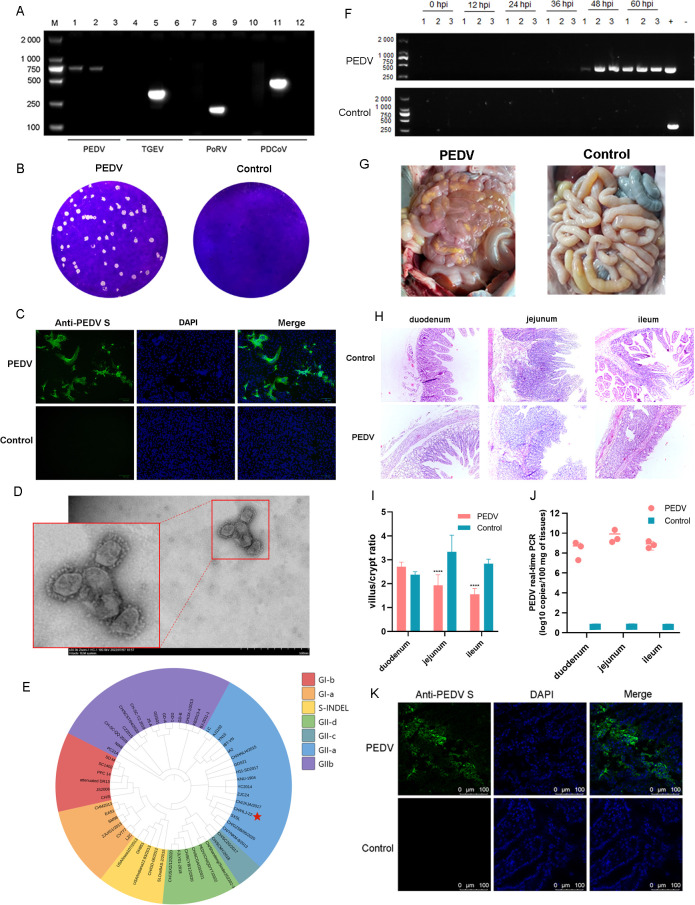
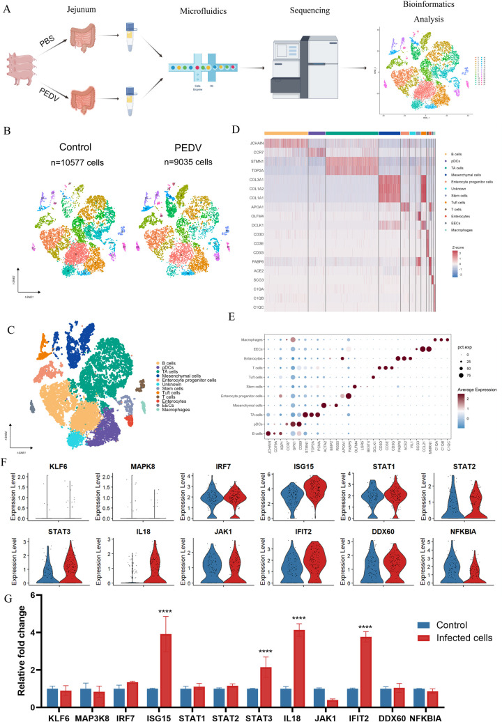
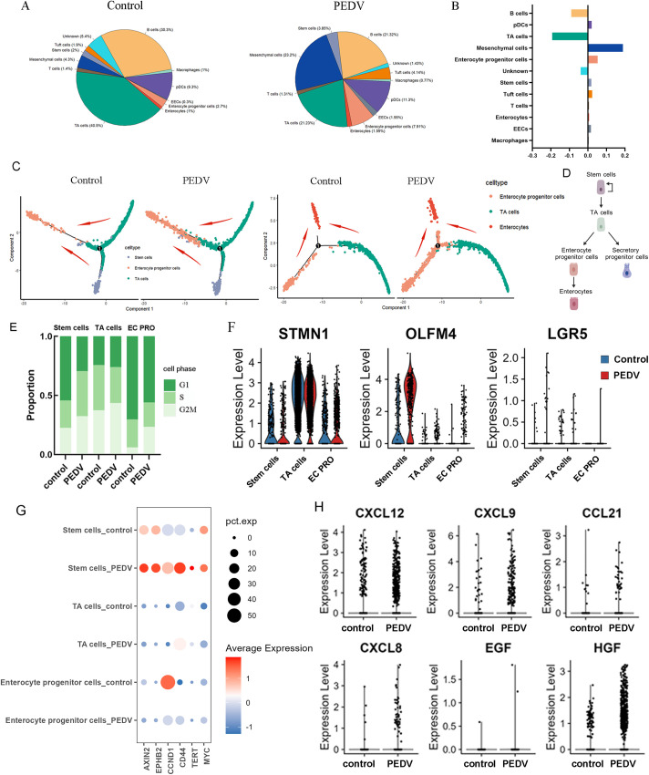
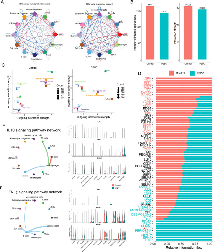
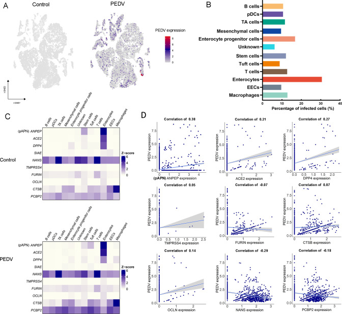
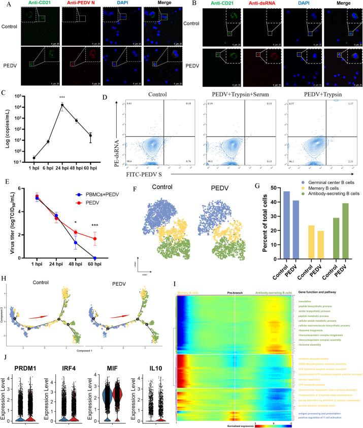
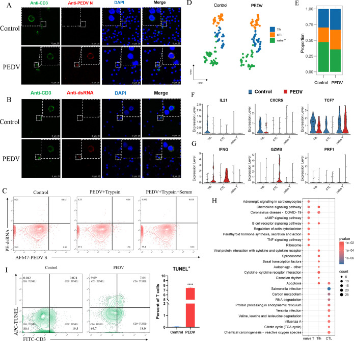
Porcine epidemic diarrhea virus (PEDV) causes severe intestinal damage and high mortality in neonatal piglets. The continuous emergence of new strains has brought new challenges to prevention and control. In this study, we isolated and characterized a prevalent PEDV virulent strain and analyzed 19,612 jejunal cells from PEDV-infected and control piglets using single-cell sequencing, revealing significant changes in cellular composition, gene expression, and intercellular communication. In response to PEDV infection, epithelial repair was enhanced through increased proliferation and differentiation of stem cells, transit-amplifying (TA) cells, and intestinal progenitor cells into enterocytes. Additionally, PEDV disrupted intercellular communication, compromising epithelial functionality while triggering immune responses, with IFN-γ and IL-10 signaling activation acting as critical regulators of immune balance and tissue homeostasis. Beyond enterocytes, viral genes were detected in various other cell types. Further experiments confirmed that PEDV could initiate replication in B and T lymphocytes but was unable to produce infectious progeny, with T cells additionally undergoing virus-induced apoptosis. These findings provide new insights into PEDV tropism, immune evasion, and epithelial repair, revealing complex host-pathogen interactions that shape disease progression and tissue regeneration, thereby contributing to a better understanding of enteric coronavirus pathogenesis.IMPORTANCEThe persistent circulation of porcine epidemic diarrhea virus (PEDV) poses a major threat to the swine industry, with emerging strains complicating prevention and control efforts. Currently, no effective measures completely prevent virus transmission, highlighting the need to understand PEDV-host interactions. In this study, we isolated a prevalent virulent strain and used single-cell sequencing to identify new PEDV-infected cell types and explore the complex interplay between the host and PEDV. These findings provide essential insights into viral pathogenesis and facilitate the design of targeted antiviral interventions.

Keywords: PEDV; host-pathogen interactions; intestinal regeneration; lymphocyte infection; single-cell RNA-seq.


Conflict of interest statement

The authors declare no conflict of interest.

Figures

Fig 1
Fig 1
Isolation and identification of the CH/HLJ-22 strain. (A) Detection of PEDV by PCR in tissue samples from pig farms. (B) Plaque morphology of the isolated PEDV strain. (C) Cytopathic effects (CPEs) of PEDV in Vero cells. (D) Image of PEDV particles by electron microscopy. (E) Genotyping of the PEDV based on the S gene. (F) Detection of PEDV in feces in PEDV-infected and control piglets. (G) The pathogenicity of PEDV in 3-day-old piglets. (H) Histopathological examination of intestinal tissues from piglets infected with CH/HLJ-22 or treated with DMEM. (I) Ratio of villus height to crypt depth in different experimental groups across three intestinal segments. The ratio was calculated by measuring the villus height and crypt depth in tissue sections from the control and PEDV-infected groups in the duodenum, jejunum, and ileum. Significant differences between the groups are indicated by asterisks (P < 0.05). Data are presented as the mean ± SEM (n = 5 per group). (J) Viral copies in the small intestine tract tissues of the PEDV-infected and control piglets. (K) Immunofluorescent detection of PEDV antigen in the jejunum of PEDV-infected piglets.
Fig 2
Fig 2
Single-cell transcriptomic analysis reveals the impact of PEDV on jejunal tissues in piglets. (A) Overview of the experimental design and bioinformatics analysis workflow. (B) tSNE of 19,612 intestinal cells from uninfected and infected piglets. (C) tSNE plot showing cell types identified in the control and PEDV-infected group. (D) Heatmap indicating the differential expression of genes specific to cell types in intestinal structural and immune cells. Colors ranging from blue to red indicate gene expression levels from low to high. (E) Dot plot showing the mean expression of marker genes used to classify cell clusters. The color intensity represents gene expression levels, whereas dot size indicates the fraction of cells expressing the gene within each cell type. (F) Vlnplot showing the expression levels of selected genes in enterocytes from control (blue) and PEDV-infected (red) groups, based on single-cell RNA-seq data. (G) qRT-PCR analysis of the same set of genes in porcine ileal epithelial cells (IPI-2I cells) confirmed their transcriptional changes upon PEDV infection. Results are shown as relative fold changes compared with controls. Data represent mean ± SD from three independent experiments. ****P < 0.0001 (unpaired two-tailed Student’s t-test)
Fig 3
Fig 3
PEDV infection enhances stem cell differentiation. (A) Pie chart showing the proportion of each cell type in PEDV-infected and control groups. (B) Bar chart displaying the percentage difference in each cell type between infected and control groups. (C) Differentiation pseudotime trajectory analysis of stem cells, TA cells, and enterocyte progenitor cells, and the red arrow indicates the direction of differentiation. (D) This schematic diagram illustrates the differentiation trajectory. from stem cells to transit-amplifying (TA) cells and from enterocyte progenitor cells to enterocytes. (E) Histogram indicating the different cell-cycle quantity of stem cells, TA cells, and EC PRO, in PEDV infection and control groups. (F) Expression of cell cycle markers in the control and infected stem cells, TA cells, and EC PRO. (G) DotPlot indicates the expression of well-established WNT signaling pathway target genes in stem cells, TA cells, and EC PRO. (H) VlnPlot indicates the expression of chemokines and growth factors in the mesenchymal cells.
Fig 4
Fig 4
Cell-cell communications between control and PEDV-infected intestine. (A) Interaction network count plot (left) and interaction weight plot (right) of each cell type in infected compared and groups. Thicker lines indicate a higher number of interactions and greater interaction strength between cell types. Blue lines represent reduced communication in the PEDV-infected group, whereas red lines indicate increased communication relative to the control group. (B) Comparison of the total number of cell communication interactions and the intensity of interactions. (C) Scatter plot showing the primary signaling sources and targets in PEDV-infected and control groups. Colors represent cell types, and dot size reflects the number of significantly expressed receptor-ligand interactions. (D) Ranking of significant signaling pathways based on differences in overall information flow within inferred networks between PEDV infections. (E) Inferred IL-10 intercellular communication network shown in a circular plot (left). Thicker lines indicate a higher number of interactions and greater interaction strength between cell types. Expression levels of IL-10 pathway genes in each cell type are compared between PEDV-infected (blue) and control (red) groups. Normalized expression levels are visualized in a violin plot (right). (F) Inferred IFN-γ intercellular communication network shown in a circular plot (left). Thicker lines indicate a higher number of interactions and greater interaction strength between cell types. Expression levels of IFN-γ pathway genes in each cell type are compared between PEDV-infected (blue) and control (red) groups. Normalized expression levels are visualized in a violin plot (right).
Fig 5
Fig 5
Determinants of cell tropism of PEDV in the porcine intestinal tract. (A) tSNE visualization of scRNA-seq data from PEDV-infected intestinal cells. Infected cells are marked as triangles, with colors representing the corrected, targeted, and normalized expression levels of PEDV based on scRNA-seq data. (B) Proportion of PEDV-infected cells across different cell types. (C) Heatmap showing the expression levels of coronavirus receptors in control and PEDV-infected groups. (D) Correlation analysis between PEDV gene expression and coronavirus receptor expression across all infected cells.
Fig 6
Fig 6
PEDV is capable of infecting B cells yet fails to establish effective replication or affect B cell differentiation. (A) Immunofluorescence staining for PEDV N protein in B cells infected with PEDV at 24 hpi. Scale bar, 25 µm. (B) Immunofluorescence staining for dsRNA in B cells infected with PEDV at 24 hpi. Scale bar, 25 µm. (C) Quantification of PEDV subgenomic RNA levels in PEDV-infected PBMCs at different time points post-infection. (D) Infection rates of B cells at 24 hpi were determined by FACS analysis in the presence or absence of serum. (E) Replication curve of PEDV with mean of virus titers (in TCID50/mL) in supernatants from cultures of PBMCs. (F) Subclustering of B cells in the PEDV infection and control group. (G) The proportion of B cell subsets in the PEDV infection and control groups. (H) Pseudotime trajectory analysis of B cell differentiation. Predicted secretory lineage cells include GCBs, ASCs, and MBCs, with the red arrow indicating the direction of differentiation. (I) The differentially expressed genes (rows) along the pseudotime (columns) of MBCs and ASCs clusters hierarchically into three profiles. The representative gene functions and pathways of each profile are presented on the right. (J) The differential expression levels of the PRDM1, IRF4, MIF, and IL10 genes in the B cells.
Fig 7
Fig 7
The functional alterations in T cells from intestinal tissues during PEDV infection. (A) Immunofluorescence staining for PEDV N protein in T cells infected with PEDV at 24 hpi. Scale bar, 25 µm. (B) Immunofluorescence staining for dsRNA in T cells infected with PEDV at 24 hpi. Scale bar, 25 µm. (C) Infection rates of T cells at 24 hpi were determined by FACS analysis in the presence or absence of serum. (D) Subclustering of T cells in the small intestine. (E) The proportion of T cell subtypes in the PEDV infection and control groups. (F) The differential expression levels of the genes IL21, CXCR5, and TCF7 in the T cell subsets. (G) The differential expression levels of the genes IFNG, GZMB, and PRF1 in the T cell subsets. (H) The KEGG signaling pathway enrichment in T cell subclusters from PEDV-infected compared with the control group. (I) FACS analysis of apoptosis in PEDV-infected T cells. The apoptosis rate is visualized in a scatter plot (left) and quantified in a histogram (right).

Similar articles

References

    1. Fehr AR, Perlman S. 2015. Coronaviruses: an overview of their replication and pathogenesis. Methods Mol Biol 1282:1–23. doi: 10.1007/978-1-4939-2438-7_1 - DOI - PMC - PubMed
    1. Chen Y, Liu Q, Guo D. 2020. Emerging coronaviruses: genome structure, replication, and pathogenesis. J Med Virol 92:418–423. doi: 10.1002/jmv.25681 - DOI - PMC - PubMed
    1. Wang Q, Vlasova AN, Kenney SP, Saif LJ. 2019. Emerging and re-emerging coronaviruses in pigs. Curr Opin Virol 34:39–49. doi: 10.1016/j.coviro.2018.12.001 - DOI - PMC - PubMed
    1. Jung K, Saif LJ. 2017. Goblet cell depletion in small intestinal villous and crypt epithelium of conventional nursing and weaned pigs infected with porcine epidemic diarrhea virus. Res Vet Sci 110:12–15. doi: 10.1016/j.rvsc.2016.10.009 - DOI - PubMed
    1. Jung K, Eyerly B, Annamalai T, Lu Z, Saif LJ. 2015. Structural alteration of tight and adherens junctions in villous and crypt epithelium of the small and large intestine of conventional nursing piglets infected with porcine epidemic diarrhea virus. Vet Microbiol 177:373–378. doi: 10.1016/j.vetmic.2015.03.022 - DOI - PMC - PubMed

MeSH terms

LinkOut - more resources