Review
. 2025 Jul 11;26(13):e202500156.
doi: 10.1002/cbic.202500156. Epub 2025 Jun 13.

Methods for Controlling Small GTPase Activity

Affiliations
Review

Methods for Controlling Small GTPase Activity

Benjamin Faulkner et al. Chembiochem. .

Abstract

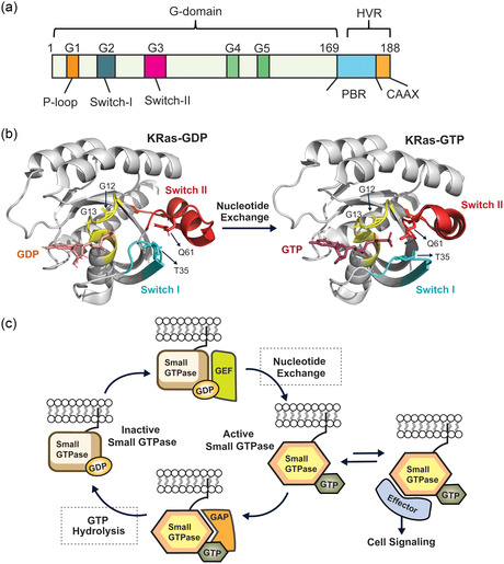
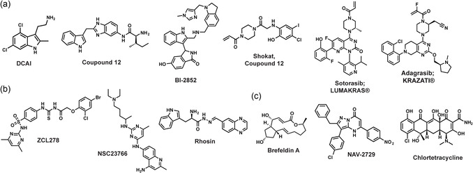
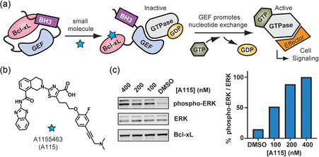
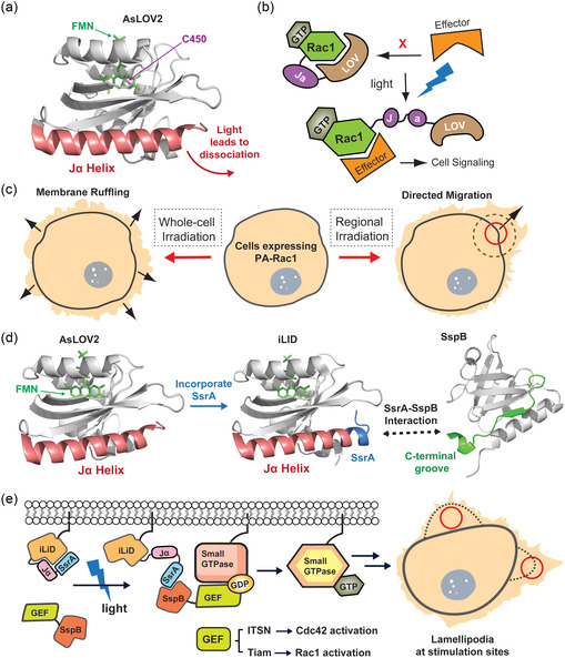
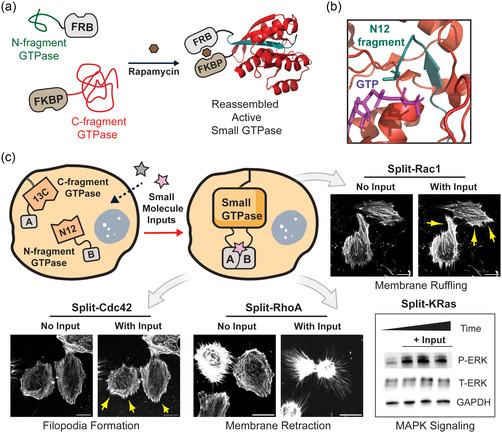
Small GTPases comprise a diverse class of signaling proteins in mammalian cells and regulate a variety of cellular processes such as cell growth, cell movement, vesicle formation, and nuclear transport. Due to their involvement in critical cellular pathways, changes in the activation state of small GTPases due to genetic mutations or alterations in gene expression can lead to human diseases. As such, the ability to control the activity of small GTPases is paramount in understanding the precise role these proteins play in human biology and in reducing their impacts on related diseases. Herein, important advances made in the development of small-molecule- and protein engineering-based strategies to control the activity of small GTPases are presented. Current approaches within each area are discussed within their historical contexts along with commentary on the importance that each technology has had on improving the ability to regulate small GTPase activity. Given this ever-evolving toolbox for controlling small GTPase signaling, continued growth in the study of this protein class is anticipated.

Keywords: inhibitors; protein engineering; signal transductions; small GTPases; split proteins.


Conflict of interest statement

The authors declare no conflict of interest.

Figures

Figure 1
Figure 1
General structure and signaling mechanism of small GTPases. a) The small GTPase superfamily shares a conserved sequence and structure, featuring both a core “GTPase domain” with five essential “G‐motifs” (G1–G5) critical for GTPase function and a hypervariable region (HVR) containing a polybasic region (PBR) and a CAAX motif, which undergoes post‐translational modifications necessary for subcellular localization. The cartoon represents the sequence from KRas. b) Crystal structures of KRas GTPases in GDP‐bound (PDB: 8ECR) and GTP‐bound (PDB: 6VJJ) states. Key domains are highlighted, including the P‐loop, Switch I, and Switch II regions. Critical residues for GDP/GTP binding, such as Gly12 and Gly13 in the P‐loop, Thr35 in Switch I, and Gln61 in Switch II, are indicated. c) The small GTPase activation cycle, showing the role of guanine nucleotide exchange factors (GEFs) in facilitating GTP loading, and GTPase‐activating proteins (GAPs) in promoting GTP hydrolysis to GDP.
Figure 2
Figure 2
Structures for small‐molecule inhibitors of small GTPases belonging to the a) Ras subfamily, b) Rho subfamily, and c) Arf subfamily.
Figure 3
Figure 3
Illustration of a small‐molecule‐regulated GEF. a) In its inactive (closed) state, the GEF is autoinhibited by an interaction between the antiapoptotic protein Bcl‐xL and a BH3 peptide. Upon the addition of a small‐molecule activator (e.g., A115), the Bcl‐xL–BH3 interaction is disrupted, allowing the GEF to catalyze the exchange of GDP for GTP on the small GTPase. b) Chemical structure of A115. c) Dose‐dependent activation of the Ras/ERK signaling pathway CIAR, a chemically inducible activator of Ras in which the GEF in panel a is SOScat. A representative Western blot shows ERK phosphorylation in HEK 293 cells expressing CIAR, treated with increasing concentrations of A115. Quantification of blot intensity from two biological replicates demonstrates a dose‐dependent control of ERK phosphorylation. Panel (c) reproduced with permission from Elsevier.[ 86 ]
Figure 4
Figure 4
Light‐activated small GTPase signaling. a) Crystal structure of AsLOV2 (PDB: 7PGX), a photoactivatable protein switch in Avena sativa phototropin1. b) Schematic representation of the PA‐Rac1 design. In the dark state, the LOV domain inhibits the interaction between Rac1 and its effector protein. Upon irradiation, the Jα‐helix unwinds, enabling Rac1 to bind its effector and become active. c) Representative cartoon of HeLa cells expressing constitutively active PA‐Rac1 and phenotypic changes induced by whole‐cell or regional illumination. d) Crystal structure of an engineered, improved light‐inducible dimerization (iLID) system (PDB: 4WF0) developed by the Kuhlmann group, compared with AsLOV2 (PDB: 7PGX). The LOV2 domain is modified at its C‐terminus with an SsrA peptide. Exposure to blue light induces unwinding of the Jα‐helix, facilitating the binding of SsrA to its partner, SspB (PDB: 1OX8). e) Schematic of the iLID‐SspB system, designed to recruit GEF proteins to activate endogenous small GTPases.
Figure 5
Figure 5
Overview of the split‐small GTPase system. a) A small GTPase is fragmented into two inactive components, each fused to rapamycin‐dependent interaction domains, FRB and FKBP. Addition of rapamycin increases the local concentration of these complementary fragments, allowing reassembly into an active GTPase (PDB: 2ODB). b) Crystal structure of the nucleotide‐binding region of Cdc42 (PDB: 2ODB), highlighting the fragmentation site between the N‐terminal fragment (green) and C‐terminal fragment (red). c) This split‐Cdc42 reassembly approach can be applied, without the need for case‐by‐case optimization to numerous small GTPases (e.g., Cdc42, Rac1, RhoA, and KRas). Each new system can be used to gate the activity of the respective small GTPase within living cells.

Similar articles

References

    1. Stryer L., Bourne H. R., Annu. Rev. Cell Dev. Biol. 1986, 2, 391. - PubMed
    1. Bourne H. R., Sanders D. A., McCormick F., Nature 1990, 348, 125. - PubMed
    1. a) Harvey J. J., Nature 1964, 204, 1104; - PubMed
    2. b) Kirsten W. H., Mayer L. A., J. Natl. Cancer Inst. 1967, 39, 311; - PubMed
    3. c) Chang E. H., Gonda M. A., Ellis R. W., Scolnick E. M., Lowy D. R., Proc. Natl. Acad. Sci. U. S. A 1982, 79, 4848. - PMC - PubMed
    1. Vetter I. R., Wittinghofer A., Science 2001, 294, 1299. - PubMed
    1. a) Leipe D. D., Wolf Y. I., Koonin E. V., Aravind L., J. Mol. Biol. 2002, 317, 41; - PubMed
    2. b) Yin G., Huang J., Petela J., Jiang H., Zhang Y., Gong S., Wu J., Liu B., Shi J., Gao Y., Signal Transduct. Target. Ther. 2023, 8, 212. - PMC - PubMed

MeSH terms

Substances

LinkOut - more resources