. 2025 Apr 14;37(1-3):2.
doi: 10.1007/s44445-025-00003-4.

Resolvin E1 and calvarial defects in rats: a comprehensive histological analysis

Affiliations

Resolvin E1 and calvarial defects in rats: a comprehensive histological analysis

Sara Alrumaih et al. Saudi Dent J. .

Abstract

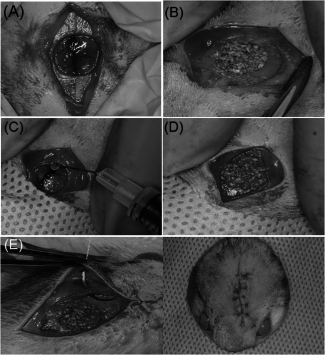
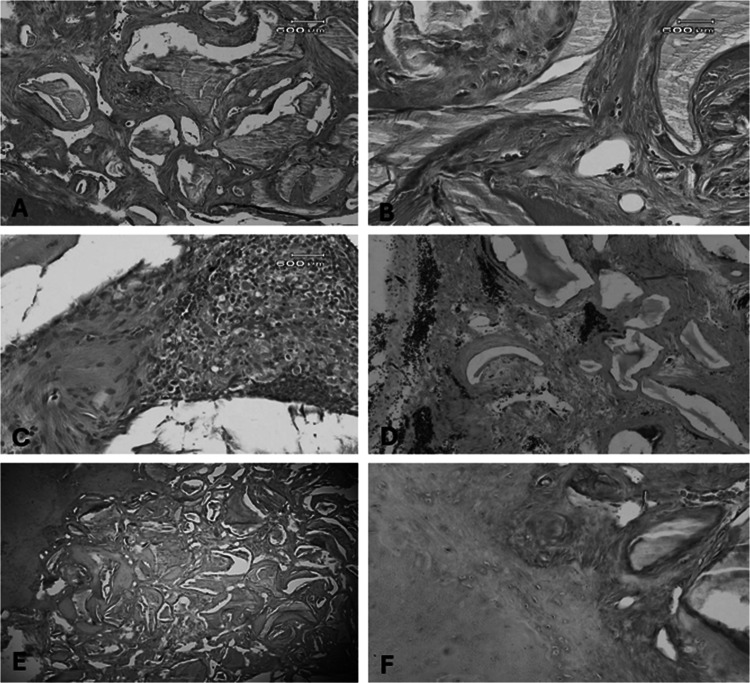
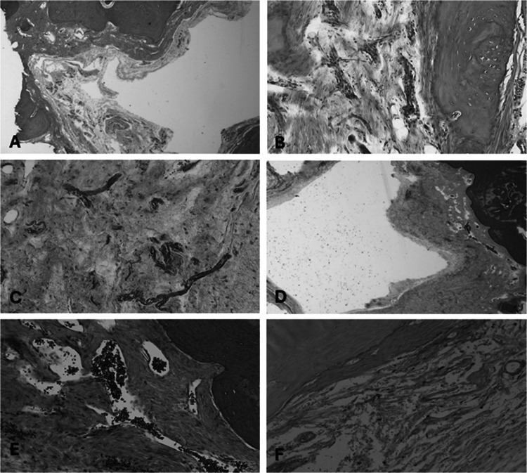
Bone loss, linked with numerous oral conditions such as periodontal diseases and periimplantitis, poses a significant challenge for dental clinicians. The current study evaluated the in vivo effects of local application of Resolvin E1 (RvE1) on bone regeneration in critical size calvarial defects in rats. Thirty female Wistar rats with 5 mm induced calvarial defects were randomly allocated into four groups: no treatment (negative control, n = 5), treatment using bovine bone grafts (positive control, n = 5), treatment using local delivery of RvE1 (n = 11) and treatment using RvE1 mixed with bovine bone graft (n = 9). After 12 weeks, the animals were sacrificed and the calvarial defects with the adjacent tissues were sectioned en-bloc and prepared for histological examination. A comprehensive qualitative and quantitative histological examination was performed to assess bone regeneration and its relation to the defect, the presence of remnant bone and RvE1 particles, the integration of the native bone with the newly formed bone, bone density and bony trabeculae, the inflammatory reaction, the connective tissue bridging in the defect, and the encapsulating fibrous tissue. Signs of neovascularization, increased cellularity, lack of the organized lamellated appearance of mature bone, disorganized arrangement of osteocytes, osteoblasts and osteoclasts were also assessed. Comparisons of the quantitative values between all studied groups indicated statistically significant differences (p ≤ 0.05) in all parameters except for the increased cellularity and granulation tissue. Histological findings indicate that RvE1 with adjunct bone graft significantly enhanced the bone formation compared to RvE1 or bovine graft alone.

Keywords: Bone graft; Bone regeneration; Calvarial bone defects; Resolvin E1.


Conflict of interest statement

Declarations. Competing interests: The authors declare no conflict of interest. Ethics and consent to participate: All procedures were performed in compliance with relevant laws and institutional guidelines and have been approved by the research ethics committee at King Saud University (KSU-SE-22–33).

Figures

Fig. 1
Fig. 1
Surgical protocol. A: The animal was placed in the prone position, and the scalp was disinfected with a povidone-iodine solution and shaved; B: A cutaneous straight incision at midline (15 mm in length) was made along the sagittal suture over the parietal bone of the scalp; C and D: The periosteum was intact after reflection of the skin; E: Creation of a full thickness standardized calvarial defect; F: Removal of full-thickness bone (including the outer and inner cortices) without damaging the dura mater
Fig. 2
Fig. 2
Treatment procedure. A: Negative control group, no treatment; B: Positive control group, placement of a bovine bone graft over the bone defect; C: Local delivery of Resolvin E1; D: Resolvin E1 in combination with bovine xenograft; E: Periosteum and flap repositioning; F: suturing using resorbable sutures
Fig. 3
Fig. 3
The en-bloc sectioned area of the surgical defect with the adjacent tissues
Fig. 4
Fig. 4
Negative Control Group – A: H&E X 40 showing incomplete closure with limited areas of new bone formation surrounded with fibrous connective tissue; B: H&E X 200 Higher magnification showing areas with severe inflammation within the wound; C: Masson Trichrome stain X 40 incomplete closure and new bone integrated with native bone surrounded with dense connective tissue fibers; D: Alizarin Red stain X 400 areas of positively stained new bone formation integrated with native bone surrounded with fibrous connective tissue around the periphery
Fig. 5
Fig. 5
Positive Control Group– A: H&E × 100 showing the defect space filled with new bone formation surrounding the graft material; B: H&E × 400 showing the new bony trabeculae embedded with osteocytes surrounded with fibrous connective tissue around the bone graft areas; C: Higher magnification showing the new bony trabeculae embedded with osteocytes filling the defect, inflammatory cells with extravasated blood cells are evident; D: Masson Trichrome stain × 400 showing blood vessels around the new bony trabeculae; E: Alizarin Red stain × 40 showing positively stained new bone formation integrated with native bone around remnants of the bone graft material; F: Higher magnification showing positively stained new bone formation integrated with native bone with an apparent line of demarcation and embedded osteocytes
Fig. 6
Fig. 6
RvE1 Group – A: H&E X 40 showing new bone formation integrated with native bone covered with dense connective tissue fibers and incomplete closure of defect; B and C: Higher magnifications showing neovascularization and angiogenesis; D: Masson Trichrome stain X 40 showing blood vessels, new bone formation integrated with native bone covered with dense connective tissue fibers, incomplete closure of defect; E; Masson Trichrome stain X 400 showing dense connective tissue fibers, increased vascularity covering new bone; F: Alizarin Red stain X 200 showing positively stained new bony trabeculae integrated with native bone surrounded with fibrous connective tissue around the periphery
Fig. 7
Fig. 7
RvE1 + BioOss Bone Graft Group – A: H & E × 40 showing defect filled with new bony trabeculae integrated with the native bone around material filling the defect; B: Higher magnification showing osteoblasts–like cells lining the newly formed bone and embedded osteocytes; C: H & E × 200—new bone formation integrated with native bone with areas of inflammation surrounded with densely hyalinized collagen fibres; D: Masson Trichrome stain × 100 bone formation around the graft material with complete closure of the defect; E: Masson Trichrome stain × 400 new bone formation around the resorption areas of the graft material and increased vascularization; F: Alizarin Red stain × 40 showing positively stained new bony trabeculae embedded with osteocytes integrated with the native bone filling the defect around remnants of the materials surrounded by connective tissue fibres

Similar articles

References

    1. Alrumaih S, Alshibani N, Alssum L, Alshehri FA, AlMayrifi MA, AlMayouf M, Bautista LS (2024) The impact of resolvin E1 on bone regeneration in critical-sized calvarial defects of rat model—a gene expression and micro-CT analysis. J Periodontal Res 59:195–203 - PubMed
    1. Birkedal-Hansen H (1993) Role of matrix metalloproteinases in human periodontal diseases. J Periodontol 64:474–484 - PubMed
    1. Brennan E, Kantharidis P, Cooper ME, Godson C (2021) Pro-resolving lipid mediators: regulators of inflammation, metabolism and kidney function. Nat Rev Nephrol 17(11):725–739 - PMC - PubMed
    1. Choi JY, Jung UW, Kim CS, Eom TK, Kang EJ, Cho KS ... Choi SH (2010) The effects of newly formed synthetic peptide on bone regeneration in rat calvarial defects. J Periodontal Implant Sci 40(1): 11–18 - PMC - PubMed
    1. El Kholy K, Freire M, Chen T, Van Dyke TE (2018) Resolvin E1 promotes bone preservation under inflammatory conditions. Front Immunol 9:1300 - PMC - PubMed

LinkOut - more resources