. 2025 Nov;398(11):15857-15871.
doi: 10.1007/s00210-025-04145-4. Epub 2025 May 21.

Anti-osteoporotic effect of sitagliptin in an osteoporosis model of ovariectomized rats: role of RUNX2 and RANKL/OPG ratio

Affiliations

Anti-osteoporotic effect of sitagliptin in an osteoporosis model of ovariectomized rats: role of RUNX2 and RANKL/OPG ratio

Salma Ahmed El-Marasy et al. Naunyn Schmiedebergs Arch Pharmacol. 2025 Nov.

Abstract

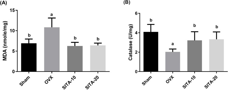
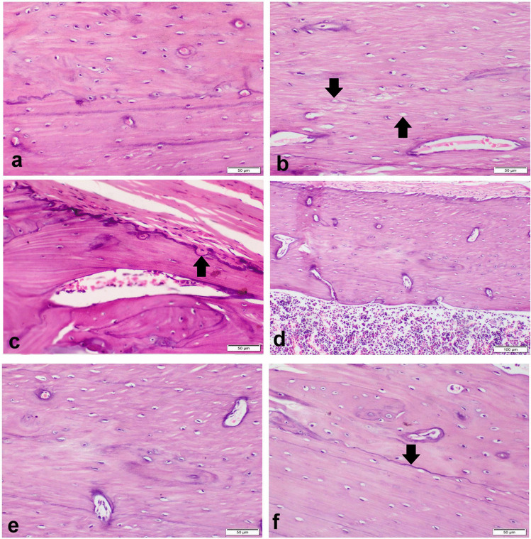
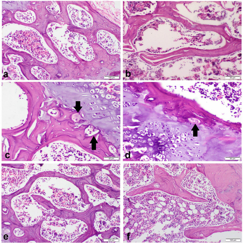
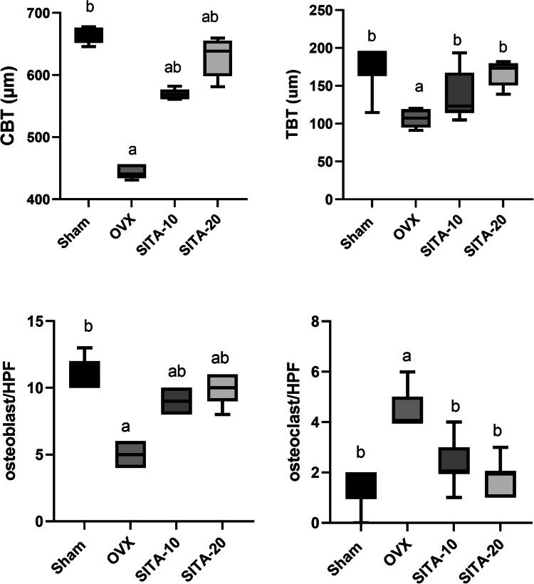
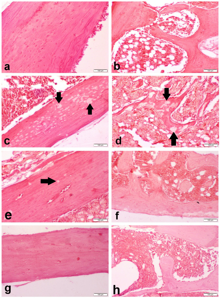
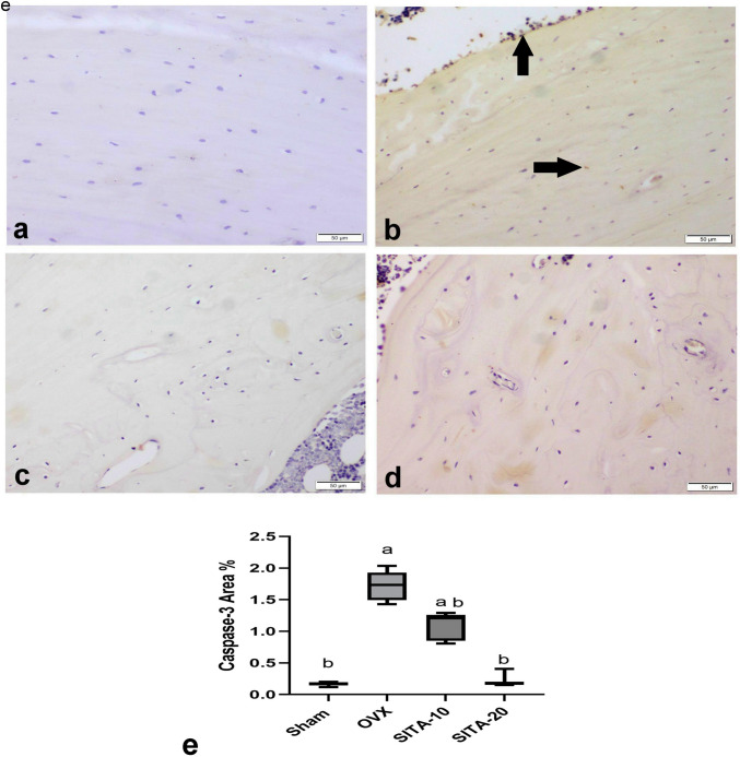
This study examines the potential anti-osteoporotic effect of sitagliptin in osteoporosis instigated by ovariectomy (OVX) in rats. Rats were assigned into 4 groups: Sham-operated, OVX group, and OVX rats orally treated with sitagliptin (10, 20 mg/kg), respectively, after 8 weeks of OVX for 4 weeks. Biochemical, real-time polymerase chain reaction, histopathological, and immunohistochemical analyses of bone resorption and formation were conducted. Sitagliptin ameliorated bone mineral density (BMD), restored calcium and phosphorus levels in OVX rats, elevated catalase and decreased malondialdehyde, reduced receptor activator of NF-κB ligand (RANKL), elevated osteoprotegerin (OPG), and reduced tartrate-resistant acid phosphatase (TRAP) femur contents. Sitagliptin mitigated variations in mRNA expressions of RUNX2 and protein kinase B (AKT) in femur tissue. Moreover, sitagliptin reduced caspase-3 protein expression and improved bone histomorphology and mechanical properties. Sitagliptin's anti-oxidant activity mediated its anti-osteoporotic effect in OVX rats via modulation of RUNX2, downregulation of RANKL/OPG, AKT pathways, apoptosis, and histomorphometry alterations revealing attenuation of osteoclastogenesis and promotion of osteoblast formation.

Keywords: Ovariectomy; RANKL/OPG; RUNX2; Rats; Sitagliptin; TRAP.


Conflict of interest statement

Declarations. Ethical approval: All animal procedures adhered to the ARRIVE guidelines and received approval from the Ethical Committee of Medical Research, National Research Centre, no. 14110112021. Consent to participate: N/A. Consent for publication: N/A. Competing interests: The authors declare no competing interests.

Figures

Fig. 1
Fig. 1
Effect of sitagliptin on A malondialdehyde and B catalase. Each bar represents the mean ± SEM of six rats in each group. avs sham rats at p ≤ 0.05, bvs OXV control rats at p ≤ 0.05. SITA-10, sitagliptin 10 mg/kg; SITA-20,sitagliptin 20 mg/kg
Fig. 2
Fig. 2
Effect sitagliptin bone mineral density of A tibia and B femur. Each bar represents the mean ± SEM of six rats in each group. avs sham rats at p ≤ 0.05, bvs OXV control rats at p ≤ 0.05. SITA-10, sitagliptin 10 mg/kg; SITA-20, sitagliptin 20 mg/kg
Fig. 3
Fig. 3
Photomicrograph of the bone shaft of the femur with H&E-stain. a Sham group showing normal architecture compact cortical bone. b OVX control group showing degenerated osteocytes with multiple empty lacunae (arrow). c OVX control group showing erosive endosteal surface (arrow). d, e SITA-10 showing a nearly normal compact bone. f SITA-20 showing a basophilic cement line (arrow) separating the borders of old and new bone matrix. SITA-10, sitagliptin 10 mg/kg; SITA-20, sitagliptin 20 mg/kg
Fig. 4
Fig. 4
Photomicrograph of cancellous bone of the femur with H&E-stain. a Sham group showing normal a network of bony trabeculae containing bone marrow. b OVX control group showing thin trabecular bones with marked widening of interconnecting spaces. c OVX control group showing erosive cavities containing osteoclasts (arrows). d, e SITA-10 normal osteocytes with regular and smooth endosteal surface. f SITA-20 showing normal cancellous bone. SITA-10, sitagliptin 10 mg/kg; SITA-20, sitagliptin 20 mg/kg
Fig. 5
Fig. 5
Morphometrical analysis of femur bone. A Cortical bone thickness. B Trabecular bone thickness. C Osteoblasts number. D Osteoclast number. Each bar represents median. avs sham rats at p ≤ 0.05, bvs OVX control rats at p ≤ 0.05. SITA-10, sitagliptin 10 mg/kg; SITA-20, sitagliptin 20 mg/kg
Fig. 6
Fig. 6
Photomicrograph of Alizarin red-stained tissue sections of the bone shaft and cancellous of the femur. a, b Sham group showing homogenous red staining calcified bone. c, d OVX control group showing multiple area of faint red decalcified bone tissues (arrow). e, f SITA-10 mg/kg showing few areas of decalcified bones (arrow). g, h SITA-20 mg/kg showing homogenous red calcified bone. SITA-10, sitagliptin 10 mg/kg; SITA-20, sitagliptin 20 mg/kg
Fig. 7
Fig. 7
Photomicrograph of MT-stained tissue sections of the bone shaft of the femur. a Sham group showing regular lamellar arrangement of blue-stained collagen fiber. b OVX control group showing marked reduction in the blue-stained collagen fiber with increase of red-stained osteoid tissue. c Sitagliptin 10 mg/kg and d sitagliptin 20 mg/kg showing marked increase in blue-stained collagen fiber. e Bar chart represents organic matrix (osteoid) area %. Values are expressed as median avs sham rats at p ≤ 0.05, bvs OXV control rats at p ≤ 0.05. SITA-10, sitagliptin 10 mg/kg; SITA-20, sitagliptin 20 mg/kg
Fig. 8
Fig. 8
Photomicrograph of Caspase-3 immunohistochemistry in shaft bone of the femur. a Sham group. b OVX control group showing Caspase-3 immune-positive cells (arrow). c SITA 10 mg/kg. d SITA 20 mg/kg. e Bar chart represents Caspase-3 immune-stained cells %. Values are expressed as median avs sham rats at p ≤ 0.05, bvs OXV control rats at p ≤ 0.05. SITA-10, sitagliptin 10 mg/kg; SITA-20, sitagliptin 20 mg/kg

References

    1. Abu-Elala NM, Abd-Elsalam RM, Marzouk MS (2015) Molecular and immunohistochemical diagnosis of Photobacterium damselae subspecies piscicida during naturally occurring disease in Egypt. J World Aquacult Soc 46:583–595
    1. Aebi H (1984) Catalase in vitro. Methods Enzymol 105:121–126. 10.1016/s0076-6879(84)05016-3 - PubMed
    1. Arafa EA, Elgendy NO, Elhemely MA, Abdelaleem EA, Mohamed WR (2023) Diosmin mitigates dexamethasone-induced osteoporosis in vivo: role of Runx2, RANKL/OPG, and oxidative stress. Biomed Pharmacother 161:114461. 10.1016/j.biopha.2023.114461 - PubMed
    1. Chen D, Wang Q, Li Y, Sun P, Kuek V, Yuan J, Yang J, Wen L, Wang H, Xu J, Chen P (2021) Notopterol Attenuates estrogen deficiency-induced osteoporosis via repressing RANKL signaling and reactive oxygen species. Front Pharmacol 12:664836. 10.3389/fphar.2021.664836 - PMC - PubMed
    1. Cheng Y, Liu P, Xiang Q, Liang J, Chen H, Zhang H, Yang L (2022b) Glucagon-like peptide-1 attenuates diabetes-associated osteoporosis in ZDF rat, possibly through the RAGE pathway. BMC Musculoskelet Disord 23:465. 10.1186/s12891-022-05396-5 - PMC - PubMed

MeSH terms