

PET/MR for predicting extranodal extension of head and neck cancer
- PMID: 40397135
- PMCID: PMC12391167
- DOI: 10.1007/s00234-025-03635-9
PET/MR for predicting extranodal extension of head and neck cancer
Abstract
Purpose: To analyze the diagnostic accuracy of multiparametric FDG-PET/MR in identifying pathologic extranodal extension (pENE) of lymph node metastases (LNM) in head and neck squamous cell carcinoma (HNSCC) patients.
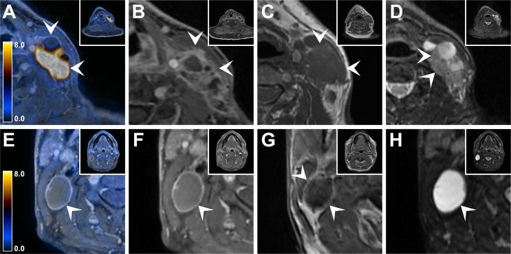
Methods and materials: Retrospective analysis of 57 HNSCC patients who underwent preoperative FDG-PET/MR imaging. PET parameters of LNM SUVmax and MTV, lymph node size as well as MR parameters flare sign, shaggy margin sign and vanishing border sign were analyzed. Histopathological assessment of neck dissection specimens served as standard of reference.
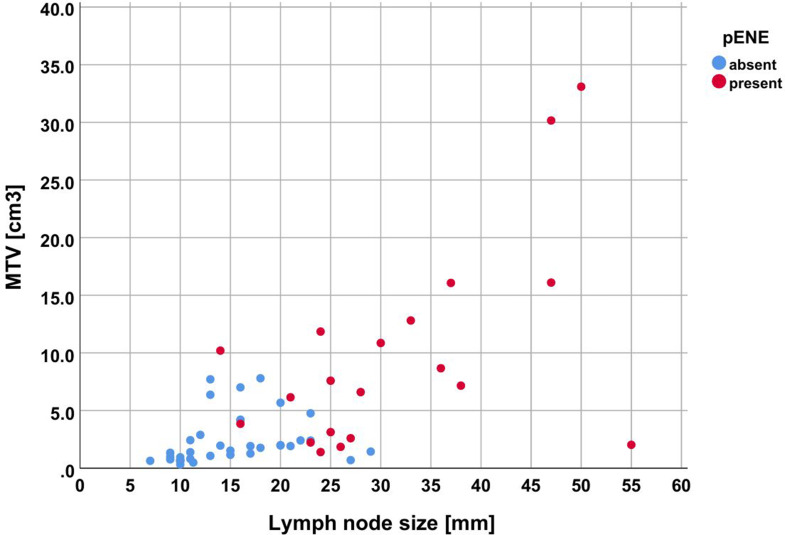
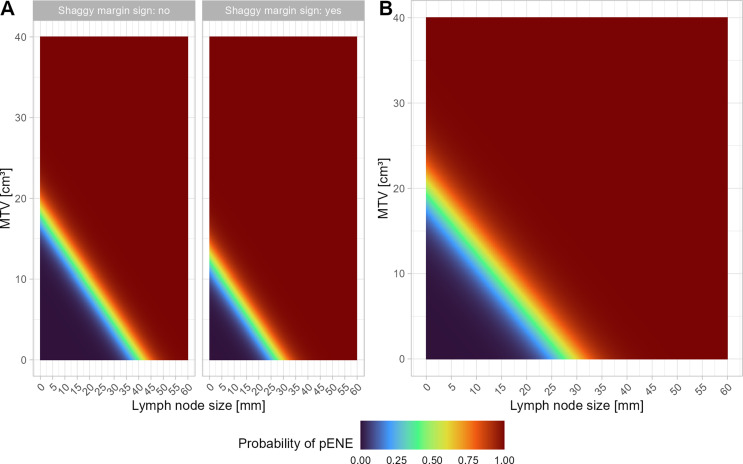
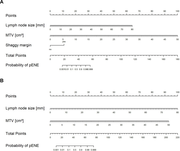
Results: A logistic regression model consisting of lymph node size (p = 0.029), shaggy margin sign (p = 0.031) and MTV (p = 0.035) proved that all three parameters significantly contributed to the prediction of pENE (χ²(3) = 54.23, p < 0.001). A second model without the reader-dependent parameter shaggy margin sign yielded similar results (χ²(2) = 45.36, p < 0.001), with every increase in lymph node size (p = 0.006) by 1 mm increasing the likelihood of pENE by a factor of 1.41 (95%-CI[1.11, 1.81]), and every increase in MTV (p = 0.023) by 1 cm3 increasing the likelihood of pENE by a factor of 1.64 (95%-CI[1.07, 2.50]). This model yielded an accuracy of 94.7% (95%-CI [85.4, 98.9]) for predicting pENE, with a specificity of 97.3% (95%-CI [85.8, 99.9]) and a sensitivity of 90.0% (95%-CI [68.3, 98.8]). Internal validation using a test dataset confirmed high accuracy of this model.
Conclusion: PET/MR-based multivariate binomial logistic regression models consisting of MTV, lymph node size and/or shaggy lymph node margins predict pENE with high accuracy.
Keywords: 2- [18F]-fluorodeoxy-D-glucose; Extranodal extension; Head and neck cancer; Hybrid imaging; PET/MR.
© 2025. The Author(s).
Conflict of interest statement
Declarations. Ethical approval: This retrospective study was performed in line with the principles of the Declaration of Helsinki. Approval for this study was obtained by the local ethics committee. Informed consent: All included patients provided written informed consent to the utilization of their medical data for research purposes. Competing interests: Dr. Stephan Skawran is supported by a grant from the Iten-Kohout Foundation, Switzerland and the Palatin-Foundation, Switzerland. Dr. Martin Huellner received grants and speaker honoraria from GE HealthCare, a fund by the Alfred and Annemarie von Sick legacy and a grant from the clinical research priority program (CRPP) Artificial Intelligence in Oncological Imaging Network of the University of Zurich.
Figures
References
-
- Hiyama T, Kuno H, Nagaki T et al (2020) Extra-nodal extension in head and neck cancer: how radiologists can help staging and treatment planning. Jpn J Radiol 38:489–506 - PubMed
-
- Joo YH, Yoo IR, Cho KJ et al (2013) Extracapsular spread and FDG PET/CT correlations in oral squamous cell carcinoma. Int J Oral Maxillofac Surg 42:158–163 - PubMed
-
- Toya R, Saito T, Matsuyama T et al (2020) Diagnostic value of FDG-PET/CT for the identification of extranodal extension in patients with head and neck squamous cell carcinoma. Anticancer Res 40:2073–2077 - PubMed
-
- Wittekindt C, Gültekin E, Weissenborn SJ et al (2005) Expression of p16 protein is associated with human papillomavirus status in tonsillar carcinomas and has implications on survival. Adv Otorhinolaryngol 62:72–80 - PubMed
MeSH terms
Substances
LinkOut - more resources
Full Text Sources
Medical
