. 2025 Jun;12(23):e2416958.
doi: 10.1002/advs.202416958. Epub 2025 May 21.

DeepCCDS: Interpretable Deep Learning Framework for Predicting Cancer Cell Drug Sensitivity through Characterizing Cancer Driver Signals

Affiliations

DeepCCDS: Interpretable Deep Learning Framework for Predicting Cancer Cell Drug Sensitivity through Characterizing Cancer Driver Signals

Jiashuo Wu et al. Adv Sci (Weinh). 2025 Jun.

Abstract

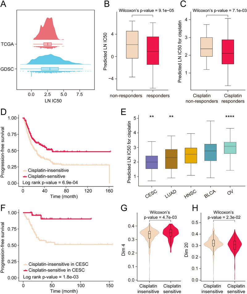
Accurate characterization of cellular states is the foundation for precise prediction of drug sensitivity in cancer cell lines, which in turn is fundamental to realizing precision oncology. However, current deep learning approaches have limitations in characterizing cellular states. They rely solely on isolated genetic markers, overlooking the complex regulatory networks and cellular mechanisms that underlie drug responses. To address this limitation, this work proposes DeepCCDS, a Deep learning framework for Cancer Cell Drug Sensitivity prediction through Characterizing Cancer Driver Signals. DeepCCDS incorporates a prior knowledge network to characterize cancer driver signals, building upon the self-supervised neural network framework. The signals can reflect key mechanisms influencing cancer cell development and drug response, enhancing the model's predictive performance and interpretability. DeepCCDS has demonstrated superior performance in predicting drug sensitivity compared to previous state-of-the-art approaches across multiple datasets. Benefiting from integrating prior knowledge, DeepCCDS exhibits powerful feature representation capabilities and interpretability. Based on these feature representations, we have identified embedding features that could potentially be used for drug screening in new indications. Further, this work demonstrates the applicability of DeepCCDS on solid tumor samples from The Cancer Genome Atlas. This work believes integrating DeepCCDS into clinical decision-making processes can potentially improve the selection of personalized treatment strategies for cancer patients.

Keywords: deep learning; drug sensitivity; feature representation; precision oncology; self‐supervised neural network.


Conflict of interest statement

The authors declare no conflict of interest.

Figures

Figure 1
Figure 1
The schematic overview of DeepCCDS.
Figure 2
Figure 2
Characterizing cancer driver signals as biological pathways. A) 38 pathways were determined based on enrichment analysis thresholds (ES > 0, FDR < 0.2) to characterize cancer driver signals. The leading‐edge subset includes cancer driver genes and other genes influenced by driver genes. B) A pathway‐gene relationship network, where each pathway is connected to its corresponding leading‐edge subset genes. The size of a pathway node indicates its degree, with larger nodes representing higher degrees.
Figure 3
Figure 3
Comprehensive performance evaluations of DeepCCDS. A–C) The correlation between predicted LN IC50 by DeepCCDS and observed LN IC50 across different datasets: (A) GDSC, (B) CCLE, and (C) NCI 60. D) Comparison of overall performance (average PCC or RMSE) in different approaches across three datasets. “Our average” refers to the mean overall performance of DeepCCDS and DeepCCDS265, while “Other average” refers to the mean overall performance of the other methods. E) The detailed PCC of different approaches in three datasets. F) Comparison of different approaches, showing the mean (bars) and standard deviation (error bars) of prediction performance for each cell line across all drugs and for each drug across all cell lines.
Figure 4
Figure 4
Analysis of embedded features generated by DeepCCDS. A,B) Comparison of (A) PCC and (B) RSEM of drug sensitivity prediction between machine learning models trained on original and embedded features. C–F) Cell distribution based on t‐SNE algorithm. The cell distribution is respectively based on original mutation features (C), original expression features (D), embedded mutation features (E), and embedded expression features (F). The cells were divided into sensitive and insensitive groups based on the quartiles of average sensitivity. G) The IG scores and activity heatmap of the top 10 pathways that are most important for the prediction of drug sensitivity.
Figure 5
Figure 5
Biological significance of mutation embedding features. A) Biological annotation of mutation embedding features. Different colored regions represent different annotation pathway categories. Annotations for dimensions 1 and 15 are highlighted with bold borders. Detailed annotation names are shown in Figure S9, Supporting Information. B) The importance (IG score) of biologically annotated feature dimensions to drug sensitivity prediction. C) Heatmap of the importance (IG scores) for each dimension associated with sensitivity of specific drugs. D) Heatmap of the correlation (PCC) between feature values across different dimensions and drug sensitivity of cells.
Figure 6
Figure 6
Correlation analysis of dimensions 1 and 15 with the specific drugs. A) The correlation between the feature values of dimension 1 and LN IC50 of cells to Vinblastine. The bar charts inside the scatter plot represent correlations within specific cell types. The red stars indicate a significant correlation between feature values in particular cell types and drug sensitivity. B) The correlation between the feature values of dimension 1 and LN IC50 of cells to Buparlisib. C) Comparison of feature values of dimension 1 across different cell types. We used a two‐sided Wilcoxon rank‐sum test to assess the differences between each group and all other patients (**** p < 1e‐4; *** 1e‐4 < p < 1e‐3; ** 1e‐3 < p < 1e‐2; * 1e‐2 < p < 5e‐2). D) The correlation between the feature values of dimension 15 and LN IC50 of cells to Trametinib. E) The correlation between the feature values of dimension 15 and LN IC50 of cells to Selumetinib. F) Comparison of feature values of dimension 15 across different cell types.
Figure 7
Figure 7
Application of DeepCCDS in solid tumor samples. A) Comparing the LN IC50 distributions of cell lines in GDSC to 25 drugs with the predicted sensitivity distributions of TCGA patients to the same 25 drugs. B) Comparing the predicted LN IC50 between all responders and non‐responders. C) Comparing the predicted LN IC50 between all responders and non‐responders of cisplatin. D) Kaplan–Meier analysis shows the PFS differences between predicted cisplatin‐sensitive and insensitive patient groups. E) Comparing predicted LN IC50 to cisplatin among patients with different types of cancer. We used a two‐sided Wilcoxon rank‐sum test to assess the differences between each group and all other patients (**** p < 1e‐4; ** 1e‐3 < p < 1e‐2). F) Kaplan–Meier analysis shows the PFS differences between predicted cisplatin‐sensitive and insensitive CESC patient groups. G,H) Comparing the feature values of dimension 4 (G) and dimension 20 (H) between predicted cisplatin‐sensitive and insensitive patient groups.

References

    1. Chawla S., Rockstroh A., Lehman M., Ratther E., Jain A., Anand A., Gupta A., Bhattacharya N., Poonia S., Rai P., Das N., Majumdar A., Jayadeva, Ahuja G., Hollier B. G., Nelson C. C., Sengupta D., Nat. Commun. 2022, 13, 5680. - PMC - PubMed
    1. Wong C. H., Siah K. W., Lo A. W., Biostatistics 2019, 20, 273. - PMC - PubMed
    1. McGranahan N., Swanton C., Cell 2017, 168, 613. - PubMed
    1. Sharifi‐Noghabi H., Zolotareva O., Collins C. C., Ester M., Bioinformatics 2019, 35, i501. - PMC - PubMed
    1. Geeleher P., Cox N. J., Huang R. S., Genome Biol. 2016, 17, 190. - PMC - PubMed

Substances

LinkOut - more resources