. 2025 Jun;12(23):e2417714.
doi: 10.1002/advs.202417714. Epub 2025 May 21.

Bioengineered Tumor-Derived Extracellular Vehicles Suppressed Colorectal Cancer Liver Metastasis and Bevacizumab Resistance

Affiliations

Bioengineered Tumor-Derived Extracellular Vehicles Suppressed Colorectal Cancer Liver Metastasis and Bevacizumab Resistance

Junjiang Wang et al. Adv Sci (Weinh). 2025 Jun.

Abstract

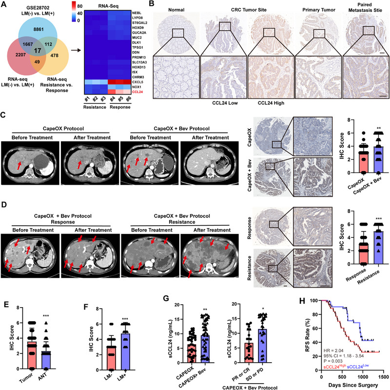
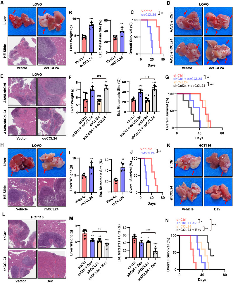
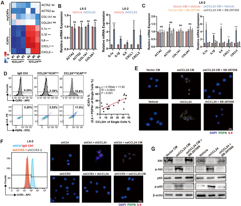
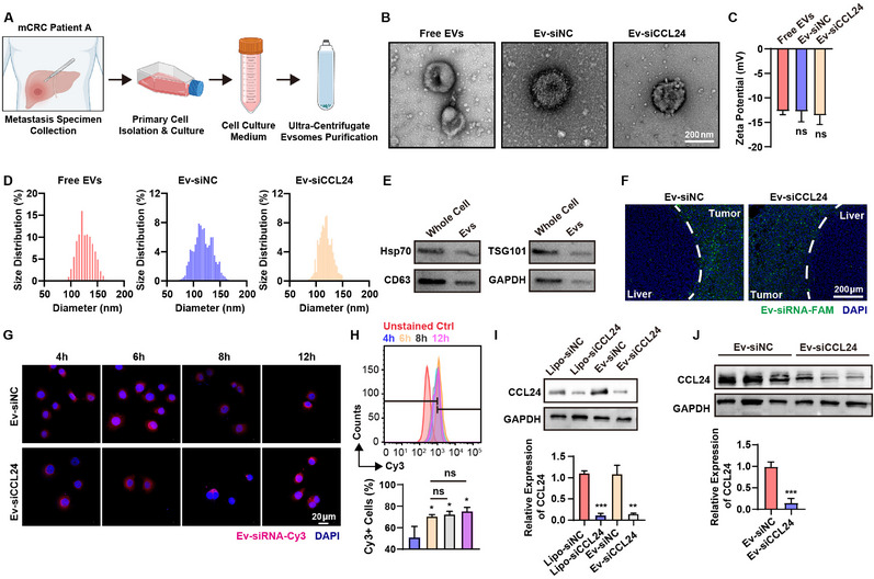
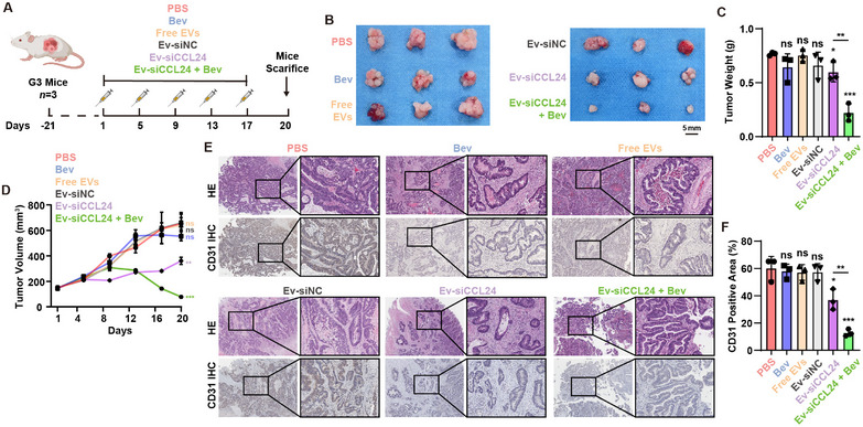
Antiangiogenic therapies, such as bevacizumab, are among the causes of cancer-related death in patients with colorectal cancer (CRC) with liver metastasis. Delivering siRNAs via primary cell originating from primary cells is a promising method for targeting CRC liver metastasis and drug resistance. Here, it is found that the expression of CCL24 is significantly upregulated in tumor tissues at the CRC liver metastasis site. In addition, CCL24 is significantly upregulated in tumor tissues from bevacizumab-resistant patients. CCL24 promotes the formation of inflammatory tumor-associated fibroblast subsets in the CRC liver metastasis microenvironment and induces resistance to bevacizumab therapy. Based on these results, a primary cell-derived extracellular vehicle delivery system is designed for the simultaneous delivery of siRNAs targeting CCL24 in the tumor microenvironment (TME). Downregulation of CCL24 in the TME by delivering bioengineered extracellular vehicles significantly increased sensitivity to antiangiogenic therapy in a CRC mouse model. A novel therapeutic target is identified for patients with CRC with liver metastasis and suggested a possible therapeutic alternative for patients with CRC with resistance to antiangiogenic therapy and distant metastasis.

Keywords: antiangiogenic therapy; colorectal cancer; engineered extracellular vehicles; liver metastasis; siRNA delivery.


Conflict of interest statement

The authors declare no conflict of interest.

Figures

Figure 1
Figure 1
The upregulated CCL24 indicated metastasic CRC Bev therapy resistance and implied poor prognosis. A) Identification of key genes for CRC in cancer metastasis and Bev therapy resistance by RNA‐sequence. The scale bar and the numbers indicates the FPKM of the genes expression. The red color implied the gene was potentially highly expression in the indicated group. The CCL24 mRNA was overexpressed in the metastasis tumor tissue and Bev‐therapy resistance tissue. B) The representative IHC photographs of the included CRC tumor tissues. Scale bar = 600µm for 20× magnification images and 100 µm for 400 × magnification images. C,D) Patients with CRC treated with Bev, whose tumor tissues highly express CCL24. CCL24 is significantly highly expressed in Bev treatment‐resistant tissues; The evaluation of therapy was measured by radiology examination. The red arrows indicate the metastatic tumors in liver. E,F) The IHC score indicated that the CCL24 was overexpressed in metastasic tumor tissue and therapy resistance tumor tissue. G) After Bev treatment, the concentration of seurm CCL24 (sCCL24) in CRC patients was significantly upregulated, and the serum CCL24 content increased in patients resistant to Bev treatment; H) The increase in sCCL24 is an independent risk factor for postoperative recurrence in CRC patients. Survival analysis was calculated using the Kaplan‐Meier method and log‐rank test. The comparison between the other two groups was calculated using an unpaired t‐test analysis; ns, not significant; *, P<0.05; **, P<0.01, ***, P<0.001 when compared to the control groups.
Figure 2
Figure 2
Tumor derived CCL24 promotes CRC distance metastasis and antiangiogenic therapy resistance in vivo. A–C) Overexpression of CCL24 in LOVO cells leads to a significant increase in tumor burden and a decrease in survival in mCRC animal models; D–G) Knocking out exogenous recombinant CCL24 chemokine, overexpression of CCL24 in LOVO cell lines significantly promotes tumor progression, increasing tumor burden in mCRC animal models, suggesting that CCL24 derived from tumor cells promotes tumor progression. H–J) Exogenous addition of circulating rhCCL24 chemokine can promote tumor progression in LOVO mCRC animal experiments, suggesting that CCL24 promotes tumor progression. K–N) Knockdown of HCT116 cell lines with high endogenous expression showed that knockdown of CCL24 in HCT116 cells significantly reduced tumor burden in mCRC animal models and enhanced the therapeutic effect of Bev treatment in mice. ns, not significant; *, P < 0.05; **, P < 0.01, ***, P < 0.001 when compared to the control groups.
Figure 3
Figure 3
HSCs differentiated as iCAFs via CCL24/CCR3/NF‐κB pathway. A) In mCRC tissues with high expression of CCL24, markers of iCAFs (IL1A, IL1B, IL6, CXCL1, CXCL5) are significantly highly expressed, indicating a high degree of iCAFs infiltration in the TME; The scale bar and the numbers indicates the FPKM of the genes expression. The red color implied the gene was potentially highly expression in the indicated group. B,C) Exogenous addition of rhCCL24 cytokine to the HSC cell line LX‐2 results in significant upregulation of iCAFs markers, while there is no significant change in myCAFs markers; D) CCL24+ cells are positively correlated with the proportion of iCAFs (IL1A+ PDPN+), suggesting that increased expression of CCL24 induces the formation of iCAFs cells in the TME. E) After exogenously adding oeCCL24 LOVO cell culture supernatant or recombinant CCL24 chemokine to the LX‐2 cell culture medium and continuing to culture for 72 h, the expression of PDPN and IL1A in LX‐2 cells is significantly upregulated. F) After knocking out CCR3 in HSC cells, exogenous addition of oeCCL24 LOVO cell culture supernatant or recombinant CCL24 chemokine does not result in high expression of CAF markers in LX‐2 cells, suggesting that CCL24 promotes the differentiation of LX‐2 into iCAFs through the CCR3 receptor. G) Adding oeCCL24 CM in LOVO cell culture supernatant to LX‐2 cells showed that the NF‐κB signaling pathway was activated. ns, not significant; *, P < 0.05; **, P < 0.01, ***, P < 0.001 when compared to the control groups.
Figure 4
Figure 4
Construction of the patient‐derived bioengineered extraceullar vehicles (EVs) and delivering the siRNAs to the tumor tissue. A) Schematic illustration of preparing primary cell culture and purifying EVs from primary cells. B) The representative TEM images of the EVs and EVs loading siRNAs. C) The zeta potential of the bioengineered EVs. D) The size distribution of the EVs. E) The western‐blot analysis was performed to verify the expression of the EV markers. F) The in vivo distribution of the bioengineered EVs in PDX mice model constructed from the same patients. The cellular uptake assays were performed by confocal microscopy (G) and flow cytometry analysis (H). Western blotting (I) and qPCR (J) assay indicated that the gene knockdown ability of the bioengineered EVs in vitro. ns, not significant; *, P < 0.05; **, P < 0.01, ***, P < 0.001 when compared to the control groups.
Figure 5
Figure 5
The anti‐tumor activity of Ev‐siRNA in CRC antiangiogenic therapy resistance in PDX mouse models. A) Schematic illustration of establishing PDX model and illustration of treatment schedule. B) The images of the resected tumors in the mice. C) The tumor weight of the mice at the endpoint of the in vivo experiments, and tumor inhibition profiles of the mice receiving different treatments. Mean ± SD (n = 3). D) The tumor volume of the mice in each group. E) The representative HE and CD31 IHC staining images among the groups. F) The estimated CD31 positive area in IHC imagews. Scale bar = 600 µm for 40× magnification images and 200 µm for 200× magnification images. ns, not significant; *, P < 0.05; **, P < 0.01, ***, P < 0.001 when compared to the control groups.
Figure 6
Figure 6
In vivo therapeutic efficacy of Exo‐siCCL24 against CRC liver metastasis. A) Schematic illustration of constructing CRC liver metastasis mice model by using the HCT116R‐mCherry cell lines and purifying EVs and the treatment protocol among the groups. B) The in vivo fluorescence of HCT116R tumor‐bearing mice at different time pointes. C) The measurement of the in vivo fluorescence intensity of HCT116R tumor‐bearing mice at different time pointes. D) The liver weight in each group at the end of the in vivo experiments. E) The estimated metastasis site in each group at the end of the in vivo experiments. H) The survival analysis of the in vivo experiments. ns, not significant; *, P < 0.05; **, P < 0.01, ***, P < 0.001 when compared to the control groups.

Similar articles

References

    1. a) Siegel R. L., Wagle N. S., Cercek A., Smith R. A., Jemal A., CA Cancer J. Clin. 2023, 73, 233; - PubMed
    2. b) Kuipers E. J., Grady W. M., Lieberman D., Seufferlein T., Sung J. J., Boelens P. G., van de Velde C. J., Watanabe T., Nat. Rev. Dis. Prim. 2015, 1, 15065. - PMC - PubMed
    1. a) Biller L. H., Schrag D., JAMA, J. Am. Med. Assoc. 2021, 325, 669; - PubMed
    2. b) Canellas‐Socias A., Sancho E., Batlle E., Nat. Rev. Gastroenterol. Hepatol. 2024, 21, 609. - PubMed
    1. Shin A. E., Giancotti F. G., Rustgi A. K., Trends Pharmacol. Sci. 2023, 44, 222. - PMC - PubMed
    1. a) Giuliante F., Vigano L., De Rose A. M., Mirza D. F., Lapointe R., Kaiser G., Barroso E., Ferrero A., Isoniemi H., Lopez‐Ben S., Popescu I., Ouellet J. F., Hubert C., Regimbeau J. M., Lin J. K., Skipenko O. G., Ardito F., Adam R., Ann. Surg. Oncol. 2021, 28, 8198; - PMC - PubMed
    2. b) Leone N., Arolfo S., Spadi R., Fortunato M. R., Passera R., Morino M., Int. J. Colorectal. Dis. 2023, 38, 169; - PubMed
    3. c) Shepherdson M., Kilburn D., Ullah S., Price T., Karapetis C. S., Nguyen P., Townsend A., Padbury R., Piantadosi C., Maddern G., Carruthers S., Roder D., Sorich M., Roy A. C., ANZ J. Surg. 2023, 93, 1847. - PubMed
    1. a) Yang M., Xu Z., Mi M., Ding Y., Pan Y., Yuan Y., Sun W., Weng S., Cancer Biol. Med. 2023, 20, 640; - PMC - PubMed
    2. b) Mi M., Weng S., Xu Z., Hu H., Wang Y., Yuan Y., Chin. J. Cancer Res. 2023, 35, 233. - PMC - PubMed

MeSH terms

LinkOut - more resources